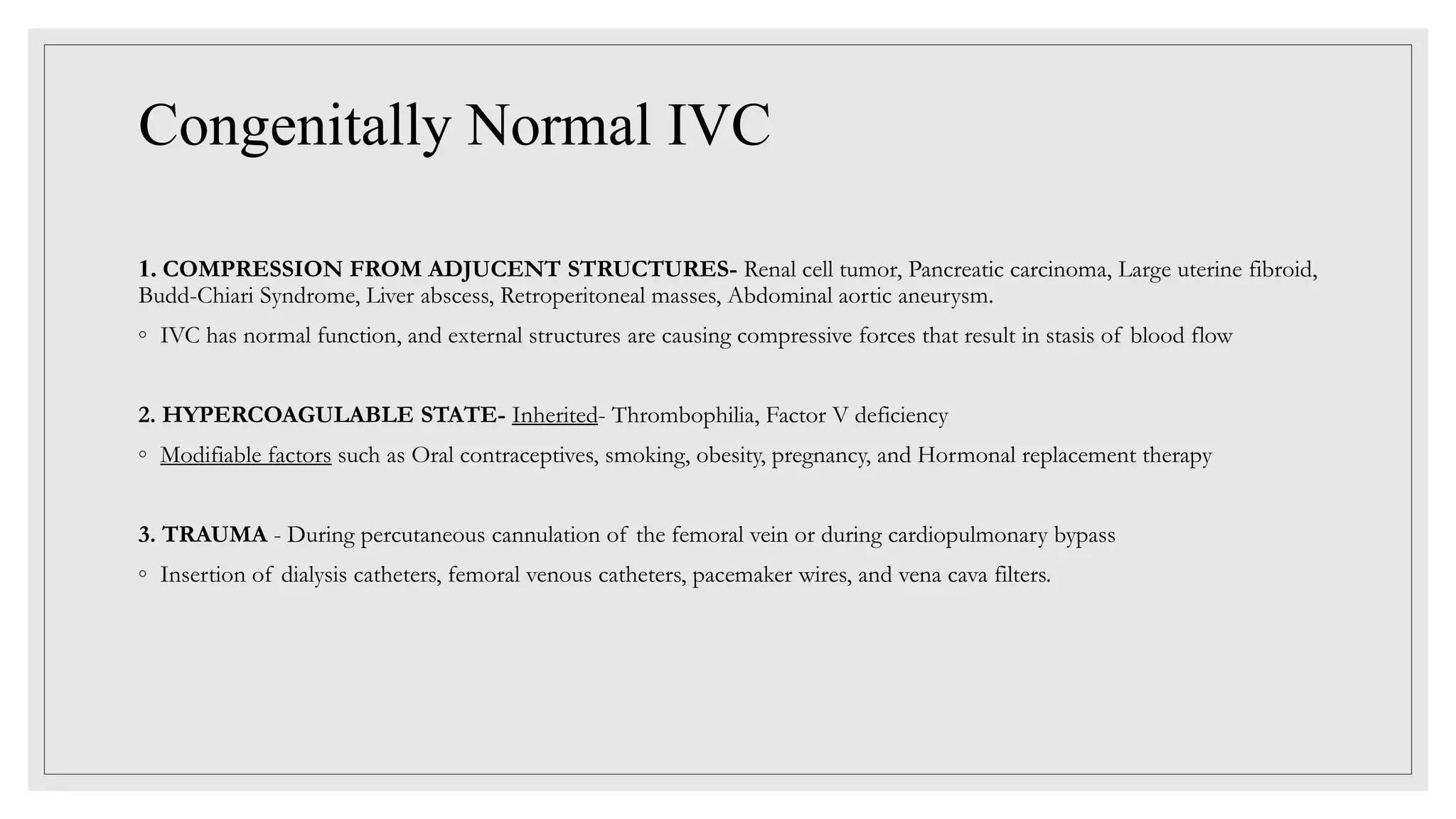
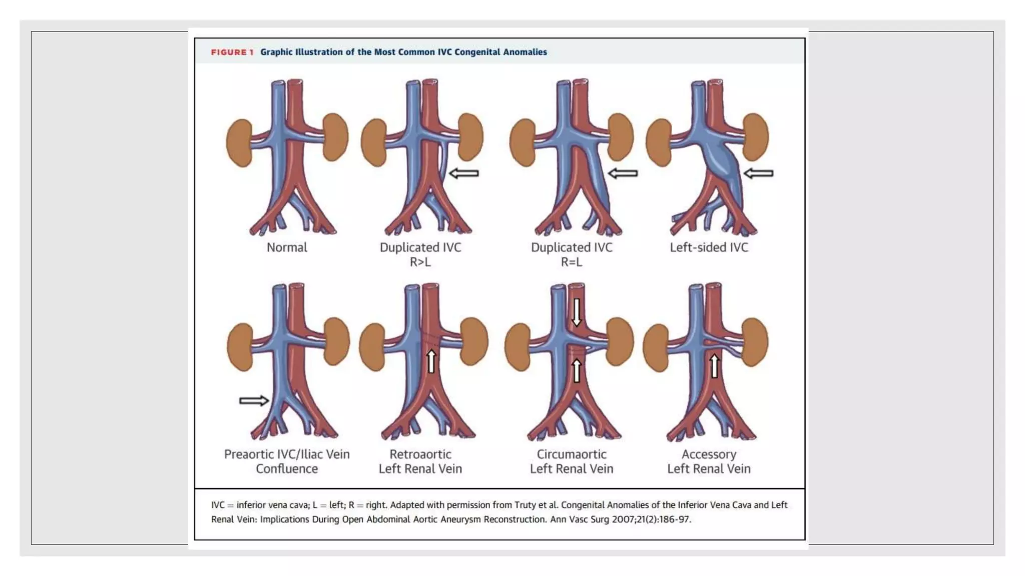
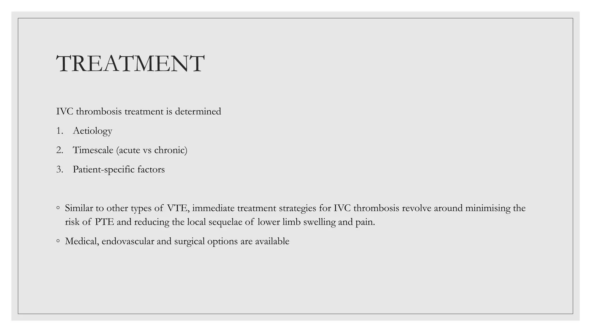
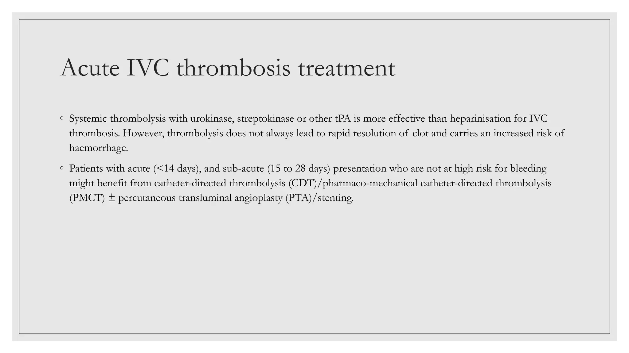
3. PATIENT PARTICULARS
◦ NAME – Mr. MA
◦ AGE/ SEX – 21/M
◦ UHID -220205906
◦ DATE OF ADMISSION- 18-02-22
◦ DATE OF DISCHARGE- 28-2-22
◦ DURATION OF HOSPITAL STAY – 10 DAYS
5. HISTORY OF PRESENTING ILLNESS
◦ Patient was apparently well 3 month back when his sister noticed distended abdominal veins on the upper
abdomen which was insidious in onset and gradually progressive in terms of distention and number of veins.
◦ Veins also appeared on the lower abdomen since 1 month.
◦ He also had lower back pain for 1 month.
◦ It was cramping in nature insidious in onset, relieved with rest and medication, increased on exertion, no radiation
or referred pain in lower limbs.
6. ◦ No h/o jaundice, abdominal distention, hematemesis, or malena.
◦ No h/o abdominal pain, feeling of lump in abdomen.
◦ No h/o distended back, chest and neck veins.
◦ No alteration in bowel/bladder activities
◦ No h/o loss of appetite/ weight loss/ sleep habits are unaffected
7. PAST/FAMILY HISTORY
◦ Non significant/ no h/o any chronic illness
PERSONAL HISTORY
• Educated till class 12th
• Works as an intern nurse at a Health care facility.
• Unmarried
• Consumes mixed diet
• Doesn’t consume alcohol/ non smoker/ no drug abuse
8. EXAMINATION
◦ VITALS AT PRESENTATION - Stable
GENERAL PHYSICAL EXAMINATION-
No pallor/icterus/ clubbing/ koilonychia/ lymphadenopathy/ oedema
No peripheral signs of hepatocellular failure were present.
9. SYSTEMIC EXAMINATION
◦ Per Abdomen- soft, Non Tender
◦ Liver was not palpable
◦ Total span of liver 10 cm
◦ Spleen was palpable 3 cm below the left coastal margin, smooth border, firm in consistency.
◦ Bowel sound present
◦ Distended tortuous abdominal veins were appreciated best in standing position in the epigastric ,hypogastric and
iliac regions. Direction of flow was below upwards in all the veins.
◦ Bilateral varicocele was appreciated more in the left side.
◦ CNS- Conscious, oriented to T/P/P; No FND
◦ Respiratory system/ CVS Examination – WNL
13. …contd..
LAB PARAMETERS VALUES
Na/ K/ Cl 139/4.1/107
Urea/ Creat 27/1.0
Ca/ P 9.1/3.4
Bilirubin (T) (mg/dL) 0.7
ALP 74
SGOT/SGPT 40/21
Total Protein/ Albumin 7.9/5
Urine r/m No pus cells/ RBC/ albumin/ sugar
14. ◦ Hep B, C and HIV serology negative
◦ Coagulogram within normal range
◦ Lipid profile normal
◦ USG WA- Chronic Portal vein thrombosis (PV size= 8mm) with formation of portal collaterals and formation of
peri-splenic and peri-gastric venous collaterals
◦ Splenomegaly
◦ Liver echotexture normal, size= 11 cm.
17. Thrombophilia profile
◦ Factor V Leiden, Anti thrombin III, Protein C and Protein S were in normal range.
◦ ANA normal
◦ Homocysteine levels WNL
18.
19. MPN extended profile
◦ JAK2 V617F mutation was positive
◦ Calreticulin, MPL, BCR-ABL were negative
20. CASE SUMMARY
20/M with, presented with H/o
◦ Distended abdominal veins for 3 months
◦ Lower back pain for 1 month
He was found to have splenomegaly, distended tortuous abdominal veins with flow below upwards
and bilateral varicocele on examination
CBC was s/o thrombocytosis
USG WA was s/o splenomegaly and Chronic Portal vein thrombosis with formation of portal
collaterals
CECT abdomen was s/o IVC thrombosis
Bone marrow and genetic testing was s/o JAK2 V617F positive ET.
Diagnosis - JAK2 V617F positive ET leading to IVC thrombosis
22. INTRODUCTION
◦ Inferior vena cava (IVC) thrombosis –disease with high morbidity
◦ Underdiagnosed
◦ IVC thrombosis should be part of the differential diagnosis for a patient with risk for a thromboembolic event
◦ The predisposing prothrombotic condition should be determined once an IVC thrombus is identified as there are
many potential causes.
23.
24. OBJECTIVES
•1. Describe the presentation of a patient
with inferior vena cava thrombosis.
2. The evaluation of a patient with
inferior vena cava thrombosis.
3. Summarize the treatment of inferior
vena cava thrombosis.
26. There are two main subgroups used to describe the
causes of IVC thrombosis:
Congenitally
normal IVC Congenitally
abnormal IVC
27. Congenitally Normal IVC
1. COMPRESSION FROM ADJUCENT STRUCTURES- Renal cell tumor, Pancreatic carcinoma, Large uterine fibroid,
Budd-Chiari Syndrome, Liver abscess, Retroperitoneal masses, Abdominal aortic aneurysm.
◦ IVC has normal function, and external structures are causing compressive forces that result in stasis of blood flow
2. HYPERCOAGULABLE STATE- Inherited- Thrombophilia, Factor V deficiency
◦ Modifiable factors such as Oral contraceptives, smoking, obesity, pregnancy, and Hormonal replacement therapy
3. TRAUMA - During percutaneous cannulation of the femoral vein or during cardiopulmonary bypass
◦ Insertion of dialysis catheters, femoral venous catheters, pacemaker wires, and vena cava filters.
28. Congenitally Abnormal IVC
◦ Congenitally abnormal IVC → turbulent blood flow →development of a thrombus
◦ Occurs in approximately 1% of the population, and within that 1%, about 60-80% develop IVC thrombosis
◦ Congenital IVC anomalies have three main anatomic configurations :
• Infrarenal - Duplicate IVC, persistent left-side IVC, preaortic IVC, absence of the infrarenal IVC
• Renal - Accessory left renal vein, retroaortic and circumaortic left renal vein
• Suprarenal - Absence of the hepatic IVC with azygos continuation, congenital caval stenosis or atresia, IVC
membranes
◦ Because of well-developed collaterals, patients with congenital IVC anomalies rarely experience symptoms-
Incidental findings on abdominal imaging
31. History and Physical
◦ Clinical presentation can vary, not pathognomonic history and physical exam
findings to diagnose IVC
◦ Typically, patients complain of leg heaviness, pain, swelling, and cramping
◦ Symptoms can also be nonspecific such as abdominal, flank or back pain.
◦ Men may present with scrotal swelling
◦ Vague nature of symptoms –delay diagnosis
◦ The presence of bilateral lower extremity edema and dilated superficial
abdominal veins suggests the possibility of an IVC thrombus.
?
?
32. EVALUATION
◦ There are currently no societal guidelines available to aid in the diagnosis and management of IVC thrombosis
◦ Multiple imaging modalities are potentially useful when IVC thrombosis is suspected.
◦ Ideal imaging modality -high diagnostic accuracy and be safe and reproducible.
33. • 1. IVC duplex ultrasound – INITIAL TEST –Non-invasive, portable and visualize collaterals.
Operator dependent, less reliable diagnosis within the abdomen because of greater difficulty in assessing venous
compressibility
◦ 2. Computer tomography (CT) venography - can also be used to visualize the location of the thrombus and as
diagnostic evaluation for the primary process (e.g., malignancy)
◦ 3. MRI (Magnetic Resonance Imaging) venography - most reliable technique for visualizing the presence,
extent and age of the thrombus. Given the poor general availability and the high cost of MRI, it should not be the
initial imaging choice
◦ 4. Direct catheter venography -definitive diagnostic method but invasive
34. Additional Imaging Considerations
1. Approximately 12% of patients with an IVC thrombus develop PTE. Therefore, dedicated CT angiogram of the
thorax may be needed.
2. If there is a concern for renal involvement, nuclear renal scanning can be used to assess for renal vascular
compromise.
3. Lumbar spine CT/MR to rule out spinal cord compression if symptoms of compressive neuropathy are present
4. Suprarenal IVC thrombus can be occasionally visible on echocardiography when detailed subcostal images are
recorded.
35. Laboratory Evaluation
◦ Gayer et al recommended that all patients with an IVC thrombosis be screened for a thrombophilic disorder. [14] In
their series, 77.7% of patients with IVC thrombosis had a positive thrombophilia screen.
◦ The decision to initiate a laboratory workup for thrombophilia is complex and difficulty, because identification of
underlying thrombophilia might not affect therapeutic strategy.
◦ In addition to the challenge of deciding who should be tested, clinicians need to be aware of when to test—or more
precisely, when not to.
Gayer, G., et al. "Congenital anomalies of the inferior vena cava revealed on CT in patients with deep vein thrombosis." American Journal of Roentgenology 180.3 (2003): 729-732.
38. STEP 1 TESTS – initial screening test, initiate anti-
coagulation after it
CBC, PT/aPTT/TT, Fibrinogen
STEP 2- Done in acute phase, on anti-coagulation
ACL, Lupus AC, ANA/dsDNA, PNH testing, Lipid
profile, MPN testing
STEP 3 – can be done if needed in acute phase since DNA
based
FVL, PTG, MTHFR mutation
STEP 4- 2 weeks after stopping anti-coagulation
AT III, Protein C, S
39. TREATMENT
IVC thrombosis treatment is determined
1. Aetiology
2. Timescale (acute vs chronic)
3. Patient-specific factors
◦ Similar to other types of VTE, immediate treatment strategies for IVC thrombosis revolve around minimising the
risk of PTE and reducing the local sequelae of lower limb swelling and pain.
◦ Medical, endovascular and surgical options are available
40. o The 2012 guidelines for antithrombotic therapy of VTE from the American College of Chest Physicians (ACCP)
recommend
LMWH or fondaparinux > IV UFH> subcutaneous UFH
o These recommendations, though largely based on lower limb DVT, include the treatment of ACUTE IVC thrombosis
o Anticoagulant therapy reduces thrombus propagation, but does not produce clot lysis, thus risking Post-thrombotic
syndrome (PTS) and Chronic venous insufficiency (CVI)
o Rapid thrombus removal is felt by many to be essential to preserve valvular function, thereby minimising CVI and PTS
o Necessary to determine whether IVC thrombosis is spontaneous (idiopathic) or secondary, as removal of thrombus will
not result in long-lasting patency if the cause (e.g. external compression) has not been corrected.
41. Acute IVC thrombosis treatment
◦ Systemic thrombolysis with urokinase, streptokinase or other tPA is more effective than heparinisation for IVC
thrombosis. However, thrombolysis does not always lead to rapid resolution of clot and carries an increased risk of
haemorrhage.
◦ Patients with acute (<14 days), and sub-acute (15 to 28 days) presentation who are not at high risk for bleeding
might benefit from catheter-directed thrombolysis (CDT)/pharmaco-mechanical catheter-directed thrombolysis
(PMCT) ± percutaneous transluminal angioplasty (PTA)/stenting.
42. Chronic IVC thrombosis
◦ Endovenous angioplasty combined with stenting along with anti-coagulation has emerged as the method of choice for chronic
thrombosis (< 28 days).
43. Thrombosis treatment in congenital IVC
anomaly
◦ Anticoagulation therapy -likely to ameliorate symptoms in patients with congenital IVC anomalies and symptomatic IVC
thrombosis.
◦ As caval anomalies remain permanent risk factors for venous stasis and thrombosis, life-long anticoagulation appears acceptable;
however, there are no explicit guidelines.
44. Continuing medical treatment in IVC
thrombosis
◦ Whether or not patients have endovascular or surgical intervention, unfractionated or LMWH treatments are the mainstays of T/T
◦ conversion after a minimum of 5 days treatment to oral therapies such as warfarin, which should be initiated early with treatment
overlap as per ACCP guidelines.
◦ Current ACCP guidelines -secondary cause is reversed w/o concurrent thrombophilia, OAC for 3 months.
◦ Co-existent or causative thrombophilia OR ir-reversible cause i.e. IVC anomaly OR t/t involves IVC stent - life-long OAC.
◦ Newer OAC such as rivaroxaban or dabigatran are not recommended as first-line, long-term anticoagulation in the 2012 ACCP
guidelines and are currently third line after warfarin and LMWH.
45.
46.
47. PROGNOSIS
◦ The overall prognosis hinges on the underlying cause of the thrombus.
◦ If a patient has a hereditary hypercoagulable disease, they are predisposed to the development of thrombi.
However, if they are compliant with their anticoagulation therapy, their prognosis will be favorable.
◦ Alternatively, if the patient is diagnosed with progressive cancer such as pancreatic cancer that has extended and
compressed the IVC, their prognosis is likely poor.
◦ Finally, there is the always a potential of pulmonary embolus
48. COMPLICATIONS
◦ If untreated, patients can suffer from a post-thrombotic syndrome, which consists of venous stasis changes leading
to ulceration in the lower extremities.
◦ Other complications include pulmonary emboli and renal ischemia due to the extension of the thrombus.
◦ Thus, if IVC thrombosis is a consideration, imaging should not be delayed.
51. TAKE HOME MESSAGE
◦ IVC thrombosis is associated with significant acute and chronic morbidity.
◦ Diagnostic challenge to the clinician and requires a high index of suspicion
◦ Pain, swelling of lower limbs, lower back pain, dilatation of superficial abdominal veins are diagnostic
indicators.
◦ Duplex USG- screening test; CT , MRI delineate IVC anatomy and highlight causes of secondary IVC
thrombosis.
◦ Where IVC thrombosis is idiopathic, especially in younger patients, a complete thrombophilia screen
should precede but not delay heparinization.
◦ In appropriate cases, locally directed thrombolysis is the most efficacious treatment, minimising long-
term complications of IVC thrombosis.
52. REFERENCES
◦ McAree BJ, O'Donnell ME, Fitzmaurice GJ, Reid JA, Spence RA, Lee B. Inferior vena cava thrombosis: a review of
current practice. Vasc Med. 2013 Feb;18(1):32-43. doi: 10.1177/1358863X12471967. PMID: 23439778
◦ Alkhouli M, Morad M, Narins CR, Raza F, Bashir R. Inferior Vena Cava Thrombosis. JACC Cardiovasc Interv. 2016 Apr
11;9(7):629-43. doi: 10.1016/j.jcin.2015.12.268. Epub 2016 Mar 4. PMID: 26952909.
◦ Hollingsworth CM, Mead T. Inferior Vena Caval Thrombosis. [Updated 2021 Jul 10]. In: StatPearls [Internet]. Treasure
Island (FL): StatPearls Publishing; 2022 Jan-. Available from: https://www.ncbi.nlm.nih.gov/books/NBK537175/
◦ https://emedicine.medscape.com/article/1933035-overview#a6
How the patient was managed and how to approach a case of IVC thrombosis will be discussed in further slides
Understanding the anatomy of the IVC and its tributaries is essential to understanding the variability in the clinical presentations of patients with IVCT.