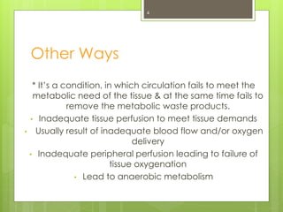
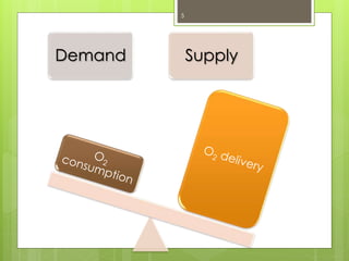
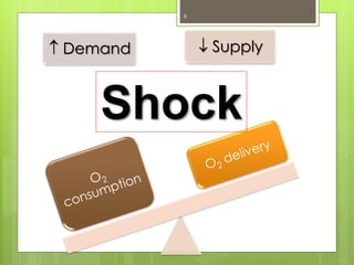
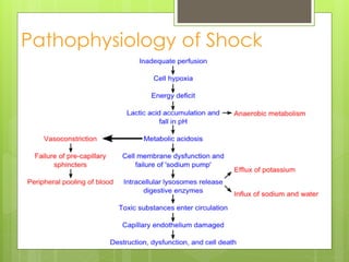
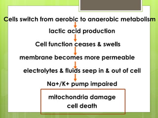
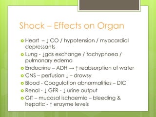
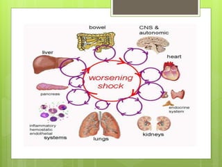
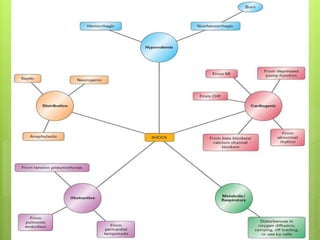
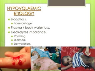
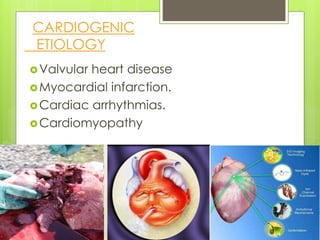
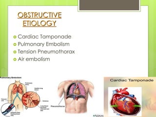
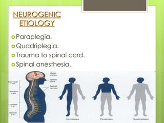
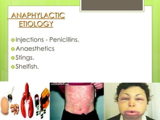
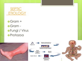
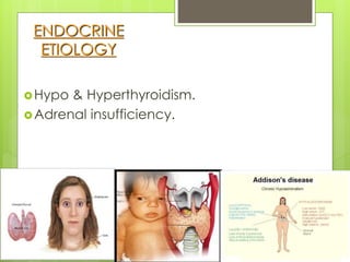
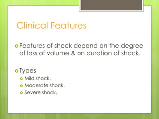
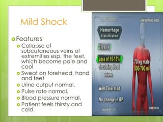
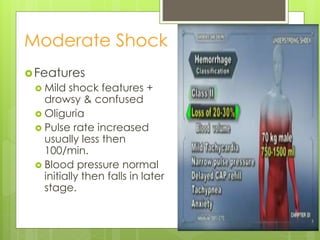
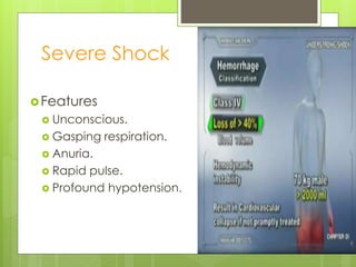
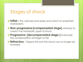
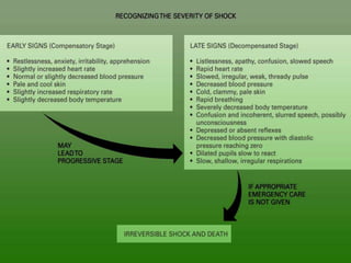
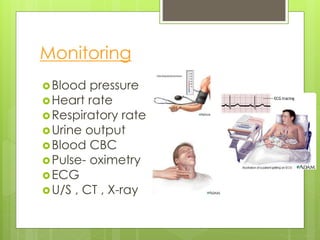
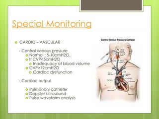
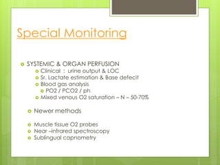
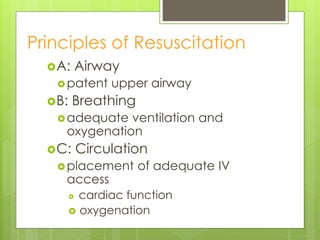
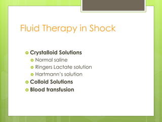
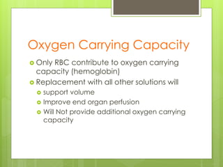
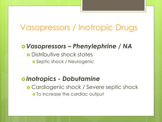
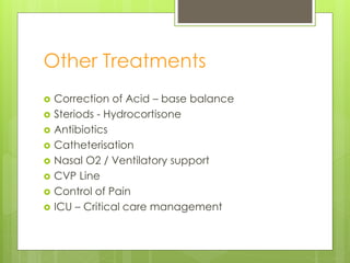
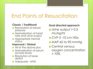
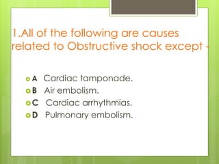
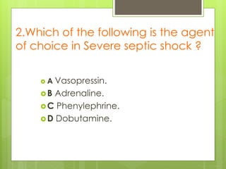
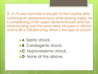
SHOCK
[ Pathophysiology,Types & Mgt ]
 Prof. Utham Murali. M.S; M.B.A
Objectives
 Definition
 Review basic physiologic aspects of shock
 Different categories with Etiology &Clinical
features
 Management aspects
Definition
Shock is a physiologic state characterized by systemic
reduction in tissue perfusion, resulting in decreased
tissue oxygen delivery.
3
Other Ways
* It’s a condition, in which circulation fails to meet the
metabolic need of the tissue & at the same time fails to
remove the metabolic waste products.
• Inadequate tissue perfusion to meet tissue demands
• Usually result of inadequate blood flow and/or oxygen
delivery
• Inadequate peripheral perfusion leading to failure of
tissue oxygenation
• Lead to anaerobic metabolism
4
5
Demand Supply
6
 Demand  Supply
Shock
Pathophysiology of Shock
Cells switch from aerobic to anaerobic metabolism
lactic acid production
Cell function ceases & swells
membrane becomes more permeable
electrolytes & fluids seep in & out of cell
Na+/K+ pump impaired
mitochondria damage
cell death
Shock – Effects on Organ
 Heart – ↓ CO / hypotension / myocardial
depressants
 Lung - ↓gas exchange / tachypnoea /
pulmonary edema
 Endocrine – ADH → ↑ reabsorption of water
 CNS – perfusion ↓ – drowsy
 Blood - Coagulation abnormalities – DIC
 Renal - ↓ GFR - ↓ urine output
 GIT – mucosal ischaemia – bleeding &
hepatic - ↑ enzyme levels
11
HYPOVOLAEMIC
ETIOLOGY
 Blood loss.
 haemorrhage
 Plasma / body water loss.
 Electrolytes imbalance.
 Vomiting.
 Diarrhea.
 Dehydration.
Valvular heart disease
Myocardial infarction.
Cardiac arrhythmias.
Cardiomyopathy
CARDIOGENIC
ETIOLOGY
OBSTRUCTIVE
ETIOLOGY
 Cardiac Tamponade
 Pulmonary Embolism
 Tension Pneumothorax
 Air embolism
NEUROGENIC
ETIOLOGY
Paraplegia.
Quadriplegia.
Trauma to spinal cord.
Spinal anesthesia.
ANAPHYLACTIC
ETIOLOGY
Injections - Penicillins.
Anaesthetics
Stings.
Shelfish.
Gram +
Gram -
Fungi / Virus
Protozoa
SEPTIC
ETIOLOGY
ENDOCRINE
ETIOLOGY
Hypo & Hyperthyroidism.
Adrenal insufficiency.
Clinical Features
Features of shock depend on the degree
of loss of volume & on duration of shock.
Types
 Mild shock.
 Moderate shock.
 Severe shock.
Mild Shock
Features
 Collapse of
subcutaneous veins of
extremities esp. the feet,
which become pale and
cool
 Sweat on forehead, hand
and feet
 Urine output normal.
 Pulse rate normal.
 Blood pressure normal.
 Patient feels thirsty and
cold.
Moderate Shock
Features
 Mild shock features +
drowsy & confused
 Oliguria
 Pulse rate increased
usually less then
100/min.
 Blood pressure normal
initially then falls in later
stage.
Severe Shock
Features
 Unconscious.
 Gasping respiration.
 Anuria.
 Rapid pulse.
 Profound hypotension.
Stages of shock
 Initial : The cells become leaky and switch to anaerobic
metabolism.
 Non-progressive:(compensated stage) Attempt to
correct the metabolic upset of shock.
 Progressive: (decompensated stage ) Eventually
the compensation will begin to fail.
 Refractory : Organs fail and the shock can no longer be
reversed.
24
SHOCK
[ Management ]
Monitoring
Blood pressure
Heart rate
Respiratory rate
Urine output
Blood CBC
Pulse- oximetry
ECG
U/S , CT , X-ray
Special Monitoring
 CARDIO – VASCULAR
- Central venous pressure
 Normal ; 5-10cmH2O,
 If CVP<5cmH2O
 Inadequacy of blood volume
 CVP>12cmH2O
 Cardiac dysfunction
- Cardiac output
 Pulmonary catheter
 Doppler ultrasound
 Pulse waveform analysis
Special Monitoring
 SYSTEMIC & ORGAN PERFUSION
 Clinical : urine output & LOC
 Sr. Lactate estimation & Base defecit
 Blood gas analysis
 PO2 / PCO2 / ph
 Mixed venous O2 saturation – N – 50-70%
 Newer methods
 Muscle tissue O2 probes
 Near –infrared spectroscopy
 Sublingual capnometry
Guidelines
 Treat the cause
 Improve Cardiac function
 Improve Tissue perfusion
Goals of Resuscitation
 Overall goal:
 increase O2 delivery
 decrease demand
Treatment
O2 content Cardiac
output
Blood
pressure
Sedation/analgesia
Principles of Resuscitation
A: Airway
patent upper airway
B: Breathing
adequate ventilation and
oxygenation
C: Circulation
placement of adequate IV
access
 cardiac function
 oxygenation
Fluid Therapy in Shock
 Crystalloid Solutions
 Normal saline
 Ringers Lactate solution
 Hartmann’s solution
 Colloid Solutions
 Blood transfusion
Oxygen Carrying Capacity
 Only RBC contribute to oxygen carrying
capacity (hemoglobin)
 Replacement with all other solutions will
 support volume
 Improve end organ perfusion
 Will Not provide additional oxygen carrying
capacity
Dynamic Fluid Response
 Infusing 250-500ml of Fluid rapidly in 5 - 10 mts.
 Responders – Improvement
 Transient responders – revert back
 Non – responders
Vasopressors / Inotropic Drugs
Vasopressors – Phenylephrine / NA
 Distributive shock states
 Septic shock / Neurogenic
Inotropics - Dobutamine
 Cardiogenic shock / Severe septic shock
 To increase the cardiac output
Other Treatments
 Correction of Acid – base balance
 Steriods - Hydrocortisone
 Antibiotics
 Catheterisation
 Nasal O2 / Ventilatory support
 CVP Line
 Control of Pain
 ICU – Critical care management
End Points of Resuscitation
Classic / Traditional
 Restoration of blood
pressure
 Normalization of heart
rate and urine output
 Appropriate mental
status
Improved / Global
 All of the above plus
 Normalization of serum
lactate levels
 Resolution of base
deficit
 Adequate - MVS
Goal directed approach
 Urine output > 0.5
mL/kg/hr
 CVP 5 -10 cm H2o
 MAP 65 to 90 mmHg
 Central venous
oxygen concentration
> 70%
Practically
Speaking….
 Know how to distinguish different types of
shock and treat accordingly.
 Look for early signs of shock.
 Monitor the patient using the HR, MAP,
mental status, urine output.
 SHOCK is not equal to hypotension.
 Start antibiotics within an hour !
 Do not wait for cultures or blood work.
1.All of the following are causes
related to Obstructive shock except -
 A Cardiac tamponade.
B Air embolism.
C Cardiac arrhythmias.
D Pulmonary embolism.
2.Which of the following is the agent
of choice in Severe septic shock ?
 A Vasopressin.
B Adrenaline.
C Phenylephrine.
D Dobutamine.
3. A 19-year-old male is brought to the hospital after
sustaining an abdominal injury while playing rugby. He
is complaining of left upper abdominal pain and has
some bruising over the same area. His pulse is 140/min
and his BP is 100/82mmHg. What is the type of shock?
 A Septic shock.
B Cardiogenic shock.
C Hypovolaemic shock.
D None of the above.
4.Which of the following is not a newer
methods for monitoring tissue perfusion -
A Muscle tissue O2 probe.
B Doppler ultrasound.
C Infrared spectroscopy.
D Sublingual capnometry.
5.Which of the following is one of the
last signs of shock ?
 A Profound hypotension.
B Tachycardia.
C Prolonged capillary refill.
D All of the above.
THANK YOU
THANK YOU . . .

shock-types-141009102815-conversion-gate01 (1).pdf

  • 1.
    SHOCK [ Pathophysiology,Types &Mgt ]  Prof. Utham Murali. M.S; M.B.A
  • 2.
    Objectives  Definition  Reviewbasic physiologic aspects of shock  Different categories with Etiology &Clinical features  Management aspects
  • 3.
    Definition Shock is aphysiologic state characterized by systemic reduction in tissue perfusion, resulting in decreased tissue oxygen delivery. 3
  • 4.
    Other Ways * It’sa condition, in which circulation fails to meet the metabolic need of the tissue & at the same time fails to remove the metabolic waste products. • Inadequate tissue perfusion to meet tissue demands • Usually result of inadequate blood flow and/or oxygen delivery • Inadequate peripheral perfusion leading to failure of tissue oxygenation • Lead to anaerobic metabolism 4
  • 5.
  • 6.
  • 7.
  • 8.
    Cells switch fromaerobic to anaerobic metabolism lactic acid production Cell function ceases & swells membrane becomes more permeable electrolytes & fluids seep in & out of cell Na+/K+ pump impaired mitochondria damage cell death
  • 9.
    Shock – Effectson Organ  Heart – ↓ CO / hypotension / myocardial depressants  Lung - ↓gas exchange / tachypnoea / pulmonary edema  Endocrine – ADH → ↑ reabsorption of water  CNS – perfusion ↓ – drowsy  Blood - Coagulation abnormalities – DIC  Renal - ↓ GFR - ↓ urine output  GIT – mucosal ischaemia – bleeding & hepatic - ↑ enzyme levels
  • 11.
  • 12.
    HYPOVOLAEMIC ETIOLOGY  Blood loss. haemorrhage  Plasma / body water loss.  Electrolytes imbalance.  Vomiting.  Diarrhea.  Dehydration.
  • 13.
    Valvular heart disease Myocardialinfarction. Cardiac arrhythmias. Cardiomyopathy CARDIOGENIC ETIOLOGY
  • 14.
    OBSTRUCTIVE ETIOLOGY  Cardiac Tamponade Pulmonary Embolism  Tension Pneumothorax  Air embolism
  • 15.
  • 16.
  • 17.
    Gram + Gram - Fungi/ Virus Protozoa SEPTIC ETIOLOGY
  • 18.
  • 19.
    Clinical Features Features ofshock depend on the degree of loss of volume & on duration of shock. Types  Mild shock.  Moderate shock.  Severe shock.
  • 20.
    Mild Shock Features  Collapseof subcutaneous veins of extremities esp. the feet, which become pale and cool  Sweat on forehead, hand and feet  Urine output normal.  Pulse rate normal.  Blood pressure normal.  Patient feels thirsty and cold.
  • 21.
    Moderate Shock Features  Mildshock features + drowsy & confused  Oliguria  Pulse rate increased usually less then 100/min.  Blood pressure normal initially then falls in later stage.
  • 22.
    Severe Shock Features  Unconscious. Gasping respiration.  Anuria.  Rapid pulse.  Profound hypotension.
  • 23.
    Stages of shock Initial : The cells become leaky and switch to anaerobic metabolism.  Non-progressive:(compensated stage) Attempt to correct the metabolic upset of shock.  Progressive: (decompensated stage ) Eventually the compensation will begin to fail.  Refractory : Organs fail and the shock can no longer be reversed.
  • 24.
  • 25.
  • 26.
    Monitoring Blood pressure Heart rate Respiratoryrate Urine output Blood CBC Pulse- oximetry ECG U/S , CT , X-ray
  • 27.
    Special Monitoring  CARDIO– VASCULAR - Central venous pressure  Normal ; 5-10cmH2O,  If CVP<5cmH2O  Inadequacy of blood volume  CVP>12cmH2O  Cardiac dysfunction - Cardiac output  Pulmonary catheter  Doppler ultrasound  Pulse waveform analysis
  • 28.
    Special Monitoring  SYSTEMIC& ORGAN PERFUSION  Clinical : urine output & LOC  Sr. Lactate estimation & Base defecit  Blood gas analysis  PO2 / PCO2 / ph  Mixed venous O2 saturation – N – 50-70%  Newer methods  Muscle tissue O2 probes  Near –infrared spectroscopy  Sublingual capnometry
  • 29.
    Guidelines  Treat thecause  Improve Cardiac function  Improve Tissue perfusion
  • 30.
    Goals of Resuscitation Overall goal:  increase O2 delivery  decrease demand Treatment O2 content Cardiac output Blood pressure Sedation/analgesia
  • 31.
    Principles of Resuscitation A:Airway patent upper airway B: Breathing adequate ventilation and oxygenation C: Circulation placement of adequate IV access  cardiac function  oxygenation
  • 32.
    Fluid Therapy inShock  Crystalloid Solutions  Normal saline  Ringers Lactate solution  Hartmann’s solution  Colloid Solutions  Blood transfusion
  • 33.
    Oxygen Carrying Capacity Only RBC contribute to oxygen carrying capacity (hemoglobin)  Replacement with all other solutions will  support volume  Improve end organ perfusion  Will Not provide additional oxygen carrying capacity
  • 34.
    Dynamic Fluid Response Infusing 250-500ml of Fluid rapidly in 5 - 10 mts.  Responders – Improvement  Transient responders – revert back  Non – responders
  • 35.
    Vasopressors / InotropicDrugs Vasopressors – Phenylephrine / NA  Distributive shock states  Septic shock / Neurogenic Inotropics - Dobutamine  Cardiogenic shock / Severe septic shock  To increase the cardiac output
  • 36.
    Other Treatments  Correctionof Acid – base balance  Steriods - Hydrocortisone  Antibiotics  Catheterisation  Nasal O2 / Ventilatory support  CVP Line  Control of Pain  ICU – Critical care management
  • 37.
    End Points ofResuscitation Classic / Traditional  Restoration of blood pressure  Normalization of heart rate and urine output  Appropriate mental status Improved / Global  All of the above plus  Normalization of serum lactate levels  Resolution of base deficit  Adequate - MVS Goal directed approach  Urine output > 0.5 mL/kg/hr  CVP 5 -10 cm H2o  MAP 65 to 90 mmHg  Central venous oxygen concentration > 70%
  • 38.
    Practically Speaking….  Know howto distinguish different types of shock and treat accordingly.  Look for early signs of shock.  Monitor the patient using the HR, MAP, mental status, urine output.  SHOCK is not equal to hypotension.  Start antibiotics within an hour !  Do not wait for cultures or blood work.
  • 40.
    1.All of thefollowing are causes related to Obstructive shock except -  A Cardiac tamponade. B Air embolism. C Cardiac arrhythmias. D Pulmonary embolism.
  • 41.
    2.Which of thefollowing is the agent of choice in Severe septic shock ?  A Vasopressin. B Adrenaline. C Phenylephrine. D Dobutamine.
  • 42.
    3. A 19-year-oldmale is brought to the hospital after sustaining an abdominal injury while playing rugby. He is complaining of left upper abdominal pain and has some bruising over the same area. His pulse is 140/min and his BP is 100/82mmHg. What is the type of shock?  A Septic shock. B Cardiogenic shock. C Hypovolaemic shock. D None of the above.
  • 43.
    4.Which of thefollowing is not a newer methods for monitoring tissue perfusion - A Muscle tissue O2 probe. B Doppler ultrasound. C Infrared spectroscopy. D Sublingual capnometry.
  • 44.
    5.Which of thefollowing is one of the last signs of shock ?  A Profound hypotension. B Tachycardia. C Prolonged capillary refill. D All of the above.
  • 45.