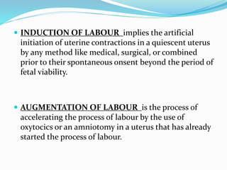
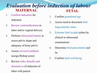
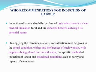
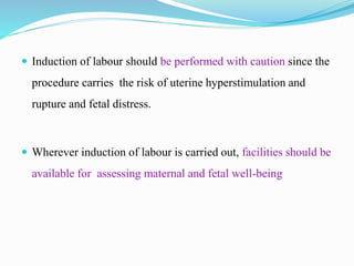
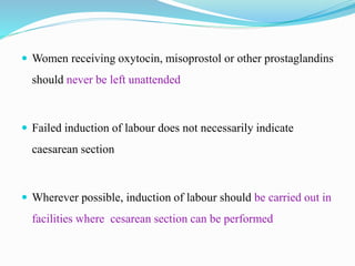
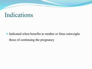
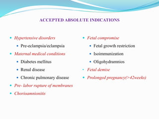
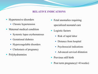
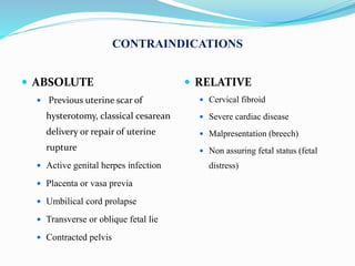
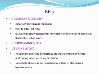
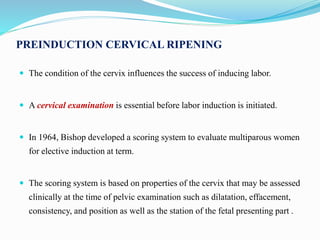
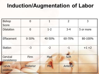
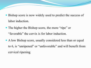
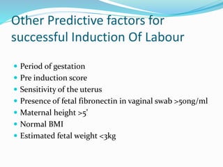
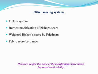
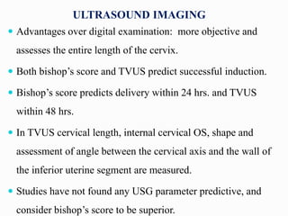
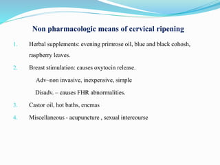
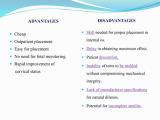
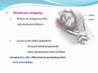
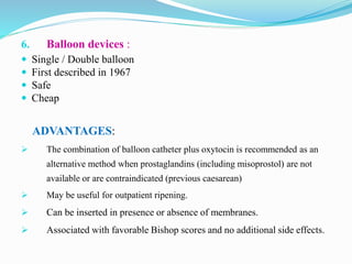
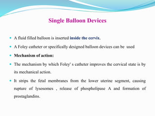
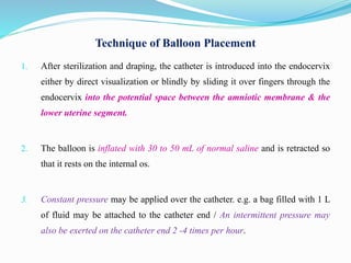
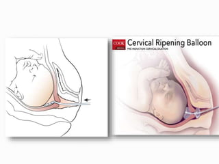
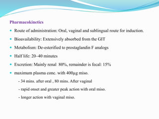
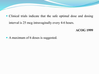
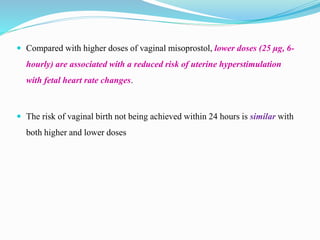
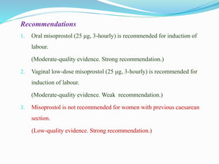
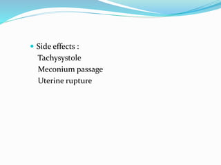
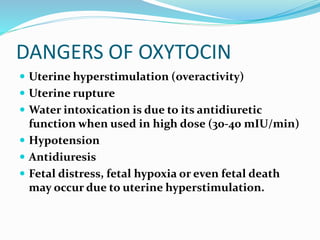
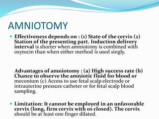
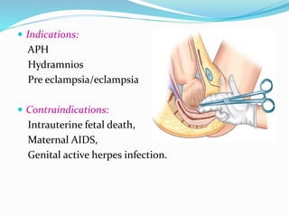
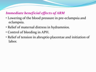
INDUCTION
OF LABOUR
 INDUCTION OF LABOUR implies the artificial
initiation of uterine contractions in a quiescent uterus
by any method like medical, surgical, or combined
prior to their spontaneous onsent beyond the period of
fetal viability.
 AUGMENTATION OF LABOUR is the process of
accelerating the process of labour by the use of
oxytocics or an amniotomy in a uterus that has already
started the process of labour.
Evaluation before induction of labour
MATERNAL FETAL
1. Confirm indication for
induction
2. Review contraindications to
labor and/or vaginal delivery
3. Perform clinical pelvimetry to
assess pelvic shape and
adequacy of bony pelvis
4. Assess cervical condition
(assign Bishop score)
5. Review risks, benefits and
alternatives of induction of
labor with patient
1. Confirm gestational age
2. Assess need to document fetal
lung maturity status
3. Estimate fetal weight (either by
clinical or ultrasound
examination)
4. Determine fetal presentation and
lie
5. Confirm fetal well-being
WHO RECOMMENDATIONS FOR INDUCTION OF
LABOUR
 Induction of labour should be performed only when there is a clear
medical indication for it and the expected benefits outweigh its
potential harms.
 In applying the recommendations, consideration must be given to
the actual condition, wishes and preferences of each woman, with
emphasis being placed on cervical status, the specific method of
induction of labour and associated conditions such as parity and
rupture of membranes.
 Induction of labour should be performed with caution since the
procedure carries the risk of uterine hyperstimulation and
rupture and fetal distress.
 Wherever induction of labour is carried out, facilities should be
available for assessing maternal and fetal well-being
 Women receiving oxytocin, misoprostol or other prostaglandins
should never be left unattended
 Failed induction of labour does not necessarily indicate
caesarean section
 Wherever possible, induction of labour should be carried out in
facilities where cesarean section can be performed
Indications
 Indicated when benefits to mother or fetus outweighs
those of continuing the pregnancy
ACCEPTED ABSOLUTE INDICATIONS
 Hypertensive disorders
 Pre-eclampsia/eclampsia
 Maternal medical conditions
 Diabetes mellitus
 Renal disease
 Chronic pulmonary disease
 Pre- labor rupture of membranes
 Chorioamnionitis
 Fetal compromise
 Fetal growth restriction
 Isoimmunization
 Oligohydramnios
• Fetal demise
 Prolonged pregnancy(>42weeks)
RELATIVE INDICATIONS
 Hypertensive disorders
 Chronic hypertension
 Maternal medical condition
 Systemic lupus erythematosus
 Gestational diabetes
 Hypercoagulable disorders
 Cholestasis of pregnancy
 Polyhydramnios
 Fetal anomalies requiring
specialized neonatal care
 Logistic factors
 Risk of rapid labor
 Distance from hospital
 Psychosocial indications
 Advanced cervical dilatation
 Previous still birth
 Post term pregnancy(>41weeks)
CONTRAINDICATIONS
 ABSOLUTE
 Previous uterine scar of
hysterotomy, classical cesarean
delivery or repair of uterine
rupture
 Active genital herpes infection
 Placenta or vasa previa
 Umbilical cord prolapse
 Transverse or oblique fetal lie
 Contracted pelvis
 RELATIVE
 Cervical fibroid
 Severe cardiac disease
 Malpresentation (breech)
 Non assuring fetal status (fetal
distress)
Risks
 CESAREAN DELIVERY
 especially increased in nulliparas
 two- to threefold risks
 rates are inversely related with favorability of the cervix at induction,
that is, the Bishop score.
 CHORIOAMNIONITIS
 UTERINE ATONY
 Postpartum atony and hemorrhage are more common in women
undergoing induction or augmentation
 Intractable atony was the indication for a third of all cesarean
hysterectomies
 Cervical ripening :
A prelude to the onset of labour whereby the cervix becomes
soft and compliant.
 This allows its shape to change from being long and closed,
to being thinned out (effaced) and starting to open (dilate).
 It either occurs naturally or as a result of physical or
pharmacological interventions
NICE 2008
Cervix
Cellular
Smooth muscle
Fibroblast
Extra cellular
Collagen I (70%)
Collagen III
(30%)
Elastin
Proteoglycans
Decorin
MECHANISM INVOLVED IN CERVICAL RIPENING
 Cervix is a complex and heterogeneous organ, that undergoes
extensive changes throughout gestation and parturition.
 Chronic process, which begins within the first trimester of pregnancy
and progressively proceeds until term
 Softens, dilates and effaces the cervix
 This remodeling process is extremely complex and involves
 properly timed biochemical cascades,
 interaction between cellular and extra cellular components, and
 infiltration by inflammatory cells.
CERVICAL
RIPENING
Enzymatic
degradation
Hormonal
influences
Increased
Hyaluronic
acid
Increased
decorin
AFFECTING ELEMENTS
 CYTOKINES –
e.g. interleukin-1β enhance the activity of collagenases
and interleukin 8,
Platelet activating factor,
monocyte chemotactic factor-1
 HORMONAL INFLUENCES –
Estrogens increases collagenases
Progesterones inhibit collagenases, hyaluronic acid & IL-8
 NITRIC OXIDE stimulates leukocytes infiltration
induce prostaglandin secretion
PREINDUCTION CERVICAL RIPENING
 The condition of the cervix influences the success of inducing labor.
 A cervical examination is essential before labor induction is initiated.
 In 1964, Bishop developed a scoring system to evaluate multiparous women
for elective induction at term.
 The scoring system is based on properties of the cervix that may be assessed
clinically at the time of pelvic examination such as dilatation, effacement,
consistency, and position as well as the station of the fetal presenting part .
 Bishop score is now widely used to predict the success of
labor induction.
 The higher the Bishop score, the more “ripe” or
“favorable” the cervix is for labor induction.
 A low Bishop score, usually considered less than or equal
to 6, is “unripened” or “unfavorable” and will benefit from
cervical ripening
Other Predictive factors for
successful Induction Of Labour
 Period of gestation
 Pre induction score
 Sensitivity of the uterus
 Presence of fetal fibronectin in vaginal swab >50ng/ml
 Maternal height >5’
 Normal BMI
 Estimated fetal weight <3kg
Other scoring systems
 Field’s system
 Burnett modification of bishops score
 Weighted Bishop’s score by Friedman
 Pelvic score by Lange
However, despite this none of the modifications have shown
improved predictability.
ULTRASOUND IMAGING
 Advantages over digital examination: more objective and
assesses the entire length of the cervix.
 Both bishop’s score and TVUS predict successful induction.
 Bishop’s score predicts delivery within 24 hrs. and TVUS
within 48 hrs.
 In TVUS cervical length, internal cervical OS, shape and
assessment of angle between the cervical axis and the wall of
the inferior uterine segment are measured.
 Studies have not found any USG parameter predictive, and
consider bishop’s score to be superior.
METHODS OF CERVICAL RIPENING
 Unfortunately, women too frequently have an indication for induction but
with an unfavorable cervix.
 As favorability or Bishop score decreases, there is an increasingly
unsuccessful induction rate.
 Methods used for cervical ripening include pharmacological preparations
and various forms of mechanical cervical distension.
Non pharmacologic means of cervical ripening
1. Herbal supplements: evening primrose oil, blue and black cohosh,
raspberry leaves.
2. Breast stimulation: causes oxytocin release.
Adv–non invasive, inexpensive, simple
Disadv. – causes FHR abnormalities.
3. Castor oil, hot baths, enemas
4. Miscellaneous - acupuncture , sexual intercourse
4. HYGROSCOPIC DILATORS:
 Natural osmotic dilators –
 Laminaria japonicum
 Laminaria digitata
 Isapgol
 Synthetic osmotic dilators
 Lamicel
 Dilapan
 They absorb endocervical and local tissue fluids, causing the device to
expand within the endocervix and provide mechanical pressure.
 cause mechanical dilation and release of prostaglandins.
 Swell up to 4 – 5 times.
 Most rapidly in first 4-6 hours but continue to swell up to 24 hours later.
ADVANTAGES DISADVANTAGES
 Cheap
 Outpatient placement
 Easy for placement
 No need for fetal monitoring
 Rapid improvement of
cervical status
 Skill needed for proper placement in
internal os.
 Delay in obtaining maximum effect.
 Patient discomfort.
 Inability of tents to be molded
without compromising mechanical
integrity.
 Lack of manufacturer specifications
for natural dilators.
 Potential for incomplete sterility.
5. Membrane stripping:
 Release of endogenous PGs.
and mechanical dilation.
 results in less labor inductions
less post dated pregnancies
more spontaneous onset of labor
- inexpensive, safe, efficacious in promoting labor
over several days
6. Balloon devices :
 Single / Double balloon
 First described in 1967
 Safe
 Cheap
ADVANTAGES:
 The combination of balloon catheter plus oxytocin is recommended as an
alternative method when prostaglandins (including misoprostol) are not
available or are contraindicated (previous caesarean)
 May be useful for outpatient ripening.
 Can be inserted in presence or absence of membranes.
 Associated with favorable Bishop scores and no additional side effects.
Single Balloon Devices
 A fluid filled balloon is inserted inside the cervix.
 A Foley catheter or specifically designed balloon devices can be used
 Mechanism of action:
 The mechanism by which Foley' s catheter improves the cervical state is by
its mechanical action.
 It strips the fetal membranes from the lower uterine segment, causing
rupture of lysosomes , release of phospholipase A and formation of
prostaglandins.
Technique of Balloon Placement
1. After sterilization and draping, the catheter is introduced into the endocervix
either by direct visualization or blindly by sliding it over fingers through the
endocervix into the potential space between the amniotic membrane & the
lower uterine segment.
2. The balloon is inflated with 30 to 50 mL of normal saline and is retracted so
that it rests on the internal os.
3. Constant pressure may be applied over the catheter. e.g. a bag filled with 1 L
of fluid may be attached to the catheter end / An intermittent pressure may
also be exerted on the catheter end 2 -4 times per hour.
4. Catheter is removed at the time of rupture of membranes or may
be expelled spontaneously which indicate a cervical dilatation of
3 - 4 Centimeters.
PHARMACOLOGICAL TECHNIQUES
 Prostaglandins
 PGE2 : Dinoprostone
 PGE1 : Misoprostol
 Oxytocin
 Others
 Estrogen
 Relaxin
 Hyaluronic acid
 Progesterone receptor antagonist
PROSTAGLANDINS
 The chemical precursor is arachidonic acid
 PGs are endogenous compounds found in the myometrium, deciduas,
and fetal membranes during pregnancy.
 Cervical production of PGE2, PGI2, PGF increases at term.
 Modulate fibroblast activity - Increase hyaluronic acid production
 Acting as chemotactic agents, Inflammatory cells further release
degradative enzymes, causing cervical ripening.
 Prostaglandins administration results in dissolution of collagen bundles and
an increase in sub mucosal water content of the cervix.
 These changes in cervical connective tissue at term are similar to
those observed in early labor.
 Unlike oxytocin, response to prostaglandins does not change throughout
gestation.
Preparations
PGE2 : Dinoprostone PGE1 : Misoprostol
 Vaginal gel : Prepidil, Cerviprime
 Removable tampon : Cervidil
 Vaginal pessary : Prostin E2
 Misoprost
 Cytotec
Cervical Ripening
Alter the extracellular ground substance
of the cervix
increases the activity of collagenase in
the cervix.
Increase in elastase, glycosaminoglycan, dermatan
sulfate, and hyaluronic acid levels in the cervix.
A relaxation of cervical smooth muscle facilitates
dilation.
Increase in intracellular calcium levels,
~ contraction of myometrial muscle
Prostaglandin E2: (Dinoprostone)
 PROSTAGLANDIN E2 (DINOPROSTONE):
 CERVIPRIME GEL - is commonly used for cervical ripening .
 is available in a 2.5-mL syringe for an intracervical application of 0.5 mg of
dinoprostone.
 With the woman supine, the tip of a pre-filled syringe is placed intracervically,
and the gel is deposited just below the internal cervical os.
 After application she remains reclined for at least 30 minutes. Doses may be
repeated every 6 hours, with a maximum of three doses recommended in 24
hours.
Intracervical placement
 Dinoprostone should only be administered at hospital.
 Continuous Uterine activity & FHR monitoring.
 If optimal response is not achieved by 6 hours, another dose can be
administered. The maximum allowed dose is 3 doses be administered per
24 hours.
 Oxytocin should not be initiated until 6 to12 hours after the last dose
because of the potential for uterine hyperstimulation with concurrent
oxytocin and prostaglandin administration.
Cervidil placed in posterior vaginal fornix
 Vaginal insert containing 10 mg of dinoprostone in a timed-release
formulation. The vaginal insert administers the medication at 0.3 mg/h
and may be left in place for up to 12 hours.
 ADVANTAGE: the insert may be removed with the onset of active
labor, rupture of membranes, or with the development of uterine
hyperstimulation.
PGE2 can cause
Uterine hyperstimulation, Fetal distress and Cesarean section.
Uterine hyperstimulation :
Uterine hyperstimulation (defined as contraction frequency
being more than five in 10 minutes or contractions
exceeding 2 minutes in duration)
- More common with intra vaginal application.
- Rapidly reversed with terbutaline or removal of insert.
- Hence fetal heart rate monitoring is needed for 2 hours following
single dose and longer if contractions persist after that.
Systemic effect
 Nausea
 Vomiting
 Diarrhea
 Caution in
 Glaucoma
 hepatic and renal disease
 Asthma
Misoprostol
 Dosing
 25 mcg
 50 mcg
 Very cheap
 Easy to store
Pharmacokinetics
 Route of administration: Oral, vaginal and sublingual route for induction.
 Bioavailability: Extensively absorbed from the GIT
 Metabolism: De-esterified to prostaglandin F analogs
 Half life: 20–40 minutes
 Excretion: Mainly renal 80%, remainder is fecal: 15%
 maximum plasma conc. with 400µg miso.
- 34 mins. after oral , 80 mins. After vaginal
- rapid onset and greater peak action with oral miso.
- longer action with vaginal miso.
 Clinical trials indicate that the safe optimal dose and dosing
interval is 25 mcg intravaginally every 4-6 hours.
ACOG 1999
 A maximum of 6 doses is suggested.
 Compared with higher doses of vaginal misoprostol, lower doses (25 μg, 6-
hourly) are associated with a reduced risk of uterine hyperstimulation
with fetal heart rate changes.
 The risk of vaginal birth not being achieved within 24 hours is similar with
both higher and lower doses
Recommendations
1. Oral misoprostol (25 μg, 3-hourly) is recommended for induction of
labour.
(Moderate-quality evidence. Strong recommendation.)
2. Vaginal low-dose misoprostol (25 μg, 3-hourly) is recommended for
induction of labour.
(Moderate-quality evidence. Weak recommendation.)
3. Misoprostol is not recommended for women with previous caesarean
section.
(Low-quality evidence. Strong recommendation.)
 Side effects :
Tachysystole
Meconium passage
Uterine rupture
OXYTOCIN
 It’s a nonapeptide synthesized in the supraoptic and
paraventricular nuclei of the hypothalamus.
 Has a half life of 3–4 minutes and a duration of
action of approximately 20 minutes.
 It is rapidly metabolized and degraded by
oxytocinase.
MODE OF ACTION:
 Myometrial oxytocin receptor concentration increases
maximum (100-200 fold) during labor.
 Oxytocin acts through receptor and voltage mediated
calcium channels to initiate myometrial contractions.
It stimulates amniotic and decidual prostaglandin
production. Bound intracellular calcium is eventually
mobilized from the sarcoplasmic reticulum to activate
the contractile protein.The uterine contractions are
physiological i.e. causing fundal contraction with
relaxation of the cervix.
DANGERS OF OXYTOCIN
 Uterine hyperstimulation (overactivity)
 Uterine rupture
 Water intoxication is due to its antidiuretic
function when used in high dose (30-40 mIU/min)
 Hypotension
 Antidiuresis
 Fetal distress, fetal hypoxia or even fetal death
may occur due to uterine hyperstimulation.
For induction of labor
Principles:
(1) Because of safety, the oxytocin should be started
with a low dose and is escalated at an interval of 20-
30 minutes where there is no response. When the
optimal response is achieved (uterine contraction
sustained for about 45 seconds and numbering 3
contractions in 10 minutes), the administration of the
particular concentration in mU/per minute is to be
continued. This is called oxytocin titration technique.
(2) The objective of oxytocin administration is not
only to initiate effective uterine contractions but
also to maintain the normal pattern of uterine
activity till delivery and at least 30-60 minutes beyond
that.
 Convenient regime:
Because of wide variation in response, it is a sound
practice to start with a low dose (1-2 mU/min) and
to escalate by 1-2 mIU/min at every 20 min
intervals up to 8 mU/min. The patient should
preferably lie on one side or in semi-Fowler’s position
to minimize venacaval compression.
AMNIOTOMY
 Effectiveness depends on : (1) State of the cervix (2)
Station of the presenting part. Induction delivery
interval is shorter when amniotomy is combined with
oxytocin than when either method is used singly.
Advantages of amniotomy : (a) High success rate (b)
Chance to observe the amniotic fluid for blood or
meconium (c) Access to use fetal scalp electrode or
intrauterine pressure catheter or for fetal scalp blood
sampling.
 Limitation: It cannot be employed in an unfavorable
cervix (long, firm cervix with os closed). The cervix
should be at least one finger dilated.
 Indications:
APH
Hydramnios
Pre eclampsia/eclampsia
 Contraindications:
Intrauterine fetal death,
Maternal AIDS,
Genital active herpes infection.
Immediate beneficial effects of ARM
• Lowering of the blood pressure in pre-eclampsia and
eclampsia.
• Relief of maternal distress in hydramnios.
• Control of bleeding in APH.
• Relief of tension in abruptio placentae and initiation of
labor.
HAZARDS OF ARM
 Chance of umbilical cord prolapse
 Amnionitis
 Accidental injury to the placenta, cervix or uterus, fetal
parts or vasa-previa
 Liquor amnii embolism (rare).
Seminar   induction of labour

Seminar induction of labour

  • 1.
  • 2.
     INDUCTION OFLABOUR implies the artificial initiation of uterine contractions in a quiescent uterus by any method like medical, surgical, or combined prior to their spontaneous onsent beyond the period of fetal viability.  AUGMENTATION OF LABOUR is the process of accelerating the process of labour by the use of oxytocics or an amniotomy in a uterus that has already started the process of labour.
  • 3.
    Evaluation before inductionof labour MATERNAL FETAL 1. Confirm indication for induction 2. Review contraindications to labor and/or vaginal delivery 3. Perform clinical pelvimetry to assess pelvic shape and adequacy of bony pelvis 4. Assess cervical condition (assign Bishop score) 5. Review risks, benefits and alternatives of induction of labor with patient 1. Confirm gestational age 2. Assess need to document fetal lung maturity status 3. Estimate fetal weight (either by clinical or ultrasound examination) 4. Determine fetal presentation and lie 5. Confirm fetal well-being
  • 4.
    WHO RECOMMENDATIONS FORINDUCTION OF LABOUR  Induction of labour should be performed only when there is a clear medical indication for it and the expected benefits outweigh its potential harms.  In applying the recommendations, consideration must be given to the actual condition, wishes and preferences of each woman, with emphasis being placed on cervical status, the specific method of induction of labour and associated conditions such as parity and rupture of membranes.
  • 5.
     Induction oflabour should be performed with caution since the procedure carries the risk of uterine hyperstimulation and rupture and fetal distress.  Wherever induction of labour is carried out, facilities should be available for assessing maternal and fetal well-being
  • 6.
     Women receivingoxytocin, misoprostol or other prostaglandins should never be left unattended  Failed induction of labour does not necessarily indicate caesarean section  Wherever possible, induction of labour should be carried out in facilities where cesarean section can be performed
  • 7.
    Indications  Indicated whenbenefits to mother or fetus outweighs those of continuing the pregnancy
  • 8.
    ACCEPTED ABSOLUTE INDICATIONS Hypertensive disorders  Pre-eclampsia/eclampsia  Maternal medical conditions  Diabetes mellitus  Renal disease  Chronic pulmonary disease  Pre- labor rupture of membranes  Chorioamnionitis  Fetal compromise  Fetal growth restriction  Isoimmunization  Oligohydramnios • Fetal demise  Prolonged pregnancy(>42weeks)
  • 9.
    RELATIVE INDICATIONS  Hypertensivedisorders  Chronic hypertension  Maternal medical condition  Systemic lupus erythematosus  Gestational diabetes  Hypercoagulable disorders  Cholestasis of pregnancy  Polyhydramnios  Fetal anomalies requiring specialized neonatal care  Logistic factors  Risk of rapid labor  Distance from hospital  Psychosocial indications  Advanced cervical dilatation  Previous still birth  Post term pregnancy(>41weeks)
  • 10.
    CONTRAINDICATIONS  ABSOLUTE  Previousuterine scar of hysterotomy, classical cesarean delivery or repair of uterine rupture  Active genital herpes infection  Placenta or vasa previa  Umbilical cord prolapse  Transverse or oblique fetal lie  Contracted pelvis  RELATIVE  Cervical fibroid  Severe cardiac disease  Malpresentation (breech)  Non assuring fetal status (fetal distress)
  • 11.
    Risks  CESAREAN DELIVERY especially increased in nulliparas  two- to threefold risks  rates are inversely related with favorability of the cervix at induction, that is, the Bishop score.  CHORIOAMNIONITIS  UTERINE ATONY  Postpartum atony and hemorrhage are more common in women undergoing induction or augmentation  Intractable atony was the indication for a third of all cesarean hysterectomies
  • 13.
     Cervical ripening: A prelude to the onset of labour whereby the cervix becomes soft and compliant.  This allows its shape to change from being long and closed, to being thinned out (effaced) and starting to open (dilate).  It either occurs naturally or as a result of physical or pharmacological interventions NICE 2008
  • 14.
    Cervix Cellular Smooth muscle Fibroblast Extra cellular CollagenI (70%) Collagen III (30%) Elastin Proteoglycans Decorin
  • 15.
    MECHANISM INVOLVED INCERVICAL RIPENING  Cervix is a complex and heterogeneous organ, that undergoes extensive changes throughout gestation and parturition.  Chronic process, which begins within the first trimester of pregnancy and progressively proceeds until term  Softens, dilates and effaces the cervix  This remodeling process is extremely complex and involves  properly timed biochemical cascades,  interaction between cellular and extra cellular components, and  infiltration by inflammatory cells.
  • 16.
  • 17.
    AFFECTING ELEMENTS  CYTOKINES– e.g. interleukin-1β enhance the activity of collagenases and interleukin 8, Platelet activating factor, monocyte chemotactic factor-1  HORMONAL INFLUENCES – Estrogens increases collagenases Progesterones inhibit collagenases, hyaluronic acid & IL-8  NITRIC OXIDE stimulates leukocytes infiltration induce prostaglandin secretion
  • 18.
    PREINDUCTION CERVICAL RIPENING The condition of the cervix influences the success of inducing labor.  A cervical examination is essential before labor induction is initiated.  In 1964, Bishop developed a scoring system to evaluate multiparous women for elective induction at term.  The scoring system is based on properties of the cervix that may be assessed clinically at the time of pelvic examination such as dilatation, effacement, consistency, and position as well as the station of the fetal presenting part .
  • 20.
     Bishop scoreis now widely used to predict the success of labor induction.  The higher the Bishop score, the more “ripe” or “favorable” the cervix is for labor induction.  A low Bishop score, usually considered less than or equal to 6, is “unripened” or “unfavorable” and will benefit from cervical ripening
  • 21.
    Other Predictive factorsfor successful Induction Of Labour  Period of gestation  Pre induction score  Sensitivity of the uterus  Presence of fetal fibronectin in vaginal swab >50ng/ml  Maternal height >5’  Normal BMI  Estimated fetal weight <3kg
  • 22.
    Other scoring systems Field’s system  Burnett modification of bishops score  Weighted Bishop’s score by Friedman  Pelvic score by Lange However, despite this none of the modifications have shown improved predictability.
  • 23.
    ULTRASOUND IMAGING  Advantagesover digital examination: more objective and assesses the entire length of the cervix.  Both bishop’s score and TVUS predict successful induction.  Bishop’s score predicts delivery within 24 hrs. and TVUS within 48 hrs.  In TVUS cervical length, internal cervical OS, shape and assessment of angle between the cervical axis and the wall of the inferior uterine segment are measured.  Studies have not found any USG parameter predictive, and consider bishop’s score to be superior.
  • 24.
    METHODS OF CERVICALRIPENING  Unfortunately, women too frequently have an indication for induction but with an unfavorable cervix.  As favorability or Bishop score decreases, there is an increasingly unsuccessful induction rate.  Methods used for cervical ripening include pharmacological preparations and various forms of mechanical cervical distension.
  • 25.
    Non pharmacologic meansof cervical ripening 1. Herbal supplements: evening primrose oil, blue and black cohosh, raspberry leaves. 2. Breast stimulation: causes oxytocin release. Adv–non invasive, inexpensive, simple Disadv. – causes FHR abnormalities. 3. Castor oil, hot baths, enemas 4. Miscellaneous - acupuncture , sexual intercourse
  • 26.
    4. HYGROSCOPIC DILATORS: Natural osmotic dilators –  Laminaria japonicum  Laminaria digitata  Isapgol  Synthetic osmotic dilators  Lamicel  Dilapan  They absorb endocervical and local tissue fluids, causing the device to expand within the endocervix and provide mechanical pressure.  cause mechanical dilation and release of prostaglandins.  Swell up to 4 – 5 times.  Most rapidly in first 4-6 hours but continue to swell up to 24 hours later.
  • 27.
    ADVANTAGES DISADVANTAGES  Cheap Outpatient placement  Easy for placement  No need for fetal monitoring  Rapid improvement of cervical status  Skill needed for proper placement in internal os.  Delay in obtaining maximum effect.  Patient discomfort.  Inability of tents to be molded without compromising mechanical integrity.  Lack of manufacturer specifications for natural dilators.  Potential for incomplete sterility.
  • 28.
    5. Membrane stripping: Release of endogenous PGs. and mechanical dilation.  results in less labor inductions less post dated pregnancies more spontaneous onset of labor - inexpensive, safe, efficacious in promoting labor over several days
  • 29.
    6. Balloon devices:  Single / Double balloon  First described in 1967  Safe  Cheap ADVANTAGES:  The combination of balloon catheter plus oxytocin is recommended as an alternative method when prostaglandins (including misoprostol) are not available or are contraindicated (previous caesarean)  May be useful for outpatient ripening.  Can be inserted in presence or absence of membranes.  Associated with favorable Bishop scores and no additional side effects.
  • 30.
    Single Balloon Devices A fluid filled balloon is inserted inside the cervix.  A Foley catheter or specifically designed balloon devices can be used  Mechanism of action:  The mechanism by which Foley' s catheter improves the cervical state is by its mechanical action.  It strips the fetal membranes from the lower uterine segment, causing rupture of lysosomes , release of phospholipase A and formation of prostaglandins.
  • 31.
    Technique of BalloonPlacement 1. After sterilization and draping, the catheter is introduced into the endocervix either by direct visualization or blindly by sliding it over fingers through the endocervix into the potential space between the amniotic membrane & the lower uterine segment. 2. The balloon is inflated with 30 to 50 mL of normal saline and is retracted so that it rests on the internal os. 3. Constant pressure may be applied over the catheter. e.g. a bag filled with 1 L of fluid may be attached to the catheter end / An intermittent pressure may also be exerted on the catheter end 2 -4 times per hour.
  • 32.
    4. Catheter isremoved at the time of rupture of membranes or may be expelled spontaneously which indicate a cervical dilatation of 3 - 4 Centimeters.
  • 34.
    PHARMACOLOGICAL TECHNIQUES  Prostaglandins PGE2 : Dinoprostone  PGE1 : Misoprostol  Oxytocin  Others  Estrogen  Relaxin  Hyaluronic acid  Progesterone receptor antagonist
  • 35.
    PROSTAGLANDINS  The chemicalprecursor is arachidonic acid  PGs are endogenous compounds found in the myometrium, deciduas, and fetal membranes during pregnancy.  Cervical production of PGE2, PGI2, PGF increases at term.  Modulate fibroblast activity - Increase hyaluronic acid production  Acting as chemotactic agents, Inflammatory cells further release degradative enzymes, causing cervical ripening.
  • 36.
     Prostaglandins administrationresults in dissolution of collagen bundles and an increase in sub mucosal water content of the cervix.  These changes in cervical connective tissue at term are similar to those observed in early labor.  Unlike oxytocin, response to prostaglandins does not change throughout gestation.
  • 37.
    Preparations PGE2 : DinoprostonePGE1 : Misoprostol  Vaginal gel : Prepidil, Cerviprime  Removable tampon : Cervidil  Vaginal pessary : Prostin E2  Misoprost  Cytotec
  • 38.
    Cervical Ripening Alter theextracellular ground substance of the cervix increases the activity of collagenase in the cervix. Increase in elastase, glycosaminoglycan, dermatan sulfate, and hyaluronic acid levels in the cervix. A relaxation of cervical smooth muscle facilitates dilation. Increase in intracellular calcium levels, ~ contraction of myometrial muscle Prostaglandin E2: (Dinoprostone)
  • 39.
     PROSTAGLANDIN E2(DINOPROSTONE):  CERVIPRIME GEL - is commonly used for cervical ripening .  is available in a 2.5-mL syringe for an intracervical application of 0.5 mg of dinoprostone.  With the woman supine, the tip of a pre-filled syringe is placed intracervically, and the gel is deposited just below the internal cervical os.  After application she remains reclined for at least 30 minutes. Doses may be repeated every 6 hours, with a maximum of three doses recommended in 24 hours.
  • 40.
  • 41.
     Dinoprostone shouldonly be administered at hospital.  Continuous Uterine activity & FHR monitoring.  If optimal response is not achieved by 6 hours, another dose can be administered. The maximum allowed dose is 3 doses be administered per 24 hours.  Oxytocin should not be initiated until 6 to12 hours after the last dose because of the potential for uterine hyperstimulation with concurrent oxytocin and prostaglandin administration.
  • 42.
    Cervidil placed inposterior vaginal fornix
  • 44.
     Vaginal insertcontaining 10 mg of dinoprostone in a timed-release formulation. The vaginal insert administers the medication at 0.3 mg/h and may be left in place for up to 12 hours.  ADVANTAGE: the insert may be removed with the onset of active labor, rupture of membranes, or with the development of uterine hyperstimulation.
  • 45.
    PGE2 can cause Uterinehyperstimulation, Fetal distress and Cesarean section. Uterine hyperstimulation : Uterine hyperstimulation (defined as contraction frequency being more than five in 10 minutes or contractions exceeding 2 minutes in duration) - More common with intra vaginal application. - Rapidly reversed with terbutaline or removal of insert. - Hence fetal heart rate monitoring is needed for 2 hours following single dose and longer if contractions persist after that.
  • 46.
    Systemic effect  Nausea Vomiting  Diarrhea  Caution in  Glaucoma  hepatic and renal disease  Asthma
  • 47.
    Misoprostol  Dosing  25mcg  50 mcg  Very cheap  Easy to store
  • 48.
    Pharmacokinetics  Route ofadministration: Oral, vaginal and sublingual route for induction.  Bioavailability: Extensively absorbed from the GIT  Metabolism: De-esterified to prostaglandin F analogs  Half life: 20–40 minutes  Excretion: Mainly renal 80%, remainder is fecal: 15%  maximum plasma conc. with 400µg miso. - 34 mins. after oral , 80 mins. After vaginal - rapid onset and greater peak action with oral miso. - longer action with vaginal miso.
  • 49.
     Clinical trialsindicate that the safe optimal dose and dosing interval is 25 mcg intravaginally every 4-6 hours. ACOG 1999  A maximum of 6 doses is suggested.
  • 50.
     Compared withhigher doses of vaginal misoprostol, lower doses (25 μg, 6- hourly) are associated with a reduced risk of uterine hyperstimulation with fetal heart rate changes.  The risk of vaginal birth not being achieved within 24 hours is similar with both higher and lower doses
  • 51.
    Recommendations 1. Oral misoprostol(25 μg, 3-hourly) is recommended for induction of labour. (Moderate-quality evidence. Strong recommendation.) 2. Vaginal low-dose misoprostol (25 μg, 3-hourly) is recommended for induction of labour. (Moderate-quality evidence. Weak recommendation.) 3. Misoprostol is not recommended for women with previous caesarean section. (Low-quality evidence. Strong recommendation.)
  • 52.
     Side effects: Tachysystole Meconium passage Uterine rupture
  • 53.
    OXYTOCIN  It’s anonapeptide synthesized in the supraoptic and paraventricular nuclei of the hypothalamus.  Has a half life of 3–4 minutes and a duration of action of approximately 20 minutes.  It is rapidly metabolized and degraded by oxytocinase.
  • 54.
    MODE OF ACTION: Myometrial oxytocin receptor concentration increases maximum (100-200 fold) during labor.  Oxytocin acts through receptor and voltage mediated calcium channels to initiate myometrial contractions. It stimulates amniotic and decidual prostaglandin production. Bound intracellular calcium is eventually mobilized from the sarcoplasmic reticulum to activate the contractile protein.The uterine contractions are physiological i.e. causing fundal contraction with relaxation of the cervix.
  • 55.
    DANGERS OF OXYTOCIN Uterine hyperstimulation (overactivity)  Uterine rupture  Water intoxication is due to its antidiuretic function when used in high dose (30-40 mIU/min)  Hypotension  Antidiuresis  Fetal distress, fetal hypoxia or even fetal death may occur due to uterine hyperstimulation.
  • 56.
    For induction oflabor Principles: (1) Because of safety, the oxytocin should be started with a low dose and is escalated at an interval of 20- 30 minutes where there is no response. When the optimal response is achieved (uterine contraction sustained for about 45 seconds and numbering 3 contractions in 10 minutes), the administration of the particular concentration in mU/per minute is to be continued. This is called oxytocin titration technique. (2) The objective of oxytocin administration is not only to initiate effective uterine contractions but also to maintain the normal pattern of uterine activity till delivery and at least 30-60 minutes beyond that.
  • 57.
     Convenient regime: Becauseof wide variation in response, it is a sound practice to start with a low dose (1-2 mU/min) and to escalate by 1-2 mIU/min at every 20 min intervals up to 8 mU/min. The patient should preferably lie on one side or in semi-Fowler’s position to minimize venacaval compression.
  • 58.
    AMNIOTOMY  Effectiveness dependson : (1) State of the cervix (2) Station of the presenting part. Induction delivery interval is shorter when amniotomy is combined with oxytocin than when either method is used singly. Advantages of amniotomy : (a) High success rate (b) Chance to observe the amniotic fluid for blood or meconium (c) Access to use fetal scalp electrode or intrauterine pressure catheter or for fetal scalp blood sampling.  Limitation: It cannot be employed in an unfavorable cervix (long, firm cervix with os closed). The cervix should be at least one finger dilated.
  • 59.
     Indications: APH Hydramnios Pre eclampsia/eclampsia Contraindications: Intrauterine fetal death, Maternal AIDS, Genital active herpes infection.
  • 60.
    Immediate beneficial effectsof ARM • Lowering of the blood pressure in pre-eclampsia and eclampsia. • Relief of maternal distress in hydramnios. • Control of bleeding in APH. • Relief of tension in abruptio placentae and initiation of labor.
  • 61.
    HAZARDS OF ARM Chance of umbilical cord prolapse  Amnionitis  Accidental injury to the placenta, cervix or uterus, fetal parts or vasa-previa  Liquor amnii embolism (rare).