Download as PDF, PPTX

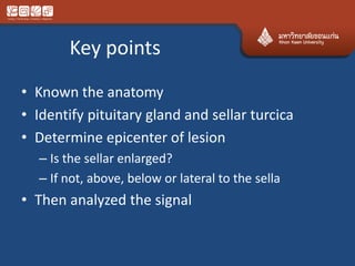
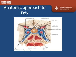

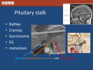
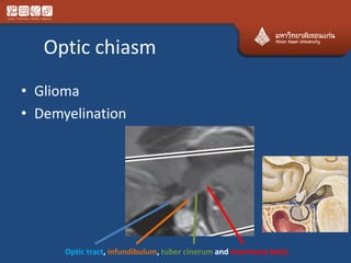
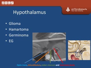
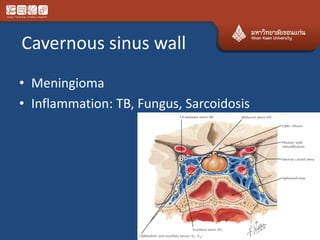
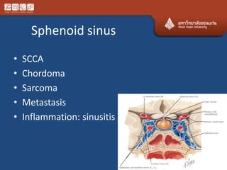
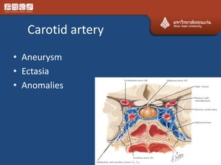
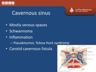
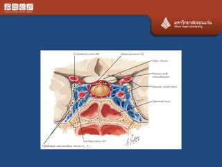
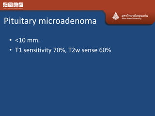
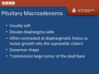
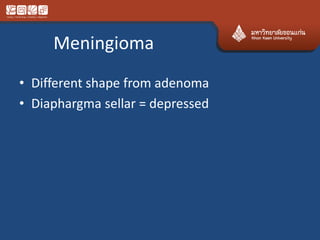
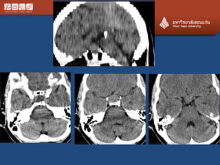
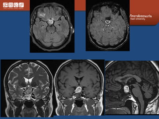
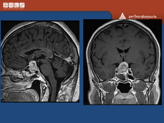
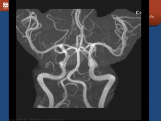
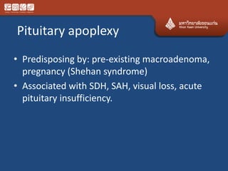
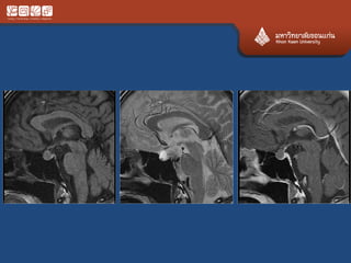

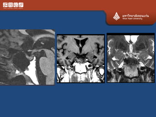
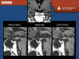
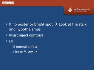

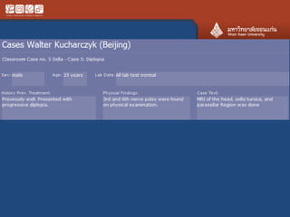
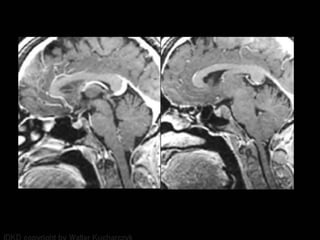
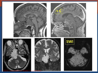
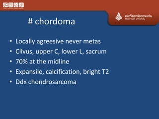
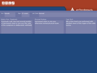


This document provides an anatomical approach and differential diagnosis for lesions in and around the pituitary gland and sellar region. It outlines key points such as identifying the pituitary gland and sellar turcica to determine if the lesion is intrasellar, suprasellar, or lateral to the sella. Common pathologies are then discussed based on their location, such as pituitary adenomas, craniopharyngiomas, meningiomas, and other lesions that can occur in the pituitary stalk, hypothalamus, cavernous sinus, sphenoid sinus, and carotid artery. Imaging characteristics and distinguishing features between pathologies are provided.


![CTEV [ clubfoot] DR ARUN LAL ,DR MOHAMED ASHRAF travancore medical college k...](https://cdn.slidesharecdn.com/ss_thumbnails/ctevclubfootdrarunlaldrmohamedashraftravancoremedicalcollegekollamkeralaindia-260208063247-18fc466c-thumbnail.jpg?width=640&height=640&fit=bounds)


