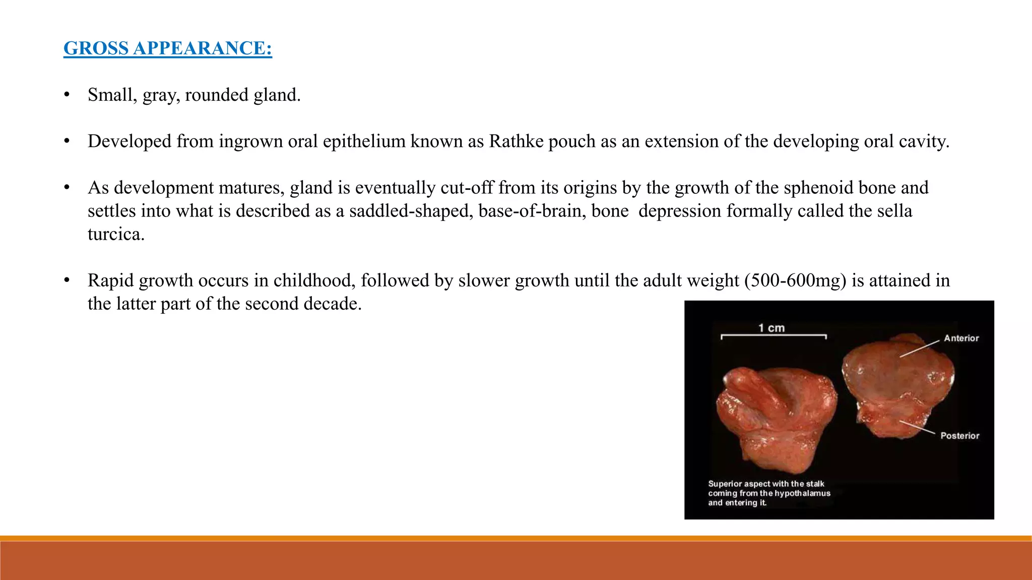
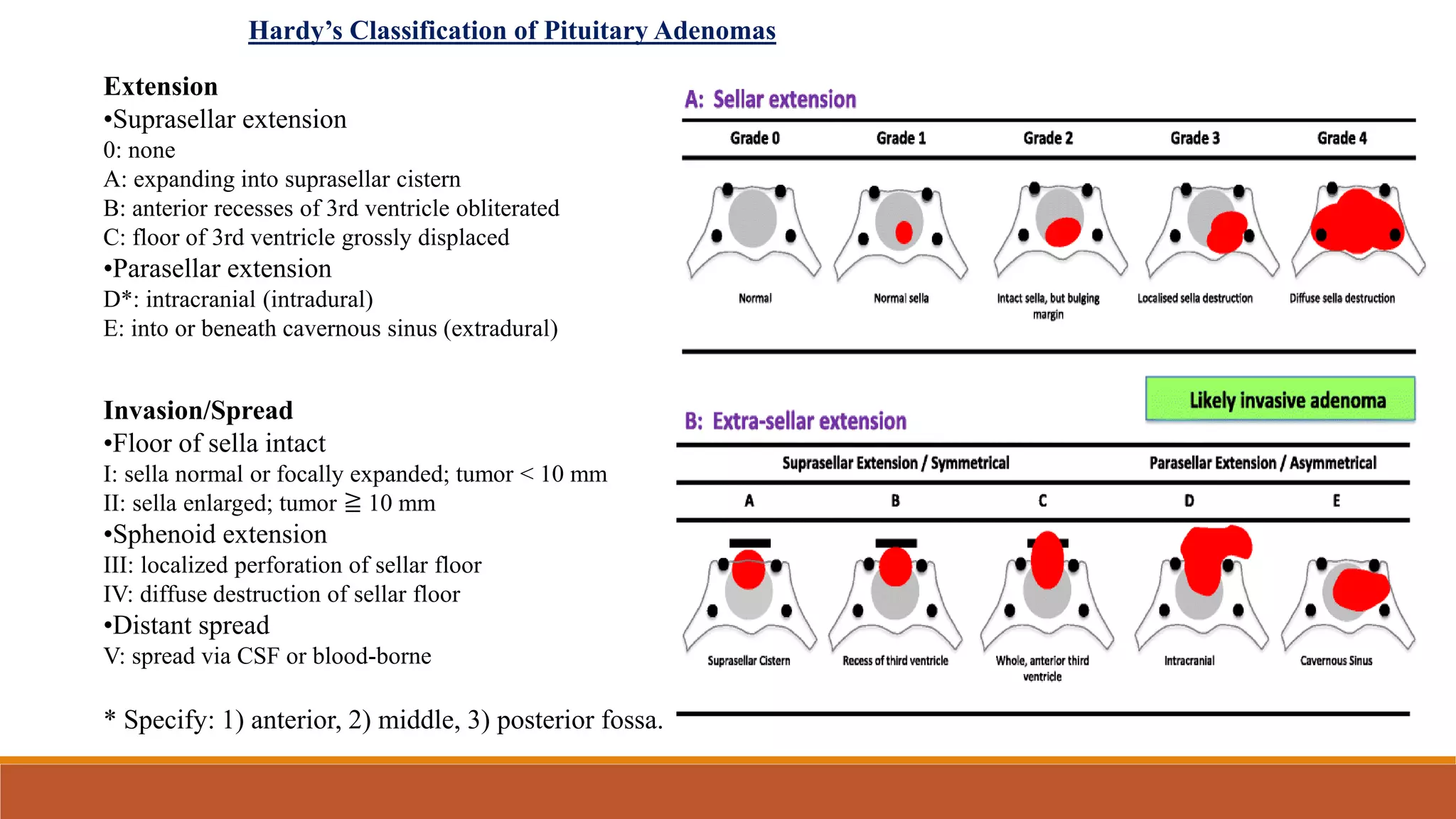
PITUITARY GLAND AND
PITUITARY TUMORS
Presenter: Dr. Monica P
MD Radiation Oncologist
CONTENTS:
• Anatomy
• Embryology
• Physiology
• Introduction of pituitary adenomas
• Incidence
• Classification
• Pathophysiology and Clinical presentation
• Diagnostic workup
• Management by : Surgery, Medical management, Radiotherapy
• Conclusion
ANATOMY:
• Pituitary gland is also known as Hypophysis cerebri
• It’s a reddish-grey, ovoid body, about 12mm in transverse and 8mm in
AP diameter, average adult weight 500mg
• It’s continuous with the infundibulum of the hypothalamus.
• It lies within the pituitary fossa of the sphenoid bone, covered
superiorly by a circular diaphragm sella of duramater.
• Diaphragma sellae is pierced centrally by an aperture for the
infundibulum and separates the anterior superior aspect of the
pituitary from optic chiasma.
• Inferiorly its separated from floor of fossa by a cavernous sinus.
• Meninges blend with the pituitary capsule and are not separate layers.
• 2 major parts of pituitary
gland:
o Neurohypophysis
o Adenohypophysis
• They differ in their
origin, structure and
function.
NEUROHYPOPHYSIS ADENOHYPOPHYSIS
It’s a diencephalic down growth connected
with the hypothalamus.
It’s an ectodermal derivative of the
stomatodeum.
Comprises of:
Median eminence
Infundibular stem
Infundibular process
Neural lobe
Comprises of:
Pars infundibularis (or Pars tuberalis)
Pars intermedia
Pars distalis
Median eminence is frequently also classified
as part of the tuber cinereum.
80% of total volume of pituitary gland
Hypophysial stalk usually refers to pars
infundibularis and infundibulum
Its an embryologic diverticulum from the
buccopharyngeal region.
It develops as a diverticulum from the floor of
the third ventricle.
Pars distalis area secretes a number of
hormones.
BLOOD SUPPLY AND INNERVATIONS:
• Hypophysis is supplied by a series hypophysial arteries from the
internal carotids.
• Maintenance and regulation of the activity of the adenohypophysis
are dependent on the blood supply by way of the hypophysial portal
system.
• Nerve fibres from the hypothalamus liberate releasing factors into
the capillary beds in the infundibulum, and these substances are then
carried by the portal vessels to the distal parts of the gland, causing
the effects relevant to the specific secretions.
• Neurohypophysis receives its main nerve supply from the
hypothalamus by way of fibres known collectively as the
hypothalamohypophysial tract.
• This tract contains two main sets of fibres:
Supraopticohypophysial tract and
Tuberohypophysial tract.
GROSS APPEARANCE:
• Small, gray, rounded gland.
• Developed from ingrown oral epithelium known as Rathke pouch as an extension of the developing oral cavity.
• As development matures, gland is eventually cut-off from its origins by the growth of the sphenoid bone and
settles into what is described as a saddled-shaped, base-of-brain, bone depression formally called the sella
turcica.
• Rapid growth occurs in childhood, followed by slower growth until the adult weight (500-600mg) is attained in
the latter part of the second decade.
DEVELOPMENT OF PITUITARY GLAND:
ANTERIOR PITUITARY GLAND:
• Constitutes the bulk of the gland’s size and weight
• Produces 6 hormones: TSH, ACTH, FSH, LH, GH & prolactin.
• These hormones serve both trophic and hormone stimulatory functions.
• It is the highly vascularized mammalian tissue, receiving about 0.8ml/g/min of blood from the portal system.
POSTERIOR PITUTARY:
• Secretes oxytocin and vasopressin.
• These are produced in the hypothalamus and then pass to the neurohypophysis via 100000 nerve fibres comprising
the hypothalamo-hypophyseal tract.
• From the posterior lobe, the hormones are released into the general circulation in response to electrical activity at
the axon endings.
INTRODUCTION:
• Most common type of pituitary tumor is pituitary adenoma.
• Most pituitary adenomas develop in adenohypophysis.
• Pituitary tumors rarely develop in neurophypophysis.
• These tumors are almost always benign and most are successfully treatable.
INCIDENCE:
• Pituitary tumors account for 12-19% of all primary brain tumors, making them 3rd most common primary brain
tumors in adults.
• It can be found in every age group with increasing incidence with the age, mostly seen after 3rd decade of life.
• These tumors are broadly classified based on whether they secrete excessive amounts of pituitary hormones or
not.
• 2/3rd of the pituitary adenomas are secreting type.
CLASSIFICATION:
PITUITARY ADENOMAS
Non-Functioning PA [NFPA]
Functioning PA [FPA] Macro adenomas [ ≥10mm]
Micro adenomas [<10mm]
• Prolactinomas comprises 40-57% of all adenomas, followed by NFPA (28-37%), GH secreting adenomas (11-
13%) and ACTH secreting adenomas (1-2%).
• Pituitary adenomas that secrete FSH, LH, TSH are rare.
Pico adenomas [<3mm]
Giant adenomas [>4cm]
ETIOLOGY:
Due to hyperplasia to adenoma sequence (hormone dependent)
Genetic alteration (MEN1, PRKAR1A, AIP, CDKN1B)
MEN1:
• autosomal dominant
• characterized by tumors of pancreatic islet cells, parathyroid glands and pituitary glands.
• 40% will develop into pituitary adenoma, most common – prolactinomas
• mutation: 11q13
AIP:
• mutations in Aryl hydrocarbon receptor Interacting Protein are associated with familial pituitary adenomas,
usually somatotrophs or somatomammotroph type.
• present in adolescence/early adulthood
• are rarely aggressive.
PATHOPHYSIOLOGY:
• The pituitary gland sits inferior to the hypothalamus. It is surrounded caudally by the sphenoid bone in a
basketlike structure called the sella turcica, and superiorly by the optic chiasm.
• The sella turcica forces an expanding adenoma superiorly, leading to compression of the optic nerve and
headaches from mass effect.
• Additionally, destruction or compression of the pituitary gland may cause complete or partial hypopituitarism.
• Pituitary adenomas are benign tumors that arise from one of the five cell types that comprise the anterior pituitary
(lactotrophs, gonadotrophs, somatotrophs, corticotrophs, and thyrotrophs).
• Tumors rarely form from a combination of these cells. Pituitary adenomas are true neoplasms with a monoclonal
cell origin.
• Hypersecretion or diminished inhibition of the hormones of the hypothalamic-pituitary axis can lead to the
constellation of endocrine symptoms often seen in patients with pituitary adenomas.
• Secretory tumors typically exert morbidity by causing metabolic, somatic and hormonal dysregulation, in addition to
local mass effects from the tumor growth.
• Non-functioning tumors may impair neurologic function by local tumor extension beyond the confines of the sella
turcica into the adjacent cavernous sinuses, optic chiasma and optic nerves, and comprise hormonal function by
compression of the normal pituitary gland.
• Goals of treatment: to minimize the effects on endocrine and neurologic function.
Symptom Cause
Visual impairment
• Bitemporal hemianopsia
• Diminished visual acuity
• Gradual visual changes
Suprasellar extension (due to compression
of optic chiasma)
Headache Expansion of sella
Diplopia Oculomotor compression(lateral
extension)
Pituitary apoplexy Due to sudden hemorrhage
Seizures Lateral extension to temporal lobe
APPROACH TO THE
EVALUATION AND
MANAGEMENT OF
PITUITARY
INCIDENTALOMA
DIAGNOSTIC
WORKUP:
RADIOLOGICAL WORKUP:
MRI Brain with gadolinium is the best imaging modality over CT
MRI is 100% sensitive and 91% specific while CT is 17-22% sensitive for pituitary gland tumors (patel et al)
Extension
•Suprasellar extension
0: none
A: expanding into suprasellar cistern
B: anterior recesses of 3rd ventricle obliterated
C: floor of 3rd ventricle grossly displaced
•Parasellar extension
D*: intracranial (intradural)
E: into or beneath cavernous sinus (extradural)
Invasion/Spread
•Floor of sella intact
I: sella normal or focally expanded; tumor < 10 mm
II: sella enlarged; tumor ≧ 10 mm
•Sphenoid extension
III: localized perforation of sellar floor
IV: diffuse destruction of sellar floor
•Distant spread
V: spread via CSF or blood-borne
* Specify: 1) anterior, 2) middle, 3) posterior fossa.
Hardy’s Classification of Pituitary Adenomas
MANAGEMENT:
• Medical/ Observation
• Surgery
• Radiotherapy
Treatment goals:
o Restore the mass effect and reverse compression symptoms
o Restore the normal hormone function
OBSERVATION:
• In asymptomatic non secreting microadenomas
• Small asymptomatic prolactinomas : 2 -4 mm  no testing required
5-9 mm  MRI can be done once yearly
• Indications for intervention
Tumor growth on imaging
symptoms of hypersecretion
development of visual field defects
• Long term follow up with MRI and hormonal assays
SURGERY:
• Goals of surgery : Decompression and to normalize hypersecretion , With preservation of normal pituitary
function ( first line treatment )
• Transsphenoidal surgery – standard approach, better and safer than transcranial approach.
• TSS resulting in rapid reduction of tumor, 90% success if small tumor.
• It’s the initial treatment chosen for all adenomas except prolactinomas.
ROLE OF RADIATION
INDICATIONS:
• DEFINITIVE: unresectable/inoperable.
Medically unfit for surgery.
• ADJUVANT :
Recurrence after surgery, and/or refractory to medical management..
Discontinue medical management 1 month prior to RT and resume after RT completed.
Improved response when RT delivered off medical therapy (may alter cell cycle and radiosensitivity).
GOALS
• Goal is to reduce or stabilize mass effect and normalize hormone levels (takes many years).
• Excellent LC of 90% to 100% in most studies regardless of RT technique and adenoma subtype.
• Smaller tumors have improved response and lower risk of hypopituitarism.
SRS versus fractionated RT:
• SRS preferred due to faster time to hormone normalization and patient convenience.
• Fractionated RT if tumor >3 cm or <3–5 mm from chiasm due to risk of visual deficits.
• Risk for hypopituitarism is high for both modalities (20% at 5 yrs; 80% at 10–15 years).
• Panhypopituitarism occurs in 5% to 10% pts at 5 yrs.
Dose:
• SRS:
14 to 16 Gy for nonsecretory tumors;
20 Gy or higher for secretory tumors.
• Fractionated RT:
45 Gy/25 # for nonsecretory;
50.4 to 54 Gy/28 to 30 # for secretory.
SRS for non functioning pituitary adenoma
Authors Patients Mean dose (Gy) Follow-up
(months)
Tumor control
(%)
Late toxicity (%)
Visual Hypopituitarism
Liscak et al., 2007 140 20 60 100 0 2
Pollock et al.,
2008
62 16 64 95 at 5 years 0 27
Gopalan et al.,
2011
48 18.4 95 83 0 39
Park et al., 2011 125 13 62 94 at 5 years 0.8 24
Starke et al., 2012 140 18 50 97 at 5 years 12.8 30.3
Runge et al., 2012 61 13 83 98 0 9.8
Wilson et al.,
2012
51 14 50 100 at 5 years 0 NA
Sheehan et al.,
2013
512 16 36 95 at 5 years 6.6 21
Lee et al., 2014 41 12 48 85 at 10 years 2.4 24.4
FSRT for pituitary adenomas
Authors Type of
adenoma
Patients Mean dose
(Gy)
Follow-up
(months)
Tumor control
(%)
Late toxicity (%)
Visual Hypopituitarism
Milker-Zabel et
al., 2004
GH 20 52.2 26 100 (92a) 0 3
Paek et al., 2005 NFA, SA 68 50 30 98 at 5 years 3 6
Colin et al., 2005 NFA, SA 110 50.4 48 99 at 5 years 1.8 29 at 4 years
Minniti et al.,
2006
NFA, SA 92 45 32 98 at 5 years 1 22
Kong et al., 2007 NFA, SA 66 50.4 36.7 97 0 27.3 at 5 years
Roug et al., 2010 GH 34 54 34 91 (30a) 0 29 at 4 years
Schalin-Jantti et
al., 2010
NFA, SA 30 45 64 100 0 40
Wilson et al., 2012 NFA 67 50 60.1 93 at 5 years 1.5 7
Kopp et al., 2013 NFA, SA 37 49.4 57 91.9 5 5
Kim et al., 2013 NFA, SA 76 50.4 80 97.1 at 7 years 0 48
Fractionated radiotherapy
DRUG Hormonal
control (%)
Octreotide 20-35%
Lanreotide 20-35%
Pegvisomant 63-90%
Cabergoline 80-90%
Bromocriptine 60-80%
CONVENTIONAL RT:
o 2 Fields technique:
• by opening a 5x5 field with centre at pituitary
• Surface marking: 2.5cm anterior and superior to external auditory meatus [point of base of sella turcica/middle of
pituitary]
• Treatment volume will include pituitary gland with margin.
• Problem: high dose to temporal lobe
o 3 fields technique:
• Additional parallel opposed with a vertex beam used, to
obtain a better distribution and prevent dose to temporal
lobe, auditory structures.
• But the beam may pass through eyes.
• So, during simulation:
Anterior wedges are put below the head rest and patient is
immobilized in a position where the neck is in forward flexion
position and is simulated. So the 2 lateral fields and 3rd field
from superior aspect bypasses the eyes
CONFORMAL RT PLANNING:
• GTV : residual tumor
• CTV: GTV+ 5mm
• PTV: CTV + 2-5mm
• 3DCRT
• IMRT and VMAT are commonly used now.
• Dose: 45-50Gy
With conventional fractionation, overall 10yr control rates are in the range of 85%. However, hormonal
activity normalization is less robust.
• For SRS: optic nerve/optic chiasma is the deciding factors.
Its best suited for small tumors (< 3cms) and 2-5mm away from optic apparatus.
CYBERKNIFE
GAMMA KNIFE
SIDE-EFFECTS OF THE THERAPY:
• Hypopituitarism warranting lifelong supplementation
• Optic or other cranial neuropathies
• Second malignancy (risk of developing meningioma/glioma in 2% at 20yrs)
CONCLUSION:
• Adequate medical knowledge and understanding of the pituitary gland include understanding multiple
feedback mechanisms required for the hormone normal regulatory processes of the human body.
• Knowledge of therapy mechanisms for abnormalities plays an essential role for medical intervention,
especially for the appropriate use of radiotherapy.
• Stereotactic irradiation remains an effective treatment modality for patients with both nonfunctioning and
secreting pituitary adenomas after unsuccessful surgery and/or resistant to medical therapy.
• Both SRS and FSRT provide excellent tumor control in the range of 85–95% at 5–10 years, with
normalization of hormone hypersecretion in more than 50% of patients.
• Hypopituitarism represents the most commonly reported late complication of treatment, whereas the
reported incidence of other late effect radiation complications are low.
PITUITARY TUMORS.pptx

PITUITARY TUMORS.pptx

  • 1.
    PITUITARY GLAND AND PITUITARYTUMORS Presenter: Dr. Monica P MD Radiation Oncologist
  • 2.
    CONTENTS: • Anatomy • Embryology •Physiology • Introduction of pituitary adenomas • Incidence • Classification • Pathophysiology and Clinical presentation • Diagnostic workup • Management by : Surgery, Medical management, Radiotherapy • Conclusion
  • 3.
    ANATOMY: • Pituitary glandis also known as Hypophysis cerebri • It’s a reddish-grey, ovoid body, about 12mm in transverse and 8mm in AP diameter, average adult weight 500mg • It’s continuous with the infundibulum of the hypothalamus. • It lies within the pituitary fossa of the sphenoid bone, covered superiorly by a circular diaphragm sella of duramater. • Diaphragma sellae is pierced centrally by an aperture for the infundibulum and separates the anterior superior aspect of the pituitary from optic chiasma. • Inferiorly its separated from floor of fossa by a cavernous sinus. • Meninges blend with the pituitary capsule and are not separate layers.
  • 4.
    • 2 majorparts of pituitary gland: o Neurohypophysis o Adenohypophysis • They differ in their origin, structure and function. NEUROHYPOPHYSIS ADENOHYPOPHYSIS It’s a diencephalic down growth connected with the hypothalamus. It’s an ectodermal derivative of the stomatodeum. Comprises of: Median eminence Infundibular stem Infundibular process Neural lobe Comprises of: Pars infundibularis (or Pars tuberalis) Pars intermedia Pars distalis Median eminence is frequently also classified as part of the tuber cinereum. 80% of total volume of pituitary gland Hypophysial stalk usually refers to pars infundibularis and infundibulum Its an embryologic diverticulum from the buccopharyngeal region. It develops as a diverticulum from the floor of the third ventricle. Pars distalis area secretes a number of hormones.
  • 5.
    BLOOD SUPPLY ANDINNERVATIONS: • Hypophysis is supplied by a series hypophysial arteries from the internal carotids. • Maintenance and regulation of the activity of the adenohypophysis are dependent on the blood supply by way of the hypophysial portal system. • Nerve fibres from the hypothalamus liberate releasing factors into the capillary beds in the infundibulum, and these substances are then carried by the portal vessels to the distal parts of the gland, causing the effects relevant to the specific secretions. • Neurohypophysis receives its main nerve supply from the hypothalamus by way of fibres known collectively as the hypothalamohypophysial tract. • This tract contains two main sets of fibres: Supraopticohypophysial tract and Tuberohypophysial tract.
  • 6.
    GROSS APPEARANCE: • Small,gray, rounded gland. • Developed from ingrown oral epithelium known as Rathke pouch as an extension of the developing oral cavity. • As development matures, gland is eventually cut-off from its origins by the growth of the sphenoid bone and settles into what is described as a saddled-shaped, base-of-brain, bone depression formally called the sella turcica. • Rapid growth occurs in childhood, followed by slower growth until the adult weight (500-600mg) is attained in the latter part of the second decade.
  • 7.
  • 9.
    ANTERIOR PITUITARY GLAND: •Constitutes the bulk of the gland’s size and weight • Produces 6 hormones: TSH, ACTH, FSH, LH, GH & prolactin. • These hormones serve both trophic and hormone stimulatory functions. • It is the highly vascularized mammalian tissue, receiving about 0.8ml/g/min of blood from the portal system. POSTERIOR PITUTARY: • Secretes oxytocin and vasopressin. • These are produced in the hypothalamus and then pass to the neurohypophysis via 100000 nerve fibres comprising the hypothalamo-hypophyseal tract. • From the posterior lobe, the hormones are released into the general circulation in response to electrical activity at the axon endings.
  • 11.
    INTRODUCTION: • Most commontype of pituitary tumor is pituitary adenoma. • Most pituitary adenomas develop in adenohypophysis. • Pituitary tumors rarely develop in neurophypophysis. • These tumors are almost always benign and most are successfully treatable.
  • 12.
    INCIDENCE: • Pituitary tumorsaccount for 12-19% of all primary brain tumors, making them 3rd most common primary brain tumors in adults. • It can be found in every age group with increasing incidence with the age, mostly seen after 3rd decade of life. • These tumors are broadly classified based on whether they secrete excessive amounts of pituitary hormones or not. • 2/3rd of the pituitary adenomas are secreting type.
  • 13.
    CLASSIFICATION: PITUITARY ADENOMAS Non-Functioning PA[NFPA] Functioning PA [FPA] Macro adenomas [ ≥10mm] Micro adenomas [<10mm] • Prolactinomas comprises 40-57% of all adenomas, followed by NFPA (28-37%), GH secreting adenomas (11- 13%) and ACTH secreting adenomas (1-2%). • Pituitary adenomas that secrete FSH, LH, TSH are rare. Pico adenomas [<3mm] Giant adenomas [>4cm]
  • 14.
    ETIOLOGY: Due to hyperplasiato adenoma sequence (hormone dependent) Genetic alteration (MEN1, PRKAR1A, AIP, CDKN1B) MEN1: • autosomal dominant • characterized by tumors of pancreatic islet cells, parathyroid glands and pituitary glands. • 40% will develop into pituitary adenoma, most common – prolactinomas • mutation: 11q13 AIP: • mutations in Aryl hydrocarbon receptor Interacting Protein are associated with familial pituitary adenomas, usually somatotrophs or somatomammotroph type. • present in adolescence/early adulthood • are rarely aggressive.
  • 15.
    PATHOPHYSIOLOGY: • The pituitarygland sits inferior to the hypothalamus. It is surrounded caudally by the sphenoid bone in a basketlike structure called the sella turcica, and superiorly by the optic chiasm. • The sella turcica forces an expanding adenoma superiorly, leading to compression of the optic nerve and headaches from mass effect. • Additionally, destruction or compression of the pituitary gland may cause complete or partial hypopituitarism. • Pituitary adenomas are benign tumors that arise from one of the five cell types that comprise the anterior pituitary (lactotrophs, gonadotrophs, somatotrophs, corticotrophs, and thyrotrophs). • Tumors rarely form from a combination of these cells. Pituitary adenomas are true neoplasms with a monoclonal cell origin. • Hypersecretion or diminished inhibition of the hormones of the hypothalamic-pituitary axis can lead to the constellation of endocrine symptoms often seen in patients with pituitary adenomas.
  • 18.
    • Secretory tumorstypically exert morbidity by causing metabolic, somatic and hormonal dysregulation, in addition to local mass effects from the tumor growth. • Non-functioning tumors may impair neurologic function by local tumor extension beyond the confines of the sella turcica into the adjacent cavernous sinuses, optic chiasma and optic nerves, and comprise hormonal function by compression of the normal pituitary gland. • Goals of treatment: to minimize the effects on endocrine and neurologic function. Symptom Cause Visual impairment • Bitemporal hemianopsia • Diminished visual acuity • Gradual visual changes Suprasellar extension (due to compression of optic chiasma) Headache Expansion of sella Diplopia Oculomotor compression(lateral extension) Pituitary apoplexy Due to sudden hemorrhage Seizures Lateral extension to temporal lobe
  • 19.
    APPROACH TO THE EVALUATIONAND MANAGEMENT OF PITUITARY INCIDENTALOMA
  • 20.
  • 22.
    RADIOLOGICAL WORKUP: MRI Brainwith gadolinium is the best imaging modality over CT MRI is 100% sensitive and 91% specific while CT is 17-22% sensitive for pituitary gland tumors (patel et al)
  • 23.
    Extension •Suprasellar extension 0: none A:expanding into suprasellar cistern B: anterior recesses of 3rd ventricle obliterated C: floor of 3rd ventricle grossly displaced •Parasellar extension D*: intracranial (intradural) E: into or beneath cavernous sinus (extradural) Invasion/Spread •Floor of sella intact I: sella normal or focally expanded; tumor < 10 mm II: sella enlarged; tumor ≧ 10 mm •Sphenoid extension III: localized perforation of sellar floor IV: diffuse destruction of sellar floor •Distant spread V: spread via CSF or blood-borne * Specify: 1) anterior, 2) middle, 3) posterior fossa. Hardy’s Classification of Pituitary Adenomas
  • 24.
    MANAGEMENT: • Medical/ Observation •Surgery • Radiotherapy Treatment goals: o Restore the mass effect and reverse compression symptoms o Restore the normal hormone function
  • 28.
    OBSERVATION: • In asymptomaticnon secreting microadenomas • Small asymptomatic prolactinomas : 2 -4 mm  no testing required 5-9 mm  MRI can be done once yearly • Indications for intervention Tumor growth on imaging symptoms of hypersecretion development of visual field defects • Long term follow up with MRI and hormonal assays
  • 29.
    SURGERY: • Goals ofsurgery : Decompression and to normalize hypersecretion , With preservation of normal pituitary function ( first line treatment ) • Transsphenoidal surgery – standard approach, better and safer than transcranial approach. • TSS resulting in rapid reduction of tumor, 90% success if small tumor. • It’s the initial treatment chosen for all adenomas except prolactinomas.
  • 30.
    ROLE OF RADIATION INDICATIONS: •DEFINITIVE: unresectable/inoperable. Medically unfit for surgery. • ADJUVANT : Recurrence after surgery, and/or refractory to medical management.. Discontinue medical management 1 month prior to RT and resume after RT completed. Improved response when RT delivered off medical therapy (may alter cell cycle and radiosensitivity). GOALS • Goal is to reduce or stabilize mass effect and normalize hormone levels (takes many years). • Excellent LC of 90% to 100% in most studies regardless of RT technique and adenoma subtype. • Smaller tumors have improved response and lower risk of hypopituitarism.
  • 31.
    SRS versus fractionatedRT: • SRS preferred due to faster time to hormone normalization and patient convenience. • Fractionated RT if tumor >3 cm or <3–5 mm from chiasm due to risk of visual deficits. • Risk for hypopituitarism is high for both modalities (20% at 5 yrs; 80% at 10–15 years). • Panhypopituitarism occurs in 5% to 10% pts at 5 yrs. Dose: • SRS: 14 to 16 Gy for nonsecretory tumors; 20 Gy or higher for secretory tumors. • Fractionated RT: 45 Gy/25 # for nonsecretory; 50.4 to 54 Gy/28 to 30 # for secretory.
  • 32.
    SRS for nonfunctioning pituitary adenoma Authors Patients Mean dose (Gy) Follow-up (months) Tumor control (%) Late toxicity (%) Visual Hypopituitarism Liscak et al., 2007 140 20 60 100 0 2 Pollock et al., 2008 62 16 64 95 at 5 years 0 27 Gopalan et al., 2011 48 18.4 95 83 0 39 Park et al., 2011 125 13 62 94 at 5 years 0.8 24 Starke et al., 2012 140 18 50 97 at 5 years 12.8 30.3 Runge et al., 2012 61 13 83 98 0 9.8 Wilson et al., 2012 51 14 50 100 at 5 years 0 NA Sheehan et al., 2013 512 16 36 95 at 5 years 6.6 21 Lee et al., 2014 41 12 48 85 at 10 years 2.4 24.4
  • 33.
    FSRT for pituitaryadenomas Authors Type of adenoma Patients Mean dose (Gy) Follow-up (months) Tumor control (%) Late toxicity (%) Visual Hypopituitarism Milker-Zabel et al., 2004 GH 20 52.2 26 100 (92a) 0 3 Paek et al., 2005 NFA, SA 68 50 30 98 at 5 years 3 6 Colin et al., 2005 NFA, SA 110 50.4 48 99 at 5 years 1.8 29 at 4 years Minniti et al., 2006 NFA, SA 92 45 32 98 at 5 years 1 22 Kong et al., 2007 NFA, SA 66 50.4 36.7 97 0 27.3 at 5 years Roug et al., 2010 GH 34 54 34 91 (30a) 0 29 at 4 years Schalin-Jantti et al., 2010 NFA, SA 30 45 64 100 0 40 Wilson et al., 2012 NFA 67 50 60.1 93 at 5 years 1.5 7 Kopp et al., 2013 NFA, SA 37 49.4 57 91.9 5 5 Kim et al., 2013 NFA, SA 76 50.4 80 97.1 at 7 years 0 48 Fractionated radiotherapy
  • 34.
    DRUG Hormonal control (%) Octreotide20-35% Lanreotide 20-35% Pegvisomant 63-90% Cabergoline 80-90% Bromocriptine 60-80%
  • 35.
    CONVENTIONAL RT: o 2Fields technique: • by opening a 5x5 field with centre at pituitary • Surface marking: 2.5cm anterior and superior to external auditory meatus [point of base of sella turcica/middle of pituitary] • Treatment volume will include pituitary gland with margin. • Problem: high dose to temporal lobe
  • 36.
    o 3 fieldstechnique: • Additional parallel opposed with a vertex beam used, to obtain a better distribution and prevent dose to temporal lobe, auditory structures. • But the beam may pass through eyes. • So, during simulation: Anterior wedges are put below the head rest and patient is immobilized in a position where the neck is in forward flexion position and is simulated. So the 2 lateral fields and 3rd field from superior aspect bypasses the eyes
  • 37.
    CONFORMAL RT PLANNING: •GTV : residual tumor • CTV: GTV+ 5mm • PTV: CTV + 2-5mm • 3DCRT • IMRT and VMAT are commonly used now. • Dose: 45-50Gy With conventional fractionation, overall 10yr control rates are in the range of 85%. However, hormonal activity normalization is less robust. • For SRS: optic nerve/optic chiasma is the deciding factors. Its best suited for small tumors (< 3cms) and 2-5mm away from optic apparatus.
  • 38.
  • 39.
    SIDE-EFFECTS OF THETHERAPY: • Hypopituitarism warranting lifelong supplementation • Optic or other cranial neuropathies • Second malignancy (risk of developing meningioma/glioma in 2% at 20yrs)
  • 40.
    CONCLUSION: • Adequate medicalknowledge and understanding of the pituitary gland include understanding multiple feedback mechanisms required for the hormone normal regulatory processes of the human body. • Knowledge of therapy mechanisms for abnormalities plays an essential role for medical intervention, especially for the appropriate use of radiotherapy. • Stereotactic irradiation remains an effective treatment modality for patients with both nonfunctioning and secreting pituitary adenomas after unsuccessful surgery and/or resistant to medical therapy. • Both SRS and FSRT provide excellent tumor control in the range of 85–95% at 5–10 years, with normalization of hormone hypersecretion in more than 50% of patients. • Hypopituitarism represents the most commonly reported late complication of treatment, whereas the reported incidence of other late effect radiation complications are low.

Editor's Notes

  • #4 Sella turcica/ pituitary fossa is a midline dural-lined structure in sphenoid bone. Sphenoid bone has anteriorly and posteriorly processes called tuberculum sellae and dorsum sellae. And outpouches called anterior and posterior clinoid processes.
  • #14 It may increase the hormone production from the cell it arises from, or may decrease the production of hormones from other cells.
  • #15 MEN: Multiple endocrine neoplasia PRKAR1A: Protein Kinase cAMP dependent type 1 regulatory subunit alpha (protein coding gene) CDKN1B: Cyclin dependent kinase inhibitor 1B (protein coding gene) MEN2A, 2B: medullary thyroid ca,pheochromocytoma, parathyroid gland cancer
  • #17 Dexamethasone lowers the adrenal cortical production of cortisol by 50mcg/L over 8hrs
  • #18 Oral glucose suppression test: glucose will stimulate insulin isulin will suppress the counterregulatory hormones(GH) if GH is not suppressed then it implies there is high GH.
  • #22 Other causes of prolactinemia: pregnancy, hypothyroidism, renal insufficiency
  • #26 For microadenomas/incidentalomas without symptoms tumor regrowth rate is 10%. Therefore observation with annual MRI and followup is valid option.
  • #27 Prolactinemia: seen in 20-50yrs age women, 90% are microadenomas Approximately 80% of patients will have their prolactin levels restored to normal with dopamine agonist therapy.  Repeat IGF-1 & GH levels during oral GTT about 12weeks post surgery along with MRI.
  • #35 Ketoconazole, an imidazole derivative which inhibits adrenal steroidogenesis, has been used with success for the metabolic control of Cushing's disease. Metyrapone blocks cortisol biosynthesis by acting as a reversible inhibitor of 11β-hydroxylase. This stimulates adrenocorticotropic hormone (ACTH) secretion, which in turn increases plasma 11-deoxycortisol levels. Nelson's syndrome is a potentially life-threatening condition which occurs when an adrenocorticotrophic hormone (ACTH) secreting tumour develops following therapeutic total bilateral adrenalectomy (TBA) for Cushing's disease. It involves enlargement of an existing adrenocorticotropic hormone-secreting pituitary tumor, typically an adenoma, after surgical resection of the adrenal glands. Octreotide, being a somatostatin analog, inhibits the release of GH from the pituitary gland through a process normally involved in negative feedback