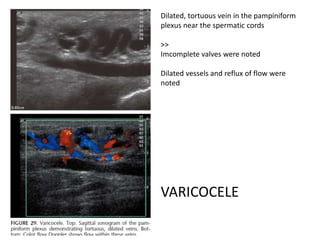
Scrotum echo 
Ultrasound Quarterly 2004;20:181-200
Seminoma 
Most common 
Most well marginated 
Most hypoechoiec 
No cyst or calcifications
Microlithiasis was thought to 
Be related to SEMINOMA 
In the patient, 
There is no obvious mass or 
hypoechoiec lesion in the echo
Embryonal cell carcinoma 
Inhomogenous 
Poorly marginated 
Cystic lesions 
>>check lab
teratoma 
Most teratoma + embryonal carcinoma 
Well defined but heterogenous texture 
Cyst: +, calcifications:+
Benign testicular condition
Cyst of tunica albuginea 
Maybe in the testis or extra-testis, 
almost less than 2cm 
Maybe multifocal
Tubular ectasia 
of the rete testis 
>> 
Multiple dilated tubular 
structure in the 
mediastinum testis 
No flow
Testicular abscess: 
Combined with UTI 
Complications of torsion, testicular 
hemorrhage, secondary to trauma 
Clinical finding: 
Fever and leukocytosis
torsion
• In the first 6 hrs, testis would become 
heterogenous hypoechoiec echo pattern 
• Nuclear flow was used but not clinical used in 
some hospital.
Testicular microlithiasis
Testis microlithiasis 
• If the calcifications more than 5 spots and 
measure 1-2mm per spots, microlithiasis is 
impressed. 
• Testis microlithiasis is related to the testis 
malignancy
Epididymitis/epididymo-orchitis
Chronic epididymo-orchitis
• Chronic epididymitis result from acute 
incomplete treatment or tuberculosis 
• Coarse calcifications and thickening of the 
tunica albuginea was noted
Spermatocele 
Sperm fluid 
accumulation: 
Sometimes occur especially in 
the post vasectomy syndrome
hydrocele 
Scrotum and peritoneum 
persistent communication 
Processus vaginalis: 
Resolved by 1.5 years
Dilated, tortuous vein in the pampiniform 
plexus near the spermatic cords 
>> 
Imcomplete valves were noted 
Dilated vessels and reflux of flow were 
noted 
VARICOCELE
SCROTAL HERNIA

Scrotum echo