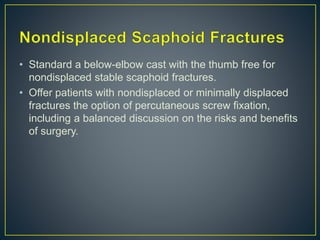
• Scaphoid comes from the
Greek word “skaphos”
meaning boat, a reference to
the shape of the bone.
• The position of the scaphoid
on the radial side of the
wrist, as the proximal
extension of the thumb ray,
makes it vulnerable to injury.
• It is located at a 45-degree plane to the longitudinal and
horizontal axes of the wrist. It has a reduced capacity for
periosteal healing and an increased tendency for delayed
union and nonunion because just over 80% of its surface
is articular cartilage.
• There are no tendon attachments to the scaphoid.
• The scaphoid is ridged across its nonarticular dorsoradial
surface, along which the critical dorsal ridge vessels
traverse.
• Largely retrograde blood supply.
• Dorsal branch: Enters via the
small foramina along the
spiralgroove and dorsal ridge of
the scaphoid and supplies 70%
to80% of the scaphoid
proximally, including the proximal
pole.
• Volar branch: Enters via the
scaphoid tubercle and supplies
the remaining 20% to 30% of
distal scaphoid
• Male predominance is seen with a male-to-female ratio of
approximately 2.5:1.
• 90% recalling a hyperextension injury.
• 30% to 40% of SUSPECTED scaphoid fractures are not
identified on initial assessment and investigation with
standard four-view radiographs.
• Occult fractures of the scaphoid detected
10-14 days of injury.
• 2 to 7% of all fractures.
• 70 to 80% of carpal fractures.
• Waist (70%) are the commonest type in adults.
• Distal pole fractures (10-20%).
• Proximal pole fractures (5-10%).
• Tubercle fractures (5%).
• Acute scaphoid fractures can be difficult to diagnose.
• Combination of ASB tenderness, scaphoid tubercle
tenderness, and ASB pain on longitudinal compression of
the thumb generated a sensitivity of 100% and a
specificity of 74%. However, only valid for the first 24
hours.
• Specificity of ASB pain on ulnar deviation of the pronated
wrist, had a negative predictiveb value (NPV) of 100%
,negative test patients could be safely discharged.
• Dark line that
represents the dorsal
lip of the radius, and
the dorsal ridge can
seem like a fracture in
a semisupinated view.
Mach effect (a dark
line that represents the
dorsal lip of the radius)
(black arrow), a white
line that represents the
proximal side of the
scaphoid tuberosity
(white arrow).
• Use of high–spatial-resolution sonography for detecting
the suspected scaphoid fracture, with the sensitivity rising
up to 100% and the specificity as high as 91%.
• MRI is argued to be the best investigation, it was found
that the sensitivity and specificity of MRI were 100%.
• Sensitive for the
diagnosis of the
occult scaphoid
fracture .
• To exclude a
fracture and remove
the unnecessary
cast.
• Russe classification
• AO classification
• Herbert and Fisher classification
• Mayo classification
• >1 mm of fracture displacement.
• A lateral intrascaphoid angle of >35 degrees .
• Bone loss or comminution.
• Fracture malalignment.
• Proximal pole fractures.
• DISI deformity.
• Perilunate fracture-dislocation.
• A scapholunate angle more than 60° or a lunocapitate
angle more than 15° also make the fracture unstable.
• Associated injuries in 10% of all scaphoid
fractures, following a high-energy mode of
injury.
• Proximal radial fractures are most
frequently seen.
• Concomitant fracture of the distal radius
can be indicative of more serious
ligamentous disruption and carpal
instability.
• Patients with a suspected occult scaphoid fracture are
reevaluated after 1 to 2 weeks of immobilization .
• Posfracture CT/X ray.
• Further 6 weeks of immobilisation before offering
operative fixation if at 6 weeks the CT shows an
unhealed fracture.
Scaphoid Tubercle Fractures
• Avulsion injuries.
• For tubercle fractures, we recommend 3 to 4 weeks in a
splint followed by active mobilization.
• Standard a below-elbow cast with the thumb free for
nondisplaced stable scaphoid fractures.
• Offer patients with nondisplaced or minimally displaced
fractures the option of percutaneous screw fixation,
including a balanced discussion on the risks and benefits
of surgery.
• Arthroscopic-assisted fixation or ORIF of the scaphoid is
recommended if there is any gapping or angulation in the
scaphoid, even if the fracture appears stable and
impacted, because our impression is that displaced
fractures are unstable.
• Relative risk of nonunion for proximal pole fractures
(Herbert B3) was 7.5 times more when compared with
more distal fractures when all were managed
nonoperatively.
• For proximal pole fractures we recommend operative
treatment using a small open dorsal approach to check
alignment in case the fracture is unstable.
• Controversy exists regarding the management
techniques for nondisplaced or minimally displaced waist
fractures (Herbert A2, B1, and B2).
• There is an increasing body of evidence to support early
percutaneous screw fixation of these fractures.
NON OPERATIVE OPERATIVE(ORIF)
EARLY REHABILITATION LATE REHABILITATION
DELAY RETURN TO WORK EARLY RETURN TO WORK
LOW COMPLICATIONS` HIGH COMPLICATIONS
RELATIVELY
- SCAPHOTRAPEZOIDAL
RTHRITIS?
HIGH CHANCE OF DELAYED &
NONUNION
LESS CHANCE
3-20% CHANCE OF
DISPLACEMENT IN CAST
RIGID FIXATION ,Good
compression
90-95% UNION
On current evidence neither method is clearly superior, not only with surgical
management associated with improved functional outcome, a more rapid return
to function/sports/work and superior union rates, but also with a significantly
increased rate of complications.
MORE TIME EXTERNAL
IMMOBILAZTION
LESS TIME EXTERNAL
IMMOMOBILIZATION
• Simple hyperextension of the wrist can
achieve good reduction.
• Minimally invasive technique.
• Low complication rate.
• Lesser,faster time for union.
• Rapid return to function.
• Reduced scar & CRPS.
• Above elbow cast.
• Colles cast.
• Scaphoid cast.
Above-elbow casting,
casting with or without the thumb included, and casting with
the wrist in 20 degrees of flexion to 20 degrees of
extension found no significant differences in union rate,
pain, grip strength, time to union, or osteonecrosis.
• Long-arm casting is recommended.
• Immobilisation in a Colles’ cast with the wrist in 20-
degree extension is therefore recommended.
• Immobilisation of the thumb did not influence union rates
scaphoid cast, leaving the interphalangeal joint of the
thumb free.
• Immobilisation.
• Stiffness,
• Diminished grip strength, and
• Delayed return to work.
• Pseudoarthrosis ensues in approximately 4% of patients
who only have casting, and is usually associated with
vertical oblique fracture patterns (due to tilting and
shearing forces) and diastasis between bone fragments.
• Trend toward earlier operative fixation of nondisplaced
fractures TO REDUCE IMMOBILISATION period.
• Surgery may be considered if new healing activity is not
evident and if union is not apparent after a trial of cast
immobilization for about 20 weeks.
• Neglected scaphoid fractures ,100% of patients
developed radiographic osteoarthritis, mostly of the
radiocarpal joint.
ScaPHOID #
ScaPHOID #
ScaPHOID #

ScaPHOID #

  • 2.
    • Scaphoid comesfrom the Greek word “skaphos” meaning boat, a reference to the shape of the bone. • The position of the scaphoid on the radial side of the wrist, as the proximal extension of the thumb ray, makes it vulnerable to injury.
  • 3.
    • It islocated at a 45-degree plane to the longitudinal and horizontal axes of the wrist. It has a reduced capacity for periosteal healing and an increased tendency for delayed union and nonunion because just over 80% of its surface is articular cartilage. • There are no tendon attachments to the scaphoid. • The scaphoid is ridged across its nonarticular dorsoradial surface, along which the critical dorsal ridge vessels traverse.
  • 4.
    • Largely retrogradeblood supply. • Dorsal branch: Enters via the small foramina along the spiralgroove and dorsal ridge of the scaphoid and supplies 70% to80% of the scaphoid proximally, including the proximal pole. • Volar branch: Enters via the scaphoid tubercle and supplies the remaining 20% to 30% of distal scaphoid
  • 6.
    • Male predominanceis seen with a male-to-female ratio of approximately 2.5:1. • 90% recalling a hyperextension injury. • 30% to 40% of SUSPECTED scaphoid fractures are not identified on initial assessment and investigation with standard four-view radiographs. • Occult fractures of the scaphoid detected 10-14 days of injury.
  • 7.
    • 2 to7% of all fractures. • 70 to 80% of carpal fractures. • Waist (70%) are the commonest type in adults. • Distal pole fractures (10-20%). • Proximal pole fractures (5-10%). • Tubercle fractures (5%).
  • 9.
    • Acute scaphoidfractures can be difficult to diagnose. • Combination of ASB tenderness, scaphoid tubercle tenderness, and ASB pain on longitudinal compression of the thumb generated a sensitivity of 100% and a specificity of 74%. However, only valid for the first 24 hours. • Specificity of ASB pain on ulnar deviation of the pronated wrist, had a negative predictiveb value (NPV) of 100% ,negative test patients could be safely discharged.
  • 14.
    • Dark linethat represents the dorsal lip of the radius, and the dorsal ridge can seem like a fracture in a semisupinated view. Mach effect (a dark line that represents the dorsal lip of the radius) (black arrow), a white line that represents the proximal side of the scaphoid tuberosity (white arrow).
  • 15.
    • Use ofhigh–spatial-resolution sonography for detecting the suspected scaphoid fracture, with the sensitivity rising up to 100% and the specificity as high as 91%. • MRI is argued to be the best investigation, it was found that the sensitivity and specificity of MRI were 100%.
  • 18.
    • Sensitive forthe diagnosis of the occult scaphoid fracture . • To exclude a fracture and remove the unnecessary cast.
  • 20.
    • Russe classification •AO classification • Herbert and Fisher classification • Mayo classification
  • 22.
    • >1 mmof fracture displacement. • A lateral intrascaphoid angle of >35 degrees . • Bone loss or comminution. • Fracture malalignment. • Proximal pole fractures. • DISI deformity. • Perilunate fracture-dislocation. • A scapholunate angle more than 60° or a lunocapitate angle more than 15° also make the fracture unstable.
  • 26.
    • Associated injuriesin 10% of all scaphoid fractures, following a high-energy mode of injury. • Proximal radial fractures are most frequently seen. • Concomitant fracture of the distal radius can be indicative of more serious ligamentous disruption and carpal instability.
  • 27.
    • Patients witha suspected occult scaphoid fracture are reevaluated after 1 to 2 weeks of immobilization . • Posfracture CT/X ray. • Further 6 weeks of immobilisation before offering operative fixation if at 6 weeks the CT shows an unhealed fracture. Scaphoid Tubercle Fractures • Avulsion injuries. • For tubercle fractures, we recommend 3 to 4 weeks in a splint followed by active mobilization.
  • 28.
    • Standard abelow-elbow cast with the thumb free for nondisplaced stable scaphoid fractures. • Offer patients with nondisplaced or minimally displaced fractures the option of percutaneous screw fixation, including a balanced discussion on the risks and benefits of surgery.
  • 29.
    • Arthroscopic-assisted fixationor ORIF of the scaphoid is recommended if there is any gapping or angulation in the scaphoid, even if the fracture appears stable and impacted, because our impression is that displaced fractures are unstable.
  • 30.
    • Relative riskof nonunion for proximal pole fractures (Herbert B3) was 7.5 times more when compared with more distal fractures when all were managed nonoperatively. • For proximal pole fractures we recommend operative treatment using a small open dorsal approach to check alignment in case the fracture is unstable.
  • 31.
    • Controversy existsregarding the management techniques for nondisplaced or minimally displaced waist fractures (Herbert A2, B1, and B2). • There is an increasing body of evidence to support early percutaneous screw fixation of these fractures.
  • 32.
    NON OPERATIVE OPERATIVE(ORIF) EARLYREHABILITATION LATE REHABILITATION DELAY RETURN TO WORK EARLY RETURN TO WORK LOW COMPLICATIONS` HIGH COMPLICATIONS RELATIVELY - SCAPHOTRAPEZOIDAL RTHRITIS? HIGH CHANCE OF DELAYED & NONUNION LESS CHANCE 3-20% CHANCE OF DISPLACEMENT IN CAST RIGID FIXATION ,Good compression 90-95% UNION On current evidence neither method is clearly superior, not only with surgical management associated with improved functional outcome, a more rapid return to function/sports/work and superior union rates, but also with a significantly increased rate of complications. MORE TIME EXTERNAL IMMOBILAZTION LESS TIME EXTERNAL IMMOMOBILIZATION
  • 33.
    • Simple hyperextensionof the wrist can achieve good reduction. • Minimally invasive technique. • Low complication rate. • Lesser,faster time for union. • Rapid return to function. • Reduced scar & CRPS.
  • 34.
    • Above elbowcast. • Colles cast. • Scaphoid cast. Above-elbow casting, casting with or without the thumb included, and casting with the wrist in 20 degrees of flexion to 20 degrees of extension found no significant differences in union rate, pain, grip strength, time to union, or osteonecrosis.
  • 35.
    • Long-arm castingis recommended. • Immobilisation in a Colles’ cast with the wrist in 20- degree extension is therefore recommended. • Immobilisation of the thumb did not influence union rates scaphoid cast, leaving the interphalangeal joint of the thumb free.
  • 36.
    • Immobilisation. • Stiffness, •Diminished grip strength, and • Delayed return to work. • Pseudoarthrosis ensues in approximately 4% of patients who only have casting, and is usually associated with vertical oblique fracture patterns (due to tilting and shearing forces) and diastasis between bone fragments.
  • 37.
    • Trend towardearlier operative fixation of nondisplaced fractures TO REDUCE IMMOBILISATION period. • Surgery may be considered if new healing activity is not evident and if union is not apparent after a trial of cast immobilization for about 20 weeks.
  • 38.
    • Neglected scaphoidfractures ,100% of patients developed radiographic osteoarthritis, mostly of the radiocarpal joint.