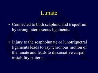
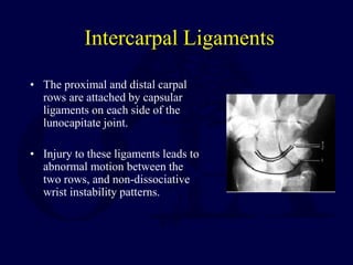
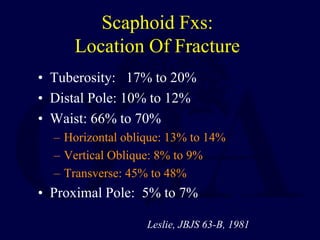
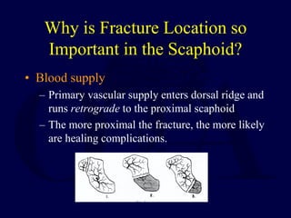
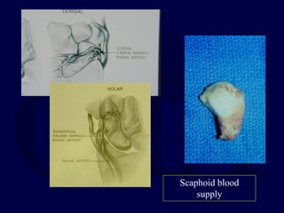
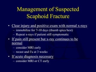
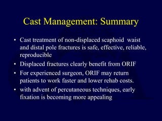
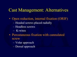
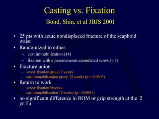
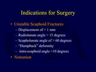
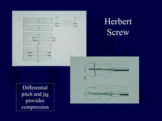
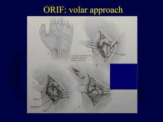
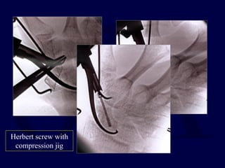
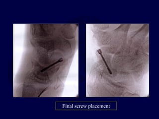
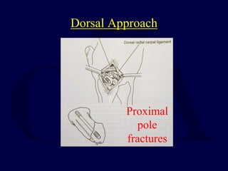
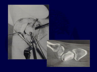
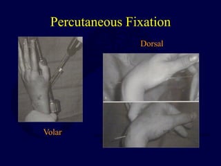
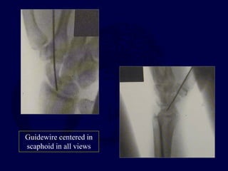
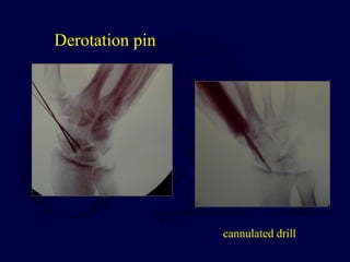
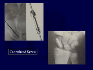
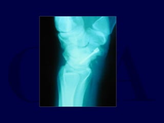
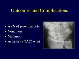
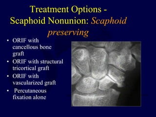
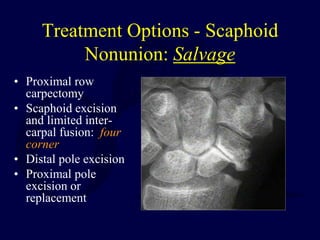
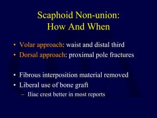
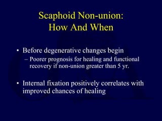
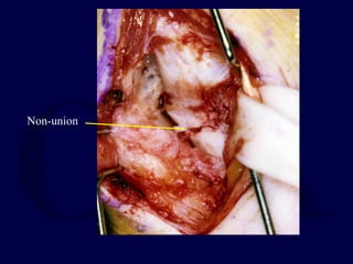
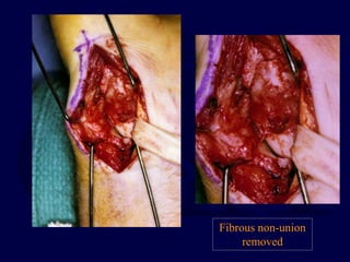
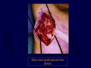
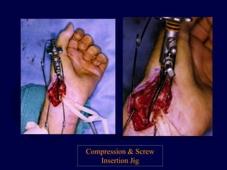
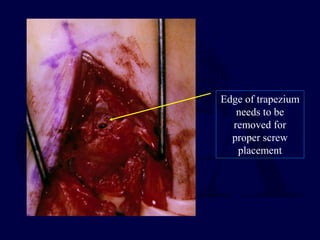
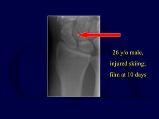
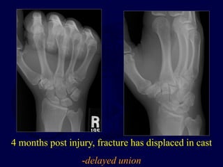
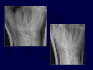
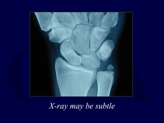
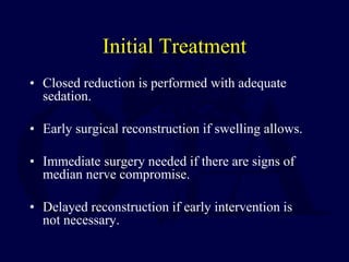
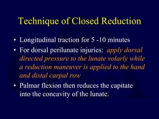
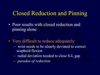
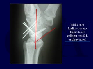
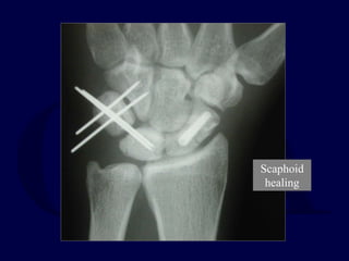
Carpal Fractures
and Dislocations
John T. Capo, MD
2nd Revision, John T. Capo, MD November 2009
Revised John T. Capo, MD, January 2006
Original Authors: Thomas F. Varecka, MD
and Andrew H. Schmidt, MD; March 2004
Anatomy of the Wrist
โ€ข Carpal bones tightly linked by capsular and
interosseous ligaments.
โ€ข Capsular (extrinsic) ligaments originate
from the radius and insert onto the carpus.
โ€ข Interosseous (intrinsic) ligaments traverse
the carpal bones.
โ€ข The lunate is the key to carpal stability.
Lunate
โ€ข Connected to both scaphoid and triquetrum
by strong interosseous ligaments.
โ€ข Injury to the scapholunate or lunotriquetral
ligaments leads to asynchronous motion of
the lunate and leads to dissociative carpal
instability patterns.
Intercarpal Ligaments
โ€ข The proximal and distal carpal
rows are attached by capsular
ligaments on each side of the
lunocapitate joint.
โ€ข Injury to these ligaments leads to
abnormal motion between the
two rows, and non-dissociative
wrist instability patterns.
Dorsal
Extrinsic
Ligaments
DIC
DRC
DRC: dorsal radio-carpal
DIC: dorsal inter-carpal
Volar
Ligaments
SL
LT
TFCC
Interosseous Ligaments:
looking dorsal to volar
Scapholunate Ligament
โ€ข Three Portions
1. Dorsal
Strongest
2. Proximal/membranous
โ€ข Capsule
3. Palmar
Imaging
โ€ข Plain radiographs: multiple views necessary:
โ€“ Anteroposterior
โ€“ Lateral
โ€“ Oblique
โ€“ Clenched-fist AP
โ€“ Radial and ulnar deviation
General Principles of Treatment
โ€ข Carefully evaluate x-rays for subtle fractures
and/or evidence of carpal instability.
โ€ข Reduce and immobilize scaphoid fractures or
perilunate injuries pending definitive treatment.
โ€ข Diagnose and appropriately treat ligament and
bony injuries.
Scaphoid Fractures
โ€œTherapy of this fracture has been characterized
by: confusion, impatience, invention,
intervention, reaction, re-evaluation and
frustration.โ€
Mazet & Hohl, JBJS, 45A, 1963
Introduction
โ€ข Scaphoid most commonly fractured carpal bone
โ€“ Incidence of scaphoid fractures estimated to be ~15%
of all wrist injuries.
โ€“ Munk, Acta Orthop Scand, 1995
โ€ข 160 scaphoid fxโ€™s among 1,052 pts. seen in E.D. for wrist
injuries.
Mechanism of Injury
โ€ข Fall on outstretched hand
โ€“ 75% to 80%
โ€ข Kick-back injury, e.g., jammed drill, etc
โ€“ 12% to 15%
โ€ข Direct Blow
โ€“ 2% to 3%
Evaluation
โ€ข History - suspect scaphoid injury in anyone
with radial wrist pain after an injury
โ€ข Physical Exam
โ€ข Imaging
Physical Findings
โ€ข โ€œSnuff boxโ€ tenderness
โ€“ scaphoid waist exposed with ulnar deviation
โ€ข Pain with palpation of scaphoid tuberosity
โ€ข Limited painful wrist ROM, especially forced
dorsiflexion
Differential Diagnosis:
radial sided wrist pain
โ€ข Scapholunate instability
โ€“ Pain and clicking in wrist
โ€“ Tender just distal to Listerโ€™s tubercle
โ€“ Positive โ€œWatsonโ€ test
โ€ข FCR tendon rupture or tendinitis
โ€ข Radial styloid fracture
โ€ข deQuervainโ€™s disease
โ€ข CMC (basal) joint arthrosis
โ€ข Radio-scaphoid arthrosis
Imaging
โ€ข X-rays
โ€“ Initial films non-diagnostic in up to 25% of
cases
โ€ข CT Scan
โ€ข MRI- most accurate
โ€ข Bone Scan โ€“ rarely used
Radiographic Imaging of
Scaphoid Fractures
โ€ข PA of wrist
โ€ข Lateral of wrist
โ€ข Scaphoid view
โ€“ PA x-ray with wrist neutral and in ulnar deviation
โ€“ elongates scaphoid to better visualize
โ€ข Pronated oblique view
Standard PA wrist view
CT scan
-In plane of scaphoid
-demonstrates subtle mal-alignment
Humpback deformity
Classification
โ€ข Typically by location:
โ€“ Proximal third
โ€“ Middle third (Waist)
โ€“ Distal Third
โ€“ Tuberosity
Scaphoid Fxs:
Location Of Fracture
โ€ข Tuberosity: 17% to 20%
โ€ข Distal Pole: 10% to 12%
โ€ข Waist: 66% to 70%
โ€“ Horizontal oblique: 13% to 14%
โ€“ Vertical Oblique: 8% to 9%
โ€“ Transverse: 45% to 48%
โ€ข Proximal Pole: 5% to 7%
Leslie, JBJS 63-B, 1981
Why is Fracture Location so
Important in the Scaphoid?
โ€ข Blood supply
โ€“ Primary vascular supply enters dorsal ridge and
runs retrograde to the proximal scaphoid
โ€“ The more proximal the fracture, the more likely
are healing complications.
Scaphoid blood
supply
Management of Suspected
Scaphoid Fracture
โ€ข Clear injury and positive exam with normal x-rays
โ€“ immobilize for 7-10 days (thumb spica best)
โ€“ Repeat x-rays if patient still symptomatic
โ€ข If pain still present but x-ray continues to be
normal
โ€“ consider MRI early
โ€“ recast and f/u at 3 weeks
โ€ข If acute diagnosis necessary
โ€“ consider MRI or CT early
Treatment Options -
Acute Injuries
โ€ข Nonoperative
โ€“ Short vs. long-arm cast
โ€“ Thumb spica vs. standard cast
โ€ข Operative
โ€“ Percutaneous pin or screw fixation
โ€“ ORIF
Indications for
Nonoperative Treatment
โ€ข Ideal indication - nondisplaced distal third
fracture
โ€ข Tuberosity fractures also heal well with
casting
โ€ข 80-90% of middle third fractures heal
โ€ข Only 60-70% of proximal third fractures
heal
โ€“ of those that do, many have deformity
Nonoperative Treatment
โ€ข Immobilize in slight flexion and slight radial
deviation.
โ€ข Initial cast: long-arm thumb spica cast for 6 weeks
โ€“ shown to lead to more rapid union and less nonunion
โ€“ Gellman et al, JBJS, 1989
โ€ข Replace with short-arm thumb spica cast until
united.
โ€ข Expected time to union:
โ€“ Distal third = 6-8 weeks
โ€“ Middle third = 8-12 weeks
โ€“ Proximal third = 12-24 weeks
Cast Management
โ€ข Cooney, CORR (1980):
โ€“ Overall, 37 / 45 (82%) acute fxโ€™s healed
โ€“ Nondisplaced fx : 27 / 27 healed
โ€ข time to union: 9.4 weeks
โ€“ Displaced fx : 10 / 13 healed (77%)
โ€ข 4 with asymptomatic malunions
Type of Cast to Use
โ€ข Gellman, JBJS-Am, (1989):
โ€“ 51 acute fxโ€™s followed prospectively
โ€“ Short- vs. long-arm cast
โ€“ LAC: n=28, 100% union
โ€ข Time to union: 9.5 weeks
โ€“ SAC: n=23, 65% union; 2 nonunions, 6
delayed unions
โ€ข Time to union: 12.7 weeks
Improved results with long arm cast
Cast Management: Summary
โ€ข Cast treatment of non-displaced scaphoid waist
and distal pole fractures is safe, effective, reliable,
reproducible
โ€ข Displaced fractures clearly benefit from ORIF
โ€ข For experienced surgeon, ORIF may return
patients to work faster and lower rehab costs.
โ€ข with advent of percutaneous techniques, early
fixation is becoming more appealing
Cast Management: Alternatives
โ€ข Open reduction, internal fixation (ORIF)
โ€“ Headed screws placed radially
โ€“ Headless screws
โ€“ K-wires
โ€ข Percutaneous fixation with cannulated
screw
โ€“ Volar approach
โ€“ Dorsal approach
Casting vs. Fixation
Bond, Shin, et al JBJS 2001
โ€ข 25 pts with acute nondisplaced fracture of the scaphoid
waist
โ€ข Randomized to either:
โ€“ cast immobilization (14)
โ€“ fixation with a percutaneous cannulated screw (11)
โ€ข Fracture union
โ€“ screw fixation group 7 weeks
โ€“ cast immobilization group 12 weeks (p = 0.0003)
โ€ข Return to work
โ€“ screw fixation 8weeks
โ€“ cast immobilization 15 weeks (p = 0.0001)
โ€ข no significant difference in ROM or grip strength at the 2
yr f/u
Indications for Surgery
โ€ข Unstable Scaphoid Fractures
โ€“ Displacement of > 1 mm
โ€“ Radiolunate angle > 15 degrees
โ€“ Scapholunate angle of > 60 degrees
โ€“ โ€œHumpbackโ€ deformity
โ€“ intra-scaphoid angle >10 degrees
โ€ข Nonunion
Herbert
Screw
Differential
pitch and jig
provides
compression
ORIF: volar approach
Herbert screw with
compression jig
Final screw placement
Dorsal Approach
Proximal
pole
fractures
Percutaneous Fixation
Volar
Dorsal
Guidewire centered in
scaphoid in all views
Derotation pin
cannulated drill
Cannulated Screw
Outcomes and Complications
โ€ข AVN of proximal pole
โ€ข Nonunion
โ€ข Malunion
โ€ข Arthritis (SNAC) wrist
Scaphoid Non-Union
โ€ข Introduction
โ€ข How does it occur?
โ€ข Should it be treated?
โ€ข Can it be treated?
โ€ข How and when should it be treated?
Treatment Options -
Scaphoid Nonunion: Scaphoid
preserving
โ€ข ORIF with
cancellous bone
graft
โ€ข ORIF with structural
tricortical graft
โ€ข ORIF with
vascularized graft
โ€ข Percutaneous
fixation alone
Treatment Options - Scaphoid
Nonunion: Salvage
โ€ข Proximal row
carpectomy
โ€ข Scaphoid excision
and limited inter-
carpal fusion: four
corner
โ€ข Distal pole excision
โ€ข Proximal pole
excision or
replacement
INITIAL FILM
AFTER 4 MONTHS
IN CAST
CT SCAN AT 4 MON. POST TREATMENT
51 y/o man presents with acute onset ulnar
sided wrist pain after playing golf
Scaphoid Nonunion: Diagnosis
โ€ข Non-union often an โ€œincidentalโ€ finding
after re-injury to wrist
โ€“ Probable disruption of a previous stable, and
therefore asymptomatic, scaphoid non-union
โ€ข Exam: tender, loss of motion, weakness
Non-union: How Does It Occur?
โ€ข Fractures at risk
โ€“ Waist fracture, especially if fracture line is
transverse to scaphoid axis (Russe)
โ€“ Displacement > 1mm associated with fracture
instability (Weber, Gellman)
โ€“ Fracture displacement occurring while in cast
(Leslie, Herbert)
โ€“ Inadequate treatment (Dias)
Non-union: How Does It Occur?
โ€ข Fractures at risk
โ€“ Disrupted vascular patterns
Gelberman, J Hand Surg, 1980
Scaphoid Non-union:
Should It Be Treated ?
โ€ข Natural history of scaphoid nonunion
suggests high incidence of wrist arthrosis
โ€“ Mack, et al., JBJS, 1984:
โ€“ 47 scaphoid nonunions, ranging from 5 to 53
yr. duration
โ€“ All developed degenerative changes
โ€“ Duration of non-union correlated with degree
of arthrosis
โ€ข 3 patterns of degeneration
Scaphoid Non-union:
Should It Be Treated ?
โ€ข Natural history of scaphoid nonunion
suggests high incidence of wrist arthrosis
โ€“ Belsky,et al., JBJS, 1985:
โ€“ 55 scaphoid non-unions, followed for longer
than 10 yrs.
โ€“ Earliest degenerative changes noted by 5 yrs.
โ€“ All had significant arthrosis by 10 yrs.
Scaphoid Non-union:
predictable pattern of arthrosis
TYPE I DJD:
N/U < 10 YR.
TYPE II DJD:
N/U ~ 15 YR.
TYPE III/IV DJD:
N/U > 25 YR.
MACK, et al., JBJS, 1984
Chronic Non-union: SNAC wrist
Scaphoid Non-union
Advanced Collapse
โ€ข Radial styloid -scaphoid
arthritis (1)
โ€ข Radius- proximal scaphoid
joint (2)
โ€ข Mid-carpal joint (3)
โ€ข Pan-carpal (4)
1
2
3
4
Scaphoid Non-union:
Should It Be Treated ?
โ€ข Natural history studies strongly suggest
scaphoid fractures left untreated lead to carpal
collapse patterns and almost 100% certainty
of developing degenerative changes
Scaphoid Non-union:
Can It Be Treated?
โ€ข Results of treatment of non-union vary
widely
โ€“ Green, J Hand Surg, 1984
โ€ข Reports results of Russe type bone grafts
โ€ข Addresses effect of avascular changes in proximal
pole
โ€ข 88% union rate; all patients with non-unions < 2yrs.
โ€ข AVN not absolute contra-indication to treatment
Scaphoid Non-union:
Can It Be Treated?
โ€ข Results of treatment of non-union vary
widely
โ€ข Schuind, et al., J Hand Surg, 1999
โ€“ Multivariate analysis of 138 surgically treated
scaphoid nonunions
โ€“ 75% healing rate
โ€“ Negative factors: duration > 5 yr.; radial
styloidectomy; dorsal approach
Scaphoid Non-union:
Can It Be Treated?
โ€ข Results of treatment of non-union vary
widely
โ€ข More recent literature reports more favorable
healing rates, up to 95% when:
โ€“ 1) deformity corrected;
โ€“ 2) iliac crest bone graft used;
โ€“ 3) rigid internal fixation employed.
Scaphoid Non-union:
How And When
โ€ข Volar approach: waist and distal third
โ€ข Dorsal approach: proximal pole fractures
โ€ข Fibrous interposition material removed
โ€ข Liberal use of bone graft
โ€“ Iliac crest better in most reports
Scaphoid Non-union:
How And When
โ€ข Before degenerative changes begin
โ€“ Poorer prognosis for healing and functional
recovery if non-union greater than 5 yr.
โ€ข Internal fixation positively correlates with
improved chances of healing
Technique: Volar ORIF
with bone graft
Exposure
โ€ข Gentle zigzag
incision directly over
the course of the
flexor carpi radialis
tendon
FCR TENDON:
stay on radial
side
Non-union
Fibrous non-union
removed
Iliac crest graft placed into
defect
Compression & Screw
Insertion Jig
Edge of trapezium
needs to be
removed for
proper screw
placement
26 y/o male,
injured skiing;
film at 10 days
4 months post injury, fracture has displaced in cast
-delayed union
18 months post ORIF,
full motion, no pain,
has returned to full
activity
Non-union: Results
โ€ข Dรผppe, JBJS-A (1994):
โ€“ 36 year follow-up of 56 fxโ€™s
โ€“ 52 acute fxโ€™s, 91% union
โ€“ 9 N/Uโ€™s: 4 primary, 5 ๏ƒข treatment
โ€ข 3 with DISI
โ€ข 5 with DJD
โ€“ ALL healed patients working
Non-union: Results
โ€ข In non-unions where stage I arthrosis is
present, ORIF gives consistently
satisfactory results.
โ€ข In nonunions > 5 yrs, achieving union is
very difficult.
โ€ข Repeat procedure for persistent non-union
has high percentage failure.
Early Non-union
Mild cystic changes, minimal collapse
Percutaneous internal fixation of selected
scaphoid non-unions with an arthroscopically
assisted dorsal approach
Slade, Geissler et al; JBJS-2003(85)
โ€ข 15 patients with early non-unions
โ€ข All cases with percutaneous screw fixation and
arthroscopic assistance
โ€ข No bone grafts used
โ€ข All scaphoids healed at average of 14 weeks
Perc screw placement- donโ€™t
over compress
Non-union: healed at 10 weeks
Non-union with Arthrosis:
Salvage
โ€ข Arthrodesis
โ€“ Intercarpal: 4 corner
โ€ข Proximal row carpectomy
โ€“ Complication rate lower
โ€ข Arthroplasty: not recommended
Non-union: Summary
โ€ข Scaphoid non-union is challenging problem
with significant risk for the wrist.
โ€ข Left untreated, scaphoid non-unions have a
near 100% rate of degenerative disease.
โ€ข If approached appropriately scaphoid
healing may be achieved
Perilunate Injuries
Mechanism of Injury
โ€ข Load applied to hand forcing the wrist into extension and
ulnar deviation
โ€ข Severe ligament injury necessary to tear the distal row
from the lunate to produce perilunate dislocation
โ€ข Injury progresses through several stages:
โ€“ usually begins radially & destabilizes thru body of scaphoid (w/
fx) or thru scapholunate interval (w/ dissociation)
โ€“ force is transmitted ulnarly thru the space of Poirier (between
lunate and capitate volarly)
โ€“ next force transmission disrupts the luno-triquetral articulation
Predictable patterns of Injury and Instability
Physical Exam
โ€ข Dorsal displacement of the carpus may be
seen
โ€ข Significant swelling common
โ€“ Evaluate for compartment syndrome
โ€ข If lunate is dislocated, median nerve
symptoms may be present
Imaging
โ€ข Note lack of โ€œcolinearityโ€
among the radius, lunate,
and capitate on the lateral
x-ray.
Imaging
โ€ข Note loss of normal carpal
โ€œarcsโ€ and abnormal
widening of the scapholunate
interval.
โ€ข Look for associated fractures
โ€œtrans-scaphoidโ€ injuries
X-ray usually
Obvious
X-ray may be subtle
Initial Treatment
โ€ข Closed reduction is performed with adequate
sedation.
โ€ข Early surgical reconstruction if swelling allows.
โ€ข Immediate surgery needed if there are signs of
median nerve compromise.
โ€ข Delayed reconstruction if early intervention is
not necessary.
Technique of Closed Reduction
โ€ข Longitudinal traction for 5 -10 minutes
โ€ข For dorsal perilunate injuries: apply dorsal
directed pressure to the lunate volarly while
a reduction maneuver is applied to the hand
and distal carpal row
โ€ข Palmar flexion then reduces the capitate
into the concavity of the lunate.
Closed Reduction and Pinning
โ€ข Poor results with closed reduction and
pinning alone
โ€ข Very difficult to reduce adequately
โ€“ wrist needs to be ulnarly deviated to correct
scaphoid flexion
โ€“ radial deviation needed to close S-L gap
โ€“ paradox of reduction
ORIF with volar and dorsal approaches
Procedure of Choice
Provisional closed
reduction
Dorsal Approach
Repair S-L ligament
Volar Approach
Lunate may be
dislocated volarly
Volar mid-carpal
ligament tear
Reduce lunate
first- may need
to temporary
pin to radius
Pin Carpus: S-L,
L-T and mid-
carpal joints
Trans-scaphoid
Perilunate Injuries
โ€ข Require reduction and fixation of the
fractured scaphoid.
โ€ข Most of these injuries best treated
โ€“ ORIF with volar and dorsal approaches
โ€“ repair of injured structures.
โ€ข Open repair supplemented by pin and screw
fixation.
Trans-scaphoid Perilunate
Dislocations
Fix scaphoid first: dorsal
approach
Pin L-T and Mid-carpal
joints
Make sure
Radius-Lunate-
Capitate are
colinear and S-L
angle restored
Scaphoid
healing
Outcome of Perilunate Injuries
โ€ข 14 cases followed for mean of 8 years
โ€ข All treated operatively (ave 6 days post-injury)
โ€“ 11 dorsal approach
โ€“ 3 combined dorsal/volar approaches
โ€ข Mayo wrist scores:
โ€“ 5 excellent
โ€“ 3 good
โ€“ 5 fair
โ€“ 1 poor
โ€ข All cases had radiographic arthrosis that did not
correlate with Mayo scores.
Herzberg & Forissier, J Hand Surg Br 27: 498-502, 2002
Perilunate Injuries Conclusion
โ€ข Perilunate fracture dislocations are high-energy
injuries
โ€ข Must recognize different injury patterns
โ€“ transcaphoid
โ€“ pure ligamentous
โ€“ trans radial-styloid
โ€ข Early open and anatomic fixation with volar and
dorsal approaches provides the best chance at a
reasonable functional result
Return to
Upper Extremity
Index
If you would like to volunteer as an author for the Resident
Slide Project or recommend updates to any of the following
slides, please send an e-mail to ota@ota.org
Bibliography
1: Forli A, Courvoisier A, Wimsey S, Corcella D, Moutet F. Perilunate Dislocations and
Transscaphoid Perilunate Fracture-Dislocations: A Retrospective Study With Minimum Ten-Year
Follow-Up. J Hand Surg Am. 2009 Nov 19.
2: Herzberg G. Perilunate and axial carpal dislocations and fracture-dislocations. J Hand Surg Am.
2008 Nov;33(9):1659-68.
3: Souer JS, Rutgers M, Andermahr J, Jupiter JB, Ring D. Perilunate fracture-dislocations of the
wrist: comparison of temporary screw versus K-wire fixation. J Hand Surg Am. 2007
Mar;32(3):318-25.
4: Knoll VD, Allan C, Trumble TE. Trans-scaphoid perilunate fracture dislocations: results of
screw fixation of the scaphoid and lunotriquetral repair with a dorsal approach. J Hand Surg Am.
2005 Nov;30(6):1145-52. Erratum in: J Hand Surg [Am]. 2006 Feb;31(2):328.
5: Trumble T, Verheyden J. Treatment of isolated perilunate and lunate dislocations with combined
dorsal and volar approach and intraosseous cerclage wire. J Hand Surg Am. 2004 May;29(3):412-
7.
6: Hildebrand KA, Ross DC, Patterson SD, Roth JH, MacDermid JC, King GJ. Dorsal perilunate
dislocations and fracture-dislocations: questionnaire, clinical, and radiographic evaluation. J Hand
Surg Am. 2000 Nov;25(6):1069-79.
7: Rettig ME, Raskin KB. Long-term assessment of proximal row carpectomy for chronic
perilunate dislocations. J Hand Surg Am. 1999 Nov;24(6):1231-6.
8: Sotereanos DG, Mitsionis GJ, Giannakopoulos PN, Tomaino MM, Herndon JH. Perilunate
dislocation and fracture dislocation: a critical analysis of the volar-dorsal approach. J Hand Surg
Am. 1997 Jan;22(1):49-56.
9: Minami A, Kaneda K. Repair and/or reconstruction of scapholunate interosseous ligament in
lunate and perilunate dislocations. J Hand Surg Am. 1993 Nov;18(6):1099-106.
10: Herzberg G, Comtet JJ, Linscheid RL, Amadio PC, Cooney WP, Stalder J. Perilunate
dislocations and fracture-dislocations: a multicenter study. J Hand Surg Am. 1993
Sep;18(5):768-79.
11: Vinnars B, Pietreanu M, Bodestedt A, Ekenstam F, Gerdin B. Nonoperative compared with
operative treatment of acute scaphoid fractures. A randomized clinical trial. J Bone Joint Surg
Am. 2008 Jun;90(6):1176-85.
12: McQueen MM, Gelbke MK, Wakefield A, Will EM, Gaebler C. Percutaneous screw
fixation versus conservative treatment for fractures of the waist of the scaphoid: a prospective
randomised study. J Bone Joint Surg Br. 2008 Jan;90(1):66-71.
13: Papaloizos MY, Fusetti C, Christen T, Nagy L, Wasserfallen JB. Minimally invasive fixation
versus conservative treatment of undisplaced scaphoid fractures: a cost-effectiveness study. J
Hand Surg Br. 2004 Apr;29(2):116-9.
14: Rettig ME, Kozin SH, Cooney WP. Open reduction and internal fixation of acute displaced
scaphoid waist fractures. J Hand Surg Am. 2001 Mar;26(2):271-6.
15: Rettig ME, Raskin KB. Retrograde compression screw fixation of acute proximal pole scaphoid
fractures. J Hand Surg Am. 1999 Nov;24(6):1206-10.
16: Robbins RR, Ridge O, Carter PR. Iliac crest bone grafting and Herbert screw fixation of
nonunions of the scaphoid with avascular proximal poles. J Hand Surg Am. 1995 Sep;20(5):818-31.
17: Fernandez DL. Anterior bone grafting and conventional lag screw fixation to treat scaphoid
nonunions. J Hand Surg Am. 1990 Jan;15(1):140-7.
18: Gellman H, Caputo RJ, Carter V, Aboulafia A, McKay M. Comparison of short and long thumb-
spica casts for non-displaced fractures of the carpal scaphoid. J Bone Joint Surg Am. 1989
Mar;71(3):354-7.
19: Bond CD, Shin AY, McBride MT, Dao KD. Percutaneous screw fixation or cast immobilization
for nondisplaced scaphoid fractures. J Bone Joint Surg Am. 2001 Apr;83-A(4):483-8.

Carpal Fractures and Dislocations.....pdf

  • 1.
    Carpal Fractures and Dislocations JohnT. Capo, MD 2nd Revision, John T. Capo, MD November 2009 Revised John T. Capo, MD, January 2006 Original Authors: Thomas F. Varecka, MD and Andrew H. Schmidt, MD; March 2004
  • 2.
    Anatomy of theWrist โ€ข Carpal bones tightly linked by capsular and interosseous ligaments. โ€ข Capsular (extrinsic) ligaments originate from the radius and insert onto the carpus. โ€ข Interosseous (intrinsic) ligaments traverse the carpal bones. โ€ข The lunate is the key to carpal stability.
  • 3.
    Lunate โ€ข Connected toboth scaphoid and triquetrum by strong interosseous ligaments. โ€ข Injury to the scapholunate or lunotriquetral ligaments leads to asynchronous motion of the lunate and leads to dissociative carpal instability patterns.
  • 4.
    Intercarpal Ligaments โ€ข Theproximal and distal carpal rows are attached by capsular ligaments on each side of the lunocapitate joint. โ€ข Injury to these ligaments leads to abnormal motion between the two rows, and non-dissociative wrist instability patterns.
  • 5.
  • 6.
  • 7.
  • 9.
    Scapholunate Ligament โ€ข ThreePortions 1. Dorsal Strongest 2. Proximal/membranous โ€ข Capsule 3. Palmar
  • 10.
    Imaging โ€ข Plain radiographs:multiple views necessary: โ€“ Anteroposterior โ€“ Lateral โ€“ Oblique โ€“ Clenched-fist AP โ€“ Radial and ulnar deviation
  • 11.
    General Principles ofTreatment โ€ข Carefully evaluate x-rays for subtle fractures and/or evidence of carpal instability. โ€ข Reduce and immobilize scaphoid fractures or perilunate injuries pending definitive treatment. โ€ข Diagnose and appropriately treat ligament and bony injuries.
  • 12.
    Scaphoid Fractures โ€œTherapy ofthis fracture has been characterized by: confusion, impatience, invention, intervention, reaction, re-evaluation and frustration.โ€ Mazet & Hohl, JBJS, 45A, 1963
  • 13.
    Introduction โ€ข Scaphoid mostcommonly fractured carpal bone โ€“ Incidence of scaphoid fractures estimated to be ~15% of all wrist injuries. โ€“ Munk, Acta Orthop Scand, 1995 โ€ข 160 scaphoid fxโ€™s among 1,052 pts. seen in E.D. for wrist injuries.
  • 14.
    Mechanism of Injury โ€ขFall on outstretched hand โ€“ 75% to 80% โ€ข Kick-back injury, e.g., jammed drill, etc โ€“ 12% to 15% โ€ข Direct Blow โ€“ 2% to 3%
  • 15.
    Evaluation โ€ข History -suspect scaphoid injury in anyone with radial wrist pain after an injury โ€ข Physical Exam โ€ข Imaging
  • 16.
    Physical Findings โ€ข โ€œSnuffboxโ€ tenderness โ€“ scaphoid waist exposed with ulnar deviation โ€ข Pain with palpation of scaphoid tuberosity โ€ข Limited painful wrist ROM, especially forced dorsiflexion
  • 17.
    Differential Diagnosis: radial sidedwrist pain โ€ข Scapholunate instability โ€“ Pain and clicking in wrist โ€“ Tender just distal to Listerโ€™s tubercle โ€“ Positive โ€œWatsonโ€ test โ€ข FCR tendon rupture or tendinitis โ€ข Radial styloid fracture โ€ข deQuervainโ€™s disease โ€ข CMC (basal) joint arthrosis โ€ข Radio-scaphoid arthrosis
  • 18.
    Imaging โ€ข X-rays โ€“ Initialfilms non-diagnostic in up to 25% of cases โ€ข CT Scan โ€ข MRI- most accurate โ€ข Bone Scan โ€“ rarely used
  • 19.
    Radiographic Imaging of ScaphoidFractures โ€ข PA of wrist โ€ข Lateral of wrist โ€ข Scaphoid view โ€“ PA x-ray with wrist neutral and in ulnar deviation โ€“ elongates scaphoid to better visualize โ€ข Pronated oblique view
  • 20.
  • 21.
    CT scan -In planeof scaphoid -demonstrates subtle mal-alignment Humpback deformity
  • 22.
    Classification โ€ข Typically bylocation: โ€“ Proximal third โ€“ Middle third (Waist) โ€“ Distal Third โ€“ Tuberosity
  • 23.
    Scaphoid Fxs: Location OfFracture โ€ข Tuberosity: 17% to 20% โ€ข Distal Pole: 10% to 12% โ€ข Waist: 66% to 70% โ€“ Horizontal oblique: 13% to 14% โ€“ Vertical Oblique: 8% to 9% โ€“ Transverse: 45% to 48% โ€ข Proximal Pole: 5% to 7% Leslie, JBJS 63-B, 1981
  • 24.
    Why is FractureLocation so Important in the Scaphoid? โ€ข Blood supply โ€“ Primary vascular supply enters dorsal ridge and runs retrograde to the proximal scaphoid โ€“ The more proximal the fracture, the more likely are healing complications.
  • 25.
  • 26.
    Management of Suspected ScaphoidFracture โ€ข Clear injury and positive exam with normal x-rays โ€“ immobilize for 7-10 days (thumb spica best) โ€“ Repeat x-rays if patient still symptomatic โ€ข If pain still present but x-ray continues to be normal โ€“ consider MRI early โ€“ recast and f/u at 3 weeks โ€ข If acute diagnosis necessary โ€“ consider MRI or CT early
  • 27.
    Treatment Options - AcuteInjuries โ€ข Nonoperative โ€“ Short vs. long-arm cast โ€“ Thumb spica vs. standard cast โ€ข Operative โ€“ Percutaneous pin or screw fixation โ€“ ORIF
  • 28.
    Indications for Nonoperative Treatment โ€ขIdeal indication - nondisplaced distal third fracture โ€ข Tuberosity fractures also heal well with casting โ€ข 80-90% of middle third fractures heal โ€ข Only 60-70% of proximal third fractures heal โ€“ of those that do, many have deformity
  • 29.
    Nonoperative Treatment โ€ข Immobilizein slight flexion and slight radial deviation. โ€ข Initial cast: long-arm thumb spica cast for 6 weeks โ€“ shown to lead to more rapid union and less nonunion โ€“ Gellman et al, JBJS, 1989 โ€ข Replace with short-arm thumb spica cast until united. โ€ข Expected time to union: โ€“ Distal third = 6-8 weeks โ€“ Middle third = 8-12 weeks โ€“ Proximal third = 12-24 weeks
  • 30.
    Cast Management โ€ข Cooney,CORR (1980): โ€“ Overall, 37 / 45 (82%) acute fxโ€™s healed โ€“ Nondisplaced fx : 27 / 27 healed โ€ข time to union: 9.4 weeks โ€“ Displaced fx : 10 / 13 healed (77%) โ€ข 4 with asymptomatic malunions
  • 31.
    Type of Castto Use โ€ข Gellman, JBJS-Am, (1989): โ€“ 51 acute fxโ€™s followed prospectively โ€“ Short- vs. long-arm cast โ€“ LAC: n=28, 100% union โ€ข Time to union: 9.5 weeks โ€“ SAC: n=23, 65% union; 2 nonunions, 6 delayed unions โ€ข Time to union: 12.7 weeks Improved results with long arm cast
  • 32.
    Cast Management: Summary โ€ขCast treatment of non-displaced scaphoid waist and distal pole fractures is safe, effective, reliable, reproducible โ€ข Displaced fractures clearly benefit from ORIF โ€ข For experienced surgeon, ORIF may return patients to work faster and lower rehab costs. โ€ข with advent of percutaneous techniques, early fixation is becoming more appealing
  • 33.
    Cast Management: Alternatives โ€ขOpen reduction, internal fixation (ORIF) โ€“ Headed screws placed radially โ€“ Headless screws โ€“ K-wires โ€ข Percutaneous fixation with cannulated screw โ€“ Volar approach โ€“ Dorsal approach
  • 34.
    Casting vs. Fixation Bond,Shin, et al JBJS 2001 โ€ข 25 pts with acute nondisplaced fracture of the scaphoid waist โ€ข Randomized to either: โ€“ cast immobilization (14) โ€“ fixation with a percutaneous cannulated screw (11) โ€ข Fracture union โ€“ screw fixation group 7 weeks โ€“ cast immobilization group 12 weeks (p = 0.0003) โ€ข Return to work โ€“ screw fixation 8weeks โ€“ cast immobilization 15 weeks (p = 0.0001) โ€ข no significant difference in ROM or grip strength at the 2 yr f/u
  • 35.
    Indications for Surgery โ€ขUnstable Scaphoid Fractures โ€“ Displacement of > 1 mm โ€“ Radiolunate angle > 15 degrees โ€“ Scapholunate angle of > 60 degrees โ€“ โ€œHumpbackโ€ deformity โ€“ intra-scaphoid angle >10 degrees โ€ข Nonunion
  • 36.
  • 37.
  • 38.
  • 39.
  • 40.
  • 42.
  • 43.
  • 44.
  • 45.
  • 47.
    Outcomes and Complications โ€ขAVN of proximal pole โ€ข Nonunion โ€ข Malunion โ€ข Arthritis (SNAC) wrist
  • 48.
    Scaphoid Non-Union โ€ข Introduction โ€ขHow does it occur? โ€ข Should it be treated? โ€ข Can it be treated? โ€ข How and when should it be treated?
  • 49.
    Treatment Options - ScaphoidNonunion: Scaphoid preserving โ€ข ORIF with cancellous bone graft โ€ข ORIF with structural tricortical graft โ€ข ORIF with vascularized graft โ€ข Percutaneous fixation alone
  • 50.
    Treatment Options -Scaphoid Nonunion: Salvage โ€ข Proximal row carpectomy โ€ข Scaphoid excision and limited inter- carpal fusion: four corner โ€ข Distal pole excision โ€ข Proximal pole excision or replacement
  • 51.
    INITIAL FILM AFTER 4MONTHS IN CAST
  • 52.
    CT SCAN AT4 MON. POST TREATMENT
  • 53.
    51 y/o manpresents with acute onset ulnar sided wrist pain after playing golf
  • 54.
    Scaphoid Nonunion: Diagnosis โ€ขNon-union often an โ€œincidentalโ€ finding after re-injury to wrist โ€“ Probable disruption of a previous stable, and therefore asymptomatic, scaphoid non-union โ€ข Exam: tender, loss of motion, weakness
  • 55.
    Non-union: How DoesIt Occur? โ€ข Fractures at risk โ€“ Waist fracture, especially if fracture line is transverse to scaphoid axis (Russe) โ€“ Displacement > 1mm associated with fracture instability (Weber, Gellman) โ€“ Fracture displacement occurring while in cast (Leslie, Herbert) โ€“ Inadequate treatment (Dias)
  • 56.
    Non-union: How DoesIt Occur? โ€ข Fractures at risk โ€“ Disrupted vascular patterns Gelberman, J Hand Surg, 1980
  • 57.
    Scaphoid Non-union: Should ItBe Treated ? โ€ข Natural history of scaphoid nonunion suggests high incidence of wrist arthrosis โ€“ Mack, et al., JBJS, 1984: โ€“ 47 scaphoid nonunions, ranging from 5 to 53 yr. duration โ€“ All developed degenerative changes โ€“ Duration of non-union correlated with degree of arthrosis โ€ข 3 patterns of degeneration
  • 58.
    Scaphoid Non-union: Should ItBe Treated ? โ€ข Natural history of scaphoid nonunion suggests high incidence of wrist arthrosis โ€“ Belsky,et al., JBJS, 1985: โ€“ 55 scaphoid non-unions, followed for longer than 10 yrs. โ€“ Earliest degenerative changes noted by 5 yrs. โ€“ All had significant arthrosis by 10 yrs.
  • 59.
    Scaphoid Non-union: predictable patternof arthrosis TYPE I DJD: N/U < 10 YR. TYPE II DJD: N/U ~ 15 YR. TYPE III/IV DJD: N/U > 25 YR. MACK, et al., JBJS, 1984
  • 60.
    Chronic Non-union: SNACwrist Scaphoid Non-union Advanced Collapse โ€ข Radial styloid -scaphoid arthritis (1) โ€ข Radius- proximal scaphoid joint (2) โ€ข Mid-carpal joint (3) โ€ข Pan-carpal (4) 1 2 3 4
  • 61.
    Scaphoid Non-union: Should ItBe Treated ? โ€ข Natural history studies strongly suggest scaphoid fractures left untreated lead to carpal collapse patterns and almost 100% certainty of developing degenerative changes
  • 62.
    Scaphoid Non-union: Can ItBe Treated? โ€ข Results of treatment of non-union vary widely โ€“ Green, J Hand Surg, 1984 โ€ข Reports results of Russe type bone grafts โ€ข Addresses effect of avascular changes in proximal pole โ€ข 88% union rate; all patients with non-unions < 2yrs. โ€ข AVN not absolute contra-indication to treatment
  • 63.
    Scaphoid Non-union: Can ItBe Treated? โ€ข Results of treatment of non-union vary widely โ€ข Schuind, et al., J Hand Surg, 1999 โ€“ Multivariate analysis of 138 surgically treated scaphoid nonunions โ€“ 75% healing rate โ€“ Negative factors: duration > 5 yr.; radial styloidectomy; dorsal approach
  • 64.
    Scaphoid Non-union: Can ItBe Treated? โ€ข Results of treatment of non-union vary widely โ€ข More recent literature reports more favorable healing rates, up to 95% when: โ€“ 1) deformity corrected; โ€“ 2) iliac crest bone graft used; โ€“ 3) rigid internal fixation employed.
  • 65.
    Scaphoid Non-union: How AndWhen โ€ข Volar approach: waist and distal third โ€ข Dorsal approach: proximal pole fractures โ€ข Fibrous interposition material removed โ€ข Liberal use of bone graft โ€“ Iliac crest better in most reports
  • 66.
    Scaphoid Non-union: How AndWhen โ€ข Before degenerative changes begin โ€“ Poorer prognosis for healing and functional recovery if non-union greater than 5 yr. โ€ข Internal fixation positively correlates with improved chances of healing
  • 67.
  • 68.
    Exposure โ€ข Gentle zigzag incisiondirectly over the course of the flexor carpi radialis tendon
  • 69.
  • 70.
  • 71.
  • 72.
    Iliac crest graftplaced into defect
  • 73.
  • 74.
    Edge of trapezium needsto be removed for proper screw placement
  • 75.
    26 y/o male, injuredskiing; film at 10 days
  • 76.
    4 months postinjury, fracture has displaced in cast -delayed union
  • 78.
    18 months postORIF, full motion, no pain, has returned to full activity
  • 79.
    Non-union: Results โ€ข Dรผppe,JBJS-A (1994): โ€“ 36 year follow-up of 56 fxโ€™s โ€“ 52 acute fxโ€™s, 91% union โ€“ 9 N/Uโ€™s: 4 primary, 5 ๏ƒข treatment โ€ข 3 with DISI โ€ข 5 with DJD โ€“ ALL healed patients working
  • 80.
    Non-union: Results โ€ข Innon-unions where stage I arthrosis is present, ORIF gives consistently satisfactory results. โ€ข In nonunions > 5 yrs, achieving union is very difficult. โ€ข Repeat procedure for persistent non-union has high percentage failure.
  • 81.
    Early Non-union Mild cysticchanges, minimal collapse
  • 82.
    Percutaneous internal fixationof selected scaphoid non-unions with an arthroscopically assisted dorsal approach Slade, Geissler et al; JBJS-2003(85) โ€ข 15 patients with early non-unions โ€ข All cases with percutaneous screw fixation and arthroscopic assistance โ€ข No bone grafts used โ€ข All scaphoids healed at average of 14 weeks
  • 83.
    Perc screw placement-donโ€™t over compress
  • 84.
  • 85.
    Non-union with Arthrosis: Salvage โ€ขArthrodesis โ€“ Intercarpal: 4 corner โ€ข Proximal row carpectomy โ€“ Complication rate lower โ€ข Arthroplasty: not recommended
  • 86.
    Non-union: Summary โ€ข Scaphoidnon-union is challenging problem with significant risk for the wrist. โ€ข Left untreated, scaphoid non-unions have a near 100% rate of degenerative disease. โ€ข If approached appropriately scaphoid healing may be achieved
  • 87.
  • 88.
    Mechanism of Injury โ€ขLoad applied to hand forcing the wrist into extension and ulnar deviation โ€ข Severe ligament injury necessary to tear the distal row from the lunate to produce perilunate dislocation โ€ข Injury progresses through several stages: โ€“ usually begins radially & destabilizes thru body of scaphoid (w/ fx) or thru scapholunate interval (w/ dissociation) โ€“ force is transmitted ulnarly thru the space of Poirier (between lunate and capitate volarly) โ€“ next force transmission disrupts the luno-triquetral articulation
  • 89.
    Predictable patterns ofInjury and Instability
  • 90.
    Physical Exam โ€ข Dorsaldisplacement of the carpus may be seen โ€ข Significant swelling common โ€“ Evaluate for compartment syndrome โ€ข If lunate is dislocated, median nerve symptoms may be present
  • 91.
    Imaging โ€ข Note lackof โ€œcolinearityโ€ among the radius, lunate, and capitate on the lateral x-ray.
  • 92.
    Imaging โ€ข Note lossof normal carpal โ€œarcsโ€ and abnormal widening of the scapholunate interval. โ€ข Look for associated fractures โ€œtrans-scaphoidโ€ injuries
  • 93.
  • 94.
  • 95.
    Initial Treatment โ€ข Closedreduction is performed with adequate sedation. โ€ข Early surgical reconstruction if swelling allows. โ€ข Immediate surgery needed if there are signs of median nerve compromise. โ€ข Delayed reconstruction if early intervention is not necessary.
  • 96.
    Technique of ClosedReduction โ€ข Longitudinal traction for 5 -10 minutes โ€ข For dorsal perilunate injuries: apply dorsal directed pressure to the lunate volarly while a reduction maneuver is applied to the hand and distal carpal row โ€ข Palmar flexion then reduces the capitate into the concavity of the lunate.
  • 97.
    Closed Reduction andPinning โ€ข Poor results with closed reduction and pinning alone โ€ข Very difficult to reduce adequately โ€“ wrist needs to be ulnarly deviated to correct scaphoid flexion โ€“ radial deviation needed to close S-L gap โ€“ paradox of reduction
  • 98.
    ORIF with volarand dorsal approaches Procedure of Choice
  • 100.
  • 101.
  • 102.
  • 103.
    Lunate may be dislocatedvolarly Volar mid-carpal ligament tear
  • 104.
    Reduce lunate first- mayneed to temporary pin to radius
  • 105.
    Pin Carpus: S-L, L-Tand mid- carpal joints
  • 106.
    Trans-scaphoid Perilunate Injuries โ€ข Requirereduction and fixation of the fractured scaphoid. โ€ข Most of these injuries best treated โ€“ ORIF with volar and dorsal approaches โ€“ repair of injured structures. โ€ข Open repair supplemented by pin and screw fixation.
  • 107.
  • 108.
    Fix scaphoid first:dorsal approach
  • 109.
    Pin L-T andMid-carpal joints
  • 110.
  • 111.
  • 112.
    Outcome of PerilunateInjuries โ€ข 14 cases followed for mean of 8 years โ€ข All treated operatively (ave 6 days post-injury) โ€“ 11 dorsal approach โ€“ 3 combined dorsal/volar approaches โ€ข Mayo wrist scores: โ€“ 5 excellent โ€“ 3 good โ€“ 5 fair โ€“ 1 poor โ€ข All cases had radiographic arthrosis that did not correlate with Mayo scores. Herzberg & Forissier, J Hand Surg Br 27: 498-502, 2002
  • 113.
    Perilunate Injuries Conclusion โ€ขPerilunate fracture dislocations are high-energy injuries โ€ข Must recognize different injury patterns โ€“ transcaphoid โ€“ pure ligamentous โ€“ trans radial-styloid โ€ข Early open and anatomic fixation with volar and dorsal approaches provides the best chance at a reasonable functional result Return to Upper Extremity Index If you would like to volunteer as an author for the Resident Slide Project or recommend updates to any of the following slides, please send an e-mail to ota@ota.org
  • 114.
    Bibliography 1: Forli A,Courvoisier A, Wimsey S, Corcella D, Moutet F. Perilunate Dislocations and Transscaphoid Perilunate Fracture-Dislocations: A Retrospective Study With Minimum Ten-Year Follow-Up. J Hand Surg Am. 2009 Nov 19. 2: Herzberg G. Perilunate and axial carpal dislocations and fracture-dislocations. J Hand Surg Am. 2008 Nov;33(9):1659-68. 3: Souer JS, Rutgers M, Andermahr J, Jupiter JB, Ring D. Perilunate fracture-dislocations of the wrist: comparison of temporary screw versus K-wire fixation. J Hand Surg Am. 2007 Mar;32(3):318-25. 4: Knoll VD, Allan C, Trumble TE. Trans-scaphoid perilunate fracture dislocations: results of screw fixation of the scaphoid and lunotriquetral repair with a dorsal approach. J Hand Surg Am. 2005 Nov;30(6):1145-52. Erratum in: J Hand Surg [Am]. 2006 Feb;31(2):328. 5: Trumble T, Verheyden J. Treatment of isolated perilunate and lunate dislocations with combined dorsal and volar approach and intraosseous cerclage wire. J Hand Surg Am. 2004 May;29(3):412- 7. 6: Hildebrand KA, Ross DC, Patterson SD, Roth JH, MacDermid JC, King GJ. Dorsal perilunate dislocations and fracture-dislocations: questionnaire, clinical, and radiographic evaluation. J Hand Surg Am. 2000 Nov;25(6):1069-79.
  • 115.
    7: Rettig ME,Raskin KB. Long-term assessment of proximal row carpectomy for chronic perilunate dislocations. J Hand Surg Am. 1999 Nov;24(6):1231-6. 8: Sotereanos DG, Mitsionis GJ, Giannakopoulos PN, Tomaino MM, Herndon JH. Perilunate dislocation and fracture dislocation: a critical analysis of the volar-dorsal approach. J Hand Surg Am. 1997 Jan;22(1):49-56. 9: Minami A, Kaneda K. Repair and/or reconstruction of scapholunate interosseous ligament in lunate and perilunate dislocations. J Hand Surg Am. 1993 Nov;18(6):1099-106. 10: Herzberg G, Comtet JJ, Linscheid RL, Amadio PC, Cooney WP, Stalder J. Perilunate dislocations and fracture-dislocations: a multicenter study. J Hand Surg Am. 1993 Sep;18(5):768-79. 11: Vinnars B, Pietreanu M, Bodestedt A, Ekenstam F, Gerdin B. Nonoperative compared with operative treatment of acute scaphoid fractures. A randomized clinical trial. J Bone Joint Surg Am. 2008 Jun;90(6):1176-85. 12: McQueen MM, Gelbke MK, Wakefield A, Will EM, Gaebler C. Percutaneous screw fixation versus conservative treatment for fractures of the waist of the scaphoid: a prospective randomised study. J Bone Joint Surg Br. 2008 Jan;90(1):66-71. 13: Papaloizos MY, Fusetti C, Christen T, Nagy L, Wasserfallen JB. Minimally invasive fixation versus conservative treatment of undisplaced scaphoid fractures: a cost-effectiveness study. J Hand Surg Br. 2004 Apr;29(2):116-9.
  • 116.
    14: Rettig ME,Kozin SH, Cooney WP. Open reduction and internal fixation of acute displaced scaphoid waist fractures. J Hand Surg Am. 2001 Mar;26(2):271-6. 15: Rettig ME, Raskin KB. Retrograde compression screw fixation of acute proximal pole scaphoid fractures. J Hand Surg Am. 1999 Nov;24(6):1206-10. 16: Robbins RR, Ridge O, Carter PR. Iliac crest bone grafting and Herbert screw fixation of nonunions of the scaphoid with avascular proximal poles. J Hand Surg Am. 1995 Sep;20(5):818-31. 17: Fernandez DL. Anterior bone grafting and conventional lag screw fixation to treat scaphoid nonunions. J Hand Surg Am. 1990 Jan;15(1):140-7. 18: Gellman H, Caputo RJ, Carter V, Aboulafia A, McKay M. Comparison of short and long thumb- spica casts for non-displaced fractures of the carpal scaphoid. J Bone Joint Surg Am. 1989 Mar;71(3):354-7. 19: Bond CD, Shin AY, McBride MT, Dao KD. Percutaneous screw fixation or cast immobilization for nondisplaced scaphoid fractures. J Bone Joint Surg Am. 2001 Apr;83-A(4):483-8.