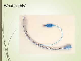
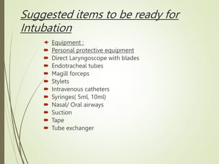
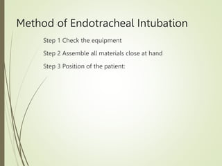
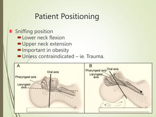
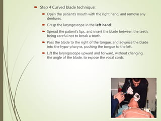
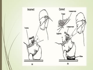
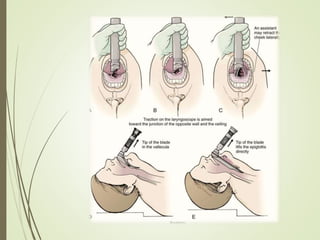
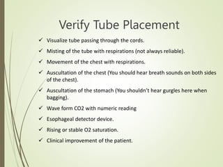
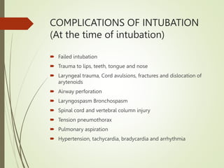
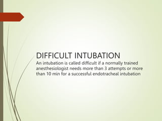
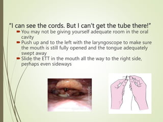
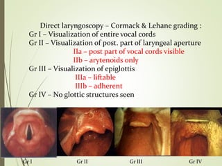
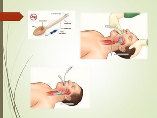
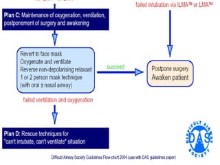
ENDOTRACHEAL
INTUBATION
DR DEEPAK SINGLA
Indications of Endotracheal
Intubation
 Airway problems: external pressures on the airway, vocal cord paralysis,
tumor, infection, and laryngospasm.
 Respiratory deficiencies: patients with poor general condition,
hypoxemic/hypercapnic respiratory insufficiency (respiratory rate less than
8 or more than 30 per minute, PO2 in blood gas less than 55 mmHg,
PCO2above 55 mmHg).
 Inadequate circulation: cardiac arrest in hypothermic and hypotensive
cases.
 Central nervous system problems: head injury, stroke, unconscious
patients, altered sensorium, raised intracranial pressure.
 Muscle weakness: (Guillain-Barre, amyotrophic lateral sclerosis,
myasthenia gravis, muscular dystrophy, acid-maltase insufficiency, phrenic
nerve injury, botulism, polymyositis, spinal cord injury, brainstem infarction,
etc.).
 Patients at risk of aspiration of the stomach contents, blood, mucus, or
secretion.
 For general anaesthesia
What is this?
Suggested items to be ready for
Intubation
 Equipment :
 Personal protective equipment
 Direct Laryngoscope with blades
 Endotracheal tubes
 Magill forceps
 Stylets
 Intravenous catheters
 Syringes( 5ml, 10ml)
 Nasal/ Oral airways
 Suction
 Tape
 Tube exchanger
Method of Endotracheal Intubation
Step 1 Check the equipment
Step 2 Assemble all materials close at hand
Step 3 Position of the patient:
Patient Positioning
Sniffing position
Lower neck flexion
Upper neck extension
Important in obesity
Unless contraindicated – ie. Trauma.
 Step 4 Curved blade technique:
 Open the patient’s mouth with the right hand, and remove any
dentures.
 Grasp the laryngoscope in the left hand.
 Spread the patient’s lips, and insert the blade between the teeth,
being careful not to break a tooth.
 Pass the blade to the right of the tongue, and advance the blade
into the hypo-pharynx, pushing the tongue to the left.
 Lift the laryngoscope upward and forward, without changing
the angle of the blade, to expose the vocal cords.
 Look for epiglottis
 If initially not found insert laryngoscope further
 If this maneuver does not work slowly pull laryngoscope
back
 Once epiglottis visualized, push laryngoscope into vallecula
and apply traction at 45 degree angle to “push” epiglottis up
and out of the way
www.int-med.uiowa.edu/Research/TLIRP/Bronchos
 Look for vocal cords or arytenoid cartilages and try to
optimize view
 (i.e. lift head, apply more traction at 45 degree
angle if necessary)
 Do not move once view is optimized!
 Insert ETT into far right aspect of mouth
 Insert ETT above and between arytenoids and through
vocal cords
 Try to visualize the ETT passing between the vocal cords
Verify Tube Placement
 Visualize tube passing through the cords.
 Misting of the tube with respirations (not always reliable).
 Movement of the chest with respirations.
 Auscultation of the chest (You should hear breath sounds on both sides
of the chest).
 Auscultation of the stomach (You shouldn’t hear gurgles here when
bagging).
 Wave form CO2 with numeric reading
 Esophageal detector device.
 Rising or stable O2 saturation.
 Clinical improvement of the patient.
COMPLICATIONS OF INTUBATION
(At the time of intubation)
 Failed intubation
 Trauma to lips, teeth, tongue and nose
 Laryngeal trauma, Cord avulsions, fractures and dislocation of
arytenoids
 Airway perforation
 Laryngospasm Bronchospasm
 Spinal cord and vertebral column injury
 Tension pneumothorax
 Pulmonary aspiration
 Hypertension, tachycardia, bradycardia and arrhythmia
COMPLICATIONS OF INTUBATION
(After intubation)
 Reasons for acute deterioration of the intubated patient:
Think DOPE
 Displacement of the tube.
 Obstruction of the tube (mucous plug, biting).
 Pneumothorax, PE, pulselessness (cardiac arrest or shock).
 Equipment failure (No oxygen, failure of the ventilator,
disconnected tubing).
DIFFICULT INTUBATION
An intubation is called difficult if a normally trained
anesthesiologist needs more than 3 attempts or more
than 10 min for a successful endotracheal intubation
Common problems
“I can’t see anything!”
Make sure tongue is swept to the left
You are probably too shallow or too deep. Even with
difficult intubations the epiglottis can be visualized
Insert laryngoscope in further looking for epiglottis
Pull laryngoscope back if this fails
“I can see the cords. But I can’t get the tube there!”
You may not be giving yourself adequate room in the oral
cavity
Push up and to the left with the laryngoscope to make sure
the mouth is still fully opened and the tongue adequately
swept away
Slide the ETT in the mouth all the way to the right side,
perhaps even sideways
“I can’t see the cords!”
Epiglottis is visualized, vocal cords are not
Removing the epiglottis partly from view is
necessary to visualize the vocal cords below
Push the end of the laryngoscope blade further
into the vallecula and “toe up”
Lifting the patient’s head with your other hand may
may improve the sniffing position and bring the
vocal cords into view
Direct laryngoscopy – Cormack & Lehane grading :
Gr I – Visualization of entire vocal cords
Gr II – Visualization of post. part of laryngeal aperture
IIa – post part of vocal cords visible
IIb – arytenoids only
Gr III – Visualization of epiglottis
IIIa – liftable
IIIb – adherent
Gr IV – No glottic structures seen
Gr I Gr II Gr III Gr IV
Rescue techniques (front of
neck access)
 Cannula cricothyroidotomy
 Surgical cricothyroidotomy
 Tracheostomy
Thank you

1092_Endotracheal-Intubation.pptx

  • 1.
  • 2.
    Indications of Endotracheal Intubation Airway problems: external pressures on the airway, vocal cord paralysis, tumor, infection, and laryngospasm.  Respiratory deficiencies: patients with poor general condition, hypoxemic/hypercapnic respiratory insufficiency (respiratory rate less than 8 or more than 30 per minute, PO2 in blood gas less than 55 mmHg, PCO2above 55 mmHg).  Inadequate circulation: cardiac arrest in hypothermic and hypotensive cases.  Central nervous system problems: head injury, stroke, unconscious patients, altered sensorium, raised intracranial pressure.  Muscle weakness: (Guillain-Barre, amyotrophic lateral sclerosis, myasthenia gravis, muscular dystrophy, acid-maltase insufficiency, phrenic nerve injury, botulism, polymyositis, spinal cord injury, brainstem infarction, etc.).  Patients at risk of aspiration of the stomach contents, blood, mucus, or secretion.  For general anaesthesia
  • 3.
  • 4.
    Suggested items tobe ready for Intubation  Equipment :  Personal protective equipment  Direct Laryngoscope with blades  Endotracheal tubes  Magill forceps  Stylets  Intravenous catheters  Syringes( 5ml, 10ml)  Nasal/ Oral airways  Suction  Tape  Tube exchanger
  • 5.
    Method of EndotrachealIntubation Step 1 Check the equipment Step 2 Assemble all materials close at hand Step 3 Position of the patient:
  • 6.
    Patient Positioning Sniffing position Lowerneck flexion Upper neck extension Important in obesity Unless contraindicated – ie. Trauma.
  • 7.
     Step 4Curved blade technique:  Open the patient’s mouth with the right hand, and remove any dentures.  Grasp the laryngoscope in the left hand.  Spread the patient’s lips, and insert the blade between the teeth, being careful not to break a tooth.  Pass the blade to the right of the tongue, and advance the blade into the hypo-pharynx, pushing the tongue to the left.  Lift the laryngoscope upward and forward, without changing the angle of the blade, to expose the vocal cords.
  • 9.
     Look forepiglottis  If initially not found insert laryngoscope further  If this maneuver does not work slowly pull laryngoscope back  Once epiglottis visualized, push laryngoscope into vallecula and apply traction at 45 degree angle to “push” epiglottis up and out of the way www.int-med.uiowa.edu/Research/TLIRP/Bronchos
  • 10.
     Look forvocal cords or arytenoid cartilages and try to optimize view  (i.e. lift head, apply more traction at 45 degree angle if necessary)  Do not move once view is optimized!  Insert ETT into far right aspect of mouth  Insert ETT above and between arytenoids and through vocal cords  Try to visualize the ETT passing between the vocal cords
  • 12.
    Verify Tube Placement Visualize tube passing through the cords.  Misting of the tube with respirations (not always reliable).  Movement of the chest with respirations.  Auscultation of the chest (You should hear breath sounds on both sides of the chest).  Auscultation of the stomach (You shouldn’t hear gurgles here when bagging).  Wave form CO2 with numeric reading  Esophageal detector device.  Rising or stable O2 saturation.  Clinical improvement of the patient.
  • 13.
    COMPLICATIONS OF INTUBATION (Atthe time of intubation)  Failed intubation  Trauma to lips, teeth, tongue and nose  Laryngeal trauma, Cord avulsions, fractures and dislocation of arytenoids  Airway perforation  Laryngospasm Bronchospasm  Spinal cord and vertebral column injury  Tension pneumothorax  Pulmonary aspiration  Hypertension, tachycardia, bradycardia and arrhythmia
  • 14.
    COMPLICATIONS OF INTUBATION (Afterintubation)  Reasons for acute deterioration of the intubated patient: Think DOPE  Displacement of the tube.  Obstruction of the tube (mucous plug, biting).  Pneumothorax, PE, pulselessness (cardiac arrest or shock).  Equipment failure (No oxygen, failure of the ventilator, disconnected tubing).
  • 15.
    DIFFICULT INTUBATION An intubationis called difficult if a normally trained anesthesiologist needs more than 3 attempts or more than 10 min for a successful endotracheal intubation
  • 16.
    Common problems “I can’tsee anything!” Make sure tongue is swept to the left You are probably too shallow or too deep. Even with difficult intubations the epiglottis can be visualized Insert laryngoscope in further looking for epiglottis Pull laryngoscope back if this fails
  • 17.
    “I can seethe cords. But I can’t get the tube there!” You may not be giving yourself adequate room in the oral cavity Push up and to the left with the laryngoscope to make sure the mouth is still fully opened and the tongue adequately swept away Slide the ETT in the mouth all the way to the right side, perhaps even sideways
  • 18.
    “I can’t seethe cords!” Epiglottis is visualized, vocal cords are not Removing the epiglottis partly from view is necessary to visualize the vocal cords below Push the end of the laryngoscope blade further into the vallecula and “toe up” Lifting the patient’s head with your other hand may may improve the sniffing position and bring the vocal cords into view
  • 19.
    Direct laryngoscopy –Cormack & Lehane grading : Gr I – Visualization of entire vocal cords Gr II – Visualization of post. part of laryngeal aperture IIa – post part of vocal cords visible IIb – arytenoids only Gr III – Visualization of epiglottis IIIa – liftable IIIb – adherent Gr IV – No glottic structures seen Gr I Gr II Gr III Gr IV
  • 24.
    Rescue techniques (frontof neck access)  Cannula cricothyroidotomy  Surgical cricothyroidotomy  Tracheostomy
  • 25.