Respiratory system (Structure).pptx
•Download as PPTX, PDF•
8 likes•15,241 views

this content will be beneficial for b.sc. nursing students 1st year.

Recommended
More Related Content
What's hot
What's hot (20)
Similar to Respiratory system (Structure).pptx
Similar to Respiratory system (Structure).pptx (20)
More from Vipin Chandran
More from Vipin Chandran (20)
Recently uploaded
Recently uploaded (20)
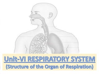
Respiratory system (Structure).pptx
- 2. Introduction: Body’s cell continually use oxygen (O2) for the metabolic reactions that release energy from nutrient molecules and produce ATP, at the same time, these reactions release carbon dioxide (CO2). This system also participates in regulating blood pH This system helps to contributes to homeostasis by providing for the exchange of gases oxygen & carbon dioxide between air, blood and tissue cells.
- 3. Structure: Respiratory system parts can be classified according to either structure or function. Structurally the respiratory system consist of two parts: 1) Upper respiratory system( Nose, Nasal cavity, Pharynx and associated structure) 2) Lower respiratory system (Larynx, Trachea, Bronchi and Lungs)
- 4. Structure: Functionally the respiratory system consist of two parts: 1) Conducting zone: (Nose, Nasal cavity, Pharynx, Larynx, Trachea, Bronchi, Bronchioles and terminal Bronchioles). 2) Respiratory zone: (Respiratory bronchioles, Alveolar ducts, Alveolar sacs, Alveoli). Branch of medicine that deals with diagnosis and treatment of disease of ear, nose & throat is called otorhinolaryngology. Pulmonologist is a specialist in the diagnosis & treatment of diseases of the lungs.
- 5. Structure's of the Respiratory System
- 6. Functions of Respiratory System: 1) Provides for gas exchange: intake of O2 for delivery to body cells and removal of Co2 produced by blood cells. 2) Helps regulate blood pH. 3) Contains receptors for sense of smell, filters inspired air, produces vocal sounds (phonation) and excrete small amount of water and heat.
- 7. 1.NOSE: It is divided into external portion and internal portion called nasal cavity. External nose is the portion of the nose visible on the face consist of bone & hyaline cartilage covered by muscle & skin and lined by mucous membrane. On the undersurface of external nose are 2 openings called External nares (nostrils). Interior structure of external nose have 3 functions (warming, moistening & filtering). next
- 8. 2.NASAL CAVITY: It is a deep hollow cavity, stretching from over the hard palate at the back to between the eye sockets. It is divided into two almost symmetrical halves by the nasal septum.
- 9. 2.NASAL CAVITY: Structure forming the boundaries of nasal cavity: 1) Roof: Formed by cribriform plate of ethmoid bone & sphenoid, frontal & nasal bones. 2) Floor: Formed by roof of mouth & consist of hard palate in front & soft palate behind. 3) Medial wall: Formed by nasal septum which consists of ethmoid & vomer posteriorly and hyaline cartilage anteriorly. 4) Lateral wall: Formed by maxilla, ethmoid bone & inferior conchae. 5) Posteriorly: Opens into nasopharynx
- 10. 2.NASAL CAVITY: Openings of Nasal cavity: 1) Nostrils opening from the exterior into the nasal cavity contains hair. 2) Posterior nares openings from the nasal cavity into the pharynx. 3) Paranasal sinuses are cavities in the bone forming the nasal cavity which contain air. The main sinuses are: (maxillary sinuses, frontal and sphenoidal sinuses & ethmoidal sinuses) 4) Nasolacrimal ducts extend from the lateral walls of the nose to the conjunctival sacs of the eyes, they drain tears from the eyes. next
- 11. 3.PHARYNX: 1) Pharynx (Throat) is 12-14cm long tube lies behind nasal and mouth cavities & larynx. It belongs to both respiratory and digestive. 2) During swallowing epiglottis covers the entrance to the larynx completely, at rest and during breathing epiglottis stand up & oesophagus is closed allowing air to pass into respiratory tract.
- 13. 3.PHARYNX: 1) Pharynx is divided into three parts: (Naso, Oro, Laryngopharynx). Nasopharynx: It lies behind the nose. Lateral structure is pharyngotympanic tube, posteriorly structure is pharyngeal tonsils (adenoids). Oropharynx: It lies behind the mouth, lateral structure is collection of lymphoid tissue called palatine tonsil, during swallowing nasal & oral part is separated by soft palate & uvula. Laryngopharynx: It extend from oropharynx above & continues as esophagus below.
- 14. Functions of Pharynx: 1) It acts as passageway for, both air and food. 2) It is useful in warming and humidifying the air. 3) The pharyngotympanic tube plays an important role in hearing. 4) The pharyngeal & laryngeal tonsils produce antibodies in response to antigen. next
- 15. 4.LARYNX: 1) Larynx or voice box lies in the neck, anterior to esophagus. At puberty it grows larger in males, prominence of the Adam’s apple and deeper voice, larynx consist of cartilages, connected by ligaments & skeletal muscles.
- 16. 4.LARYNX: 1) It is connected to hyoid bone with help of connective tissue. 2) Thyroid cartilage consist of two flat pieces of hyaline cartilage fused anteriorly to form laryngeal prominence (Adam's apple). 3) Epiglottis is elastic plate of cartilage it is attached to thyroid cartilage by ligaments, during swallowing base of tongue presses the epiglottis with this food is prevented from entering into the trachea. 4) Cricoid cartilage consist of hyaline cartilage lies below to thyroid cartilage, it separates of air tract and food tract. 5) Arytenoid cartilage two in number influence position and tension in vocal cords.
- 17. Functions of Larynx: 1) Larynx helps in production of sound. 2) Pitch (frequency) of the voice depends upon the length and tension of vocal cords. 3) Volume or loudness of voice depends upon the force with which the cord vibrate. 4) It is a passageway for air between the pharynx & trachea. 5) It protects the lower respiratory tract during swallowing. next
- 18. 5.TRACHEA: Trachea (windpipe) is a tubular passageway of air, it is about 12 cm long 2.5 cm in diameter, located anterior to esophagus, extend from larynx to 5th thoraxic vertebra, where it divide into right & left bronchi
- 19. 5.TRACHEA: Trachea is having 4 layers (mucosa, submucosa, hyaline cartilage & adventitia (composed of areolar connective tissue). Consist of 16-20 incomplete rings of hyaline cartilage resemble letter “C”. Inner wall of trachea lined by mucous membrane containing numerous mucus secreting goblet cells.
- 20. 6.BRONCHI: Trachea divides into right and left primary bronchus which goes to lungs. Point where trachea divides into left & right primary bronchi an internal ridge called “carina” is formed, its mucous membrane is very sensitive and it trigger the cough reflex. Trachea primary bronchi secondary bronchi tertiary bronchi bronchioles terminal bronchioles respiratory bronchioles alveolar ducts alveolar sacs alveoli
- 21. 7.LUNGS:
- 22. 7.LUNGS: There are two cone-shaped lungs, one lying on each side of the midline in thoracic cavity. Apex is rounded & base of lung is concave and semi- lunar in shape, it rests on diaphragm. Medial surface having hilum structures which enter and leave hilum include (1 bronchus, 1 pulmonary artery, 2 pulmonary vein, 1 bronchial artery, bronchial vein, parasympathetic & sympathetic nerves. Area between lung is mediastinum, it is occupied by heart, great vessels, trachea, bronchi, oesophagus, lymph nodes, lymph vessels & nerves. Right lung divided into 3 lobes (superior, middle & inferior), left lung into 2 lobes (superior & inferior).
- 23. 7.LUNGS: Air in the alveoli is separated from blood in pulmonary capillaries by a wall called respiratory membrane which consist of alveolar wall and capillary wall. Lungs are enveloped by pleura which has 2 layers parietal pleura (superficial layer) and visceral pleura (deep inside layer), this space is filled with approx. 2 ml of serous lubricating fluid called pleural fluid. Lungs receives blood via 2 sets of arteries (pulmonary & bronchial arteries), oxygenated blood to the heart occur by 4 pulmonary vein which drains into left atrium (heart).
- 24. MECHANISM OF RESPIRATION: Eupnoea means rhythmic breathing at rest. It consists of inspiration and expiration. Inspiration: It is a active process during which size of thoracic cavity is increased by contraction of appropriate muscles. Parietal pleura follows the expanding chest wall, expansion of lung is associated with a fall in pressure in the lung parenchyma & atmospheric air. Expiration: It is a passive process. At the end of inspiration, the muscle which contracts actively during inspiration relaxes & the elastic recoil of thoracic wall & lungs cause passive expiration.
- 25. MECHANISM OF RESPIRATION: At rest an adult breathes at a respiratory rate of 12-14 breaths per minute & the amount of air inspired or expired per breath (i.e. tidal air) is approx. 500 mL, thus 6-7 litres of air is breathed in or out of the lungs per minute called pulmonary ventilation..
- 26. MECHANISM OF INSPIRATION: 1) RIB MOVEMENTS: On inspiration ribs move upwards to assume a more horizontal position due to contraction of external intercostal muscles to cause increase in anteroposterior diameter of the chest. 2) DIAPHRAGMATIC MOVEMENTS: During inspiration as a result discharge in phrenic neuron (c3,4,5), muscle fibre contract & draw the central tendon downwards by 1.5cm in eupnoea & by 7cm in deep inspiration. This causes increase in diameter of thoracic cage, for each 1cm descent 200-300 ml air is sucked. 3) ACCESSORY MUSCLE OF INSPIRATION: Scalene, Sternocleidomastoid muscles & intrinsic muscles of larynx.
- 27. MECHANISM OF EXPIRATION: 1) CONTRACTION OF ANTERIOR ABDOMINAL WALL MUSCLES increases intra-abdominal pressure & draws the lower rib down & medially, thereby diaphragm is relaxing. 2) INTERNAL INTERCOSTAL MUSCLES: they pass obliquely downwards & posteriorly from rib to rib. On contraction they pull the upper ribs down so that ribs acquire the position 3) ACCESSORY MUSCLES OF EXPIRATION are adductor muscles of vocal cords. They begin to contract early in expiration. Their main function is protective i.e. prevent entry of food & fluid into trachea.
- 28. LUNG VOLUMES & CAPACITIES: Lung volumes & capacities can be divided into 2 major headings: 1) Static lung volumes and capacities (here time factor is not involved, therefore expressed in ml or L.) 2) Dynamic lung volumes and capacities (these are time dependent, therefore it is expressed in ml/ minute or L/minute).
- 29. STATIC LUNG VOLUMES & CAPACITIES: VOLUMES: 1) Tidal volume (TV): Is the air breathed in or out of lung during quiet respiration. Normal: 500 ml. 2) Inspiratory reserve volume (IRV): maximum volume of air which can be inspired in normal inspiration. Normal: 2000-3200 ml. 3) Expiratory reserve volume (ERV): maximum volume of air which can be expired after normal expiration. Normal: 750-1000 ml. 4) Residual volume (RV): It is the volume of air which remains in lungs after maximal expiration. Normal: 1200 ml
- 30. STATIC LUNG VOLUMES & CAPACITIES: CAPACITIES: 1) Inspiratory capacity (IC): It is the maximum volume of air which can be inspired after completing tidal expiration. It can be computed as: (TV + IRV) Normal: 2500-3700 ml. 2) Expiratory capacity (EC): It is the maximum volume of air which can be expired after completing tidal inspiration. It can be computed as: (TV + ERV), Normal: 1250-1500 ml. 3) Vital capacity (VC): vital capacity is the maximum volume of air which can be expelled from lungs by forceful effort following a maximal inspiration. It is computed as: TV+ IRV+ ERV. Normal: 4.8 litres in males & 3.2 litres in females.
- 31. STATIC LUNG VOLUMES & CAPACITIES: CAPACITIES: 4) Functional residual capacity (FRC): It is the volume of air which is contained in the lungs after completion of tidal expiration. It can be computed as: RV+ ERV, Normal: 2.5 litres. It is act as buffer & allows continuos exchange of gases to occur even during expiration, it also prevent collapse of the alveoli. 5) Total lung capacity (TLC): It is the volume of air contained in lungs after a maximal inspiration. It can be computed as: VC+ RV, Normal: 6 litres.
- 32. DYNAMIC LUNG VOLUMES & CAPACITIES: 1) Timed Vital Capacity (TVC) or Forced Vital Capacity (FVC): It is the maximum volume of air which can be breathed out as forcefully & rapidly as possible following a maximum inspiration. 2) Minute Ventilation (MV) or Pulmonary Ventilation (PV): This is the volume of air expired or inspired by the lungs in 1 minute. PV= TV x RR per minute = 500 x 12 = 6L/minute, normally.
- 33. X Y
- 34. EXCHANGE OF GASES (o2 & Co2): Exchange of gases takes place between alveoli and blood across the respiratory membrane (External respiration) and between blood and tissue across capillary membrane (Internal respiration) by the process of diffusion. Diffusion is movement of any substance (Solute) from higher concentration to lower concentration until equilibrium is established. Exchange of gases occurs when a difference in partial pressure exists. Partial pressure (PP): the pressure exerted by the individual gas in a mixture of gases.
- 36. Partial Pressure of Gases: Condition Po2 PCo2 Atmospheric air 159mmHG 0.3mmHG Alveolar air 104mmHG 40mmHG Pulmonary capillary 40mmHG 46mmHG Arterial end 95mmHG 40mmHG Tissues 40mmHG 46mmHG Pressure Gradient: Difference between partial pressure of two gases. = Po2 (Atm) – Po2 (Alveoli) = 159-104 = 55mmHG
- 38. REGULATION OF RESPIRATION: The normal rate of respiration in adults is 12- 16 breaths per minute. With tidal volume of approx. 500 ml. the rate & depth of respiration, i.e. total pulmonary ventilation can be adjusted to the requirement. This requirement is adjusted by brain by two mechanism: 1) Nervous Regulatory Mechanism 2) Chemical Regulatory Mechanism
- 39. 1. Nervous regulation of respiration: Nervous regulation of respiration is brought about by two system. They are: A. System responsible for automatic control of respiration that is located in the brain stem (upper medulla & pons). B. System responsible for voluntary control of respiration that is located in cerebral cortex.
- 40. 1. Nervous regulation of respiration: A. Automatic control of respiration: The collection of certain groups of neurons in medulla & pons constitutes medullary & pontine respiratory centres respectively. Medullary respiratory centre: Here neuron shows rhythmic discharge with varying frequencies. They are of two type. 1) Those which discharge during inspiration only, i.e. I- neurons. 2) Those which discharge during expiration only, i.e. E- neurons. They both have inhibitory connections to each other, i.e. their exist reciprocal innervation between the two.
- 41. 1. Nervous regulation of respiration: Pontine respiratory centre: 1) An area in the lower pons contains neurons which are tonically active and activate the I- neuron in medulla. This area is referred to as apneustic centre. 2) An area in the upper pons that contains both I and E neurons is called pneumotaxic centre. It inhibits the neurons of the apneustic centre in the lower pons.
- 42. 1. Nervous regulation of respiration: B. Voluntary control of respiration: Respiration can be modified both in rate/depth at will for a specific period only. For e.g. voluntary hyperventilation, breath holding and forceful inspiratory or expiratory efforts. The pathway for such a control is via corticospinal tract which originates from cerebral cortex to end on spinal neuron innervating respiratory group of muscles.
- 43. 1. Nervous regulation of respiration: A. Genesis of inspiration: Inspiratory centre (contains I-neuron) although has its own rhythmicity, is activated by apneustic centre, inspiratory centre discharges over pathways in spinal cord to C3,4,5 & T1,2. therefore inspiration starts. B. Genesis of expiration: The inspiration must be inhibited for expiration to proceed. 1) I-neurons to medulla send excitatory impulses to pneumotaxic centre which, in turn, discharges inhibitory impulses to apneustic centre.
- 44. 1. Nervous regulation of respiration: 2) The pulmonary stretch receptors in lungs which get stimulated during inspiration send inhibitory impulses via vagus (X) nerve to apneustic centre. 3) The pneumotaxic centre stimulates expiratory centre (contains E-neurons) which reciprocally inhibits inspiratory centre.
- 45. 2. Chemical regulation of respiration: The chemical regulatory mechanism adjusts ventilation in such a way that alveolar pCO2 is kept constant at normal value of 40 mmHG. It also maintains the tension of O2, Co2 and H+ of blood. These changes are mediated via respiratory chemoreceptors. The respiratory chemoreceptors are of two types: 1) Peripheral chemoreceptors 2) Medullary (or central) chemoreceptors.
- 46. 2. Chemical regulation of respiration: 1) Peripheral chemoreceptors: Carotid body near carotid artery and aortic bodies near arch of the aorta. 2) Medullary (central) chemoreceptors: It is located in the medulla near the respiratory centre, they get stimulated by H+ concentration in brain.