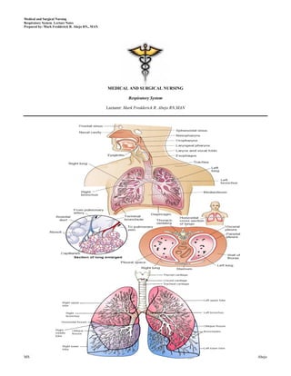
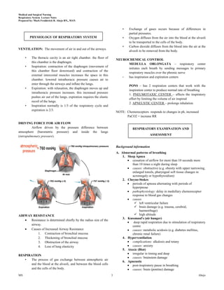
Call Girls Jayanagar Just Call 7001305949 Top Class Call Girl Service Available
Respiratory Nursing
1. Medical and Surgical Nursing
Respiratory System Lecture Notes
Prepared by: Mark Fredderick R. Abejo RN,, MAN
MEDICAL AND SURGICAL NURSING
Respiratory System
Lecturer: Mark Fredderick R. Abejo RN,MAN
MS 1 Abejo
2. Medical and Surgical Nursing
Respiratory System Lecture Notes
Prepared by: Mark Fredderick R. Abejo RN,, MAN
ANATOMY OF RESPIRATORY SYSTEM 2. Bronchi
Lobar Bronchi: 3 R and 2 L
Segmental Bronchi: 10 R and 8 L
Subsegmental Bronchi
OXYGENATON: the dynamic interaction of gases in the body for 3. Bronchioles
the purpose of delivering adequate oxygen essential for cellular Terminal Bronchioles
survival Respiratory Bronchioles, considered to be
the transitional passageways between the
RESPIRATORY SYSTEM MAIN FUNCTION: conducting airways and the gas exchange
GAS EXCHANGE 4. Alveoli
- functional cellular units or gas-exchange
I. Upper Respiratory Tract units of the lungs.
A. Functions - O2 and CO2 exchange takes place
1. Filtering - Made up of about 300 million
2. Warming and moistening TYPE 1 - provide structure to the alveoli
3. Humidification TYPE 2 - secrete SURFACTANT, reduces surface
B. Parts tension; increases alveoli stability & prevents their
1. Nose - made up of framework of cartilages; divided collapse
into R and L by the nasal septum. TYPE 3 – alveolar cell macrophages, destroys
2. Paranasal Sinuses – includes four pair of bony foreign material, such as bacteria
cavities that are lined with nasal mucosa and ciliated
epithelium. Lecithin
3. Tubernate Bones ( Conchae ) Sphingomyelin
4. Pharynx – muscular passageway for both food and L/S ratio indicates lung maturity
air 2:1 normal
Nasopharynx 1:2 immature lungs
Oropharynx
Laryngopharynx PULMONARY CIRCULATION
5. Tonsils and Adenoids - Provides for reoxygenation of blood and release of CO2
6. Larynx – voice production, coughing reflex PULMONARY ARTERIES, carry blood from
Made up of framework of: the heart to the lungs.
Epiglottis – valve that covers the opening
to the larynx during swallowing. PULMONARY VEINS, is a large blood vessel
Glottis – opening between the vocal cords of the circulatory system that carries blood
Hyoid bone – u shaped bone in neck from the lungs to the left atrium of the heart.
Cricoid cartilage
Thyroid cartilage, forms the Adam’s apple
Arythenoid cartilage
Speech production and cough reflex
Vocal cords
7. Trachea - consists of cartilaginous rings
Passageway of air
Site of tracheostomy (4th-6th tracheal ring)
II. Lower respiratory tract
A. Function: facilitates gas exchange
B. Parts
1. Lungs, are paired elastic structure enclosed in the
thoracic cage, which is an airtight chamber with
distensible walls.
Right – 3 lobes, 10 segments
Left – 2 lobes, 8 segments
Client post pneumonectomy affected side to promote expansion
Post lobectomy unaffected side to promote drainage
RESPIRATORY MUSCLES
Pleural cavity
- PRIMARY: diaphragm and external intercostal muscles
Parietal
Visceral - ACCESORY: sternocleidomastoid (elevated sternum),
Pleural Fluid: prevents pleural friction rub the scalene muscles (anterior, middle and posterior
(as seen in pneumonia and pleural effusion) scalene) and the nasal alae
MS 2 Abejo
3. Medical and Surgical Nursing
Respiratory System Lecture Notes
Prepared by: Mark Fredderick R. Abejo RN,, MAN
• Exchange of gases occurs because of differences in
partial pressures.
PHYSIOLOGY OF RESPIRATORY SYSTEM • Oxygen diffuses from the air into the blood at the alveoli
to be transported to the cells of the body.
• Carbon dioxide diffuses from the blood into the air at the
VENTILATION: The movement of air in and out of the airways.
alveoli to be removed from the body.
• The thoracic cavity is an air tight chamber. the floor of
NEUROCHEMICAL CONTROL
this chamber is the diaphragm.
MEDULLA OBLONGATA – respiratory center
• Inspiration: contraction of the diaphragm (movement of
initiates each breath by sending messages to primary
this chamber floor downward) and contraction of the
respiratory muscles over the phrenic nerve
external intercostal muscles increases the space in this
- has inspiration and expiration centers
chamber. lowered intrathoracic pressure causes air to
enter through the airways and inflate the lungs.
PONS – has 2 respiration centers that work with the
• Expiration: with relaxation, the diaphragm moves up and
inspiration center to produce normal rate of breathing
intrathoracic pressure increases. this increased pressure
1. PNEUMOTAXIC CENTER – affects the inspiratory
pushes air out of the lungs. expiration requires the elastic
effort by limiting the volume of air inspired
recoil of the lungs.
2. APNEUSTIC CENTER – prolongs inhalation
• Inspiration normally is 1/3 of the respiratory cycle and
expiration is 2/3.
NOTE: Chemoreceptors responds to changes in ph, increased
PaCO2 = increase RR
DRIVING FORCE FOR AIR FLOW
Airflow driven by the pressure difference between RESPIRATORY EXAMINATION AND
atmosphere (barometric pressure) and inside the lungs
(intrapulmonary pressure). ASSESSMENT
Background information
A. Abnormal patterns of breathing
1. Sleep Apnea
cessation of airflow for more than 10 seconds more
than 10 times a night during sleep
causes: obstructive (e.g. obesity with upper narrowing,
enlarged tonsils, pharyngeal soft tissue changes in
acromegaly or hypothyroidism)
2. Cheyne-Stokes
periods of apnoea alternating with periods of
hyperpnoae
pathophysiology: delay in medullary chemoreceptor
response to blood gas changes
causes
left ventricular failure
brain damage (e.g. trauma, cerebral,
haemorrhage)
AIRWAY RESISTANCE high altitude
3. Kussmaul's (air hunger)
Resistance is determined chiefly by the radius size of the
deep rapid respiration due to stimulation of respiratory
airway. centre
Causes of Increased Airway Resistance causes: metabolic acidosis (e.g. diabetes mellitus,
1. Contraction of bronchial mucosa chronic renal failure)
2. Thickening of bronchial mucosa 4. Hyperventilation
3. Obstruction of the airway complications: alkalosis and tetany
4. Loss of lung elasticity causes: anxiety
5. Ataxic (Biot)
irregular in timing and deep
RESPIRATION
causes: brainstem damage
• The process of gas exchange between atmospheric air
6. Apneustic
and the blood at the alveoli, and between the blood cells post-inspiratory pause in breathing
and the cells of the body. causes: brain (pontine) damage
MS 3 Abejo
4. Medical and Surgical Nursing
Respiratory System Lecture Notes
Prepared by: Mark Fredderick R. Abejo RN,, MAN
- asthma
7. Paradoxical - carcinoma of bronchus
the abdomen sucks with respiration (normally, it - left ventricular failure
pouches uotward due to diaphragmatic descent) - interstitial lung disease
causes: diaphragmatic paralysis - ACE inhibitors
Sputum
B. Cyanosis volume
1. Refers to blue discoloration of skin and mucous type (purulent, mucoid, mucopurulent)
membranes , is due to presence of deoxygenated presence or absence of blood?
haemoglobin in superficial blood vessels Stridor
2. Central cyanosis = abnromal amout of deoxygenated croaking noise loudest on inspiration
haemoglobin in arteries and that blue discoloration is is a sign that requires urgent attention
present in parts of body with good circulation such as causes: (obstruction of larynx, trachea or large
tongue broncus)
3. Peripheral cyanosis = occurs when blood supply to a - acute onset (minutes)
certain part of body is reduced, and the tissue extracts inhaled foreign body
more oxygen from normal from the circulating blood, e.g. acute epiglottitis
lips in cold weather are often blue, but lips are spared anaphylaxis
4. Causes of cyanosis toxic gas inhalation
Central cyanosis - gradual onset (days, weeks)
decreased arterial saturation laryngeal and pharyngeal tumours
decreased concentration of inspired oxygen: crico-arytenoid rheumatoid arthritis
high altitude bilateral vocal cord palsy
lung disease: COPD with cor pulmoale, tracheal carcinoma
massive pulmonary embolism paratracheal compression by lymph nodes
right to left cardiac shunt (cyanotic congenital post-tracheostomy or intubation
heart disease) granulomata
polycythaemia Hoarseness
haemoglobin abnromalities (rare): causes include:
methaemoglobinaemia, sulphaemoglobinaemia - laryngitis
Peripheral cyanosis - laryngeal nerve palsy associated with
all causes of central cyanosis cause peripheral carcinoma of lung
cyanosis - laryngeal carcinoma
exposure to cold
reduced cardiac output: left ventricular failure or The Hands
shock Clubbing
arterial or venous obstruction
commonly cause by respiratory disease (but NOT
Position: patient sitting over edge of bed emphysema or chronic bronchitis)
occasionally, clubbing is associated with hypertrophic
General appearance pulmonary osteoarthropathy (HPO)
look for the following characterised by periosteal inflammation at distal ends
Dyspnea of long bones, wrists, ankles, metacarpals and
normal respiratory rate < 14 each minute metatarsals
tachypnoea = rapid respiratory rate sweelling and tenderness over wrists and other
are accessory muscles being used (sternomastoids, involved areas
platysma, strap muscles of neck) - characteristically,
the accessory muscles cause elevation of shoulders
with inspiration and aid respiration by increasing
chest expansion
Cyanosis
Character of cough
ask patient to cough several times
Staining
lack of usual explosive beginning may indicate
staining of fingers - sign of cigarette smoking (caused by
vocal cord paralysis (bovine cough)
tar, not nicotine)
muffled, wheezy ineffective cough suggests airflow
Wasting and weakness
limitation
Pulse rate
very loose productive cough suggests excessive
Flapping tremor (asterixis) - unreliable sign
bronchial secretions due to:
ask patient to dorsiflex wrists and spread out fingers, with
- chronic bronchitis
arms outstretched
- pneumonia
flapping tremor may occur with severe carbon dioxide
- bronchiectasis
retention (severe chronic airflow limitation)
dry irritating cough may occur with:
- chest infection
MS 4 Abejo
5. Medical and Surgical Nursing
Respiratory System Lecture Notes
Prepared by: Mark Fredderick R. Abejo RN,, MAN
The Face Funnel chest (pectus excavatum)
developmental defect involving a localised depression
Eyes
of lower end of sternum in severe cases, lung capacity
Horner's syndrome? (constricted pupil, partial ptosis and may be restricted
loss of sweating which can be due to apical lung tumour
compressing sympathetic nerves in neck)
Nose
polpys? (associated with asthma)
engorged turbinates? (various allergic conditions)
deviated septum? (nasal obstruction)
Mouth and tongue
look for central cyanosis
evidence of upper respiratory tract infection (a reddened
pharynx and tonsillar enlargement with or without a
coating of pus) Harrison's sulcus
broken tooth - may predispose to lung abscess or innar depression of lower ribs just above costal margins
pneumonia at site of attachment of diaphragm
sinusitis is indicated by tenderness over the sinuses on causes:
palpation severe asthma in childhood
some patients with obstructive sleep apnoea will be obese rickets
with a receding chin, a small pharynx and a short thick neck Kyphosis , exaggerated forward curvature of spine
The Trachea Scoliosis , lateral bowing
Kyphoscoliosis: causes:
causes of tracheal displacement: idiopathic (80%)
toward the side of the lung lesion secondary to poliomyelitis (inflammation involving
upper lobe collapse grey matter of cord)
upper lobe fibrosis (note: severe thoracic kyphoscoliosis may reduce lung
pneumonectomy capacity and increase work of breathing)
upper mediastinal masses, such as retrosternal goitre Lesions of chest wall
tracheal tug (finger resting on trachea feels it move inferiorly scars - previous thoracic operations or chest drains for a
with each inspiration) is a sign of gross overexpansion of the previous pneumothorax or pleural effusion
chest because of airflow obstruction thoracoplasty (was once performed to remove TB, but
no longer is because of effective antituberculosis
The Chest: inspection
chemotherapy) invovled removal of large number of
Shape and symmetry of chest ribs on one side to achieve permanent collapse of
Barrel shaped affected lung
anteroposterior (AP) diameter is increased compared erythema and thickening of skin may occur in
with lateral diameter radiotherapy; there is a sharp demarcation between
causes: hyperinflation due to asthma, emphysema abnormal and normal skin
Diffuse swelling of chest wall and neck
pathophysiology: air tracking from the lungs
causes:
pneumothorax
rupture of oesopahagus
Prominent veins
cause: superior vena caval obstruction
Asymmetry of chest wall movements
Pigeon chest (pectus carinatum) assess this by inspecting from behind patient, looking
localised prominence (outward bowing of sternum and down the clavicles during moderate respiration -
costal cartilages) diminished movement indicates underlying lung disease
causes: the affected side will showed delayed or decreased
manifestation of chronic childhood illness (due to movement
repeated strong contractions of diaphragm while causes of reduced chest wall movements on one side are
thorax is still pliable) localised:
rickets localised pulmonary fibrosis
consolidation
collapse
pleural effusion
pneumothroax
causes of bilateral reduced chest wall movements are
diffuse:
chronic airflow limitation
diffuse pulmonary fibrosis
MS 5 Abejo
6. Medical and Surgical Nursing
Respiratory System Lecture Notes
Prepared by: Mark Fredderick R. Abejo RN,, MAN
The Chest: palpation cardiac dullness:
area of cardiac dullness is uaully present on left side of
chest expansion
chest
place hands firmly on chest wall with fingers extending this may decrease in emphysema or asthma
around sides of chest (fugyre 4.5)
as patient takes a big breath in, the thumbs should move The Chest: auscultation
symmetrically apart about 5 cm
breath sounds
reduced expansion on one side indicates a lesion on that
introduction
side
one should use the diaphragm of stethoscope to listen
note: lower lobe expansion is tested here; upper lobe is
to breath sound in each area, comparing each side
tested for on inspection (as above)
remember to listen high up into the axillae
apex beat
(discussed in cardiac section) remember to use bell of stethoscope to listen to lung
from above the clavicles
for respiratory diseases:
displacement toward site of lesion - can be caused by: quality of breath sounds
collapse of lower lobe normal breat sounds
are heard with stethoscope over all parts of
localised pulmonary fibrosis
chest, produced in airways rather than alveoli
displacement away from site of lesion - can be caused
(although once they had been thought to arise
by:
from alveoli (vesicles) and are therefore called
pleural effusion
vesicular sounds)
tension pneumothorax normal (vesicular) breath sounds are louder and
apex beat is often impalpable in a chest which is longer on inspiration than on expiration; and
hyperexpanded secondary to chronic airflow limitation there is no gap between the inspiratory and
vocal fremitus expiratory sounds
palpate chest wall with palm of hand while patient repeats bronchial breath sounds
"99" turbulence in large airways is heard without
front and back of chest are each palpated in 2 comparable being filtered by the alveoli, and therefore
positions with palms; in this way differences in vibration on produce a different quality; they are heard over
chest wall can be detected the trachea normally, but not over the lungs
causes of change in vocal fremitus are the same as those for are audible throughout expiration, and often
vocal resonance (see later) there is a gap between inspiration and expiration
ribs are heard over areas of consolidation since solid
gently compress chest wall anteroposteriorly and laterally lung conducts the sound of turbulence in main
localised pain suggests a rib fracture (may be secondary to airways to peripheral areas without filtering
trauma or spontaneous as a result of tumour deposition or causes include:
bone disease) - lung consolidation (lobar pneumonia) -
The Chest: percussion common
- localised pulmonary fibrosis - uncommon
with left hand on chest wall and fingers slightly separated and - pleural effusion (above the fluid) -
aligned with ribs, the middle finger is pressed firmly against uncommon
the chest; pad of right middle finger is used to strike firmly the - collapsed lung (e.g. adjacent to a pleural
middle phalanx of middle finger of left hand effusion) - uncommon
percussion of symmetrical areas of: amphoric sound = when breath sounds over a
anterior (chest) large cavity have an exaggerated bronchial
posterior (back) (ask patient to move elbows forward quality)
across the front of chest - this rotates the scapulae intensity of breath sounds
anteriorly, i.e. moves it out of the way) causes of reduced breath sounds include:
axillary region (side) chronic airflow limitation (especially
supraclavicular fossa emphysema)
percussion over a solid structure (e.g. liver, consolidated lung) pleural effusion
produces a dull note pneumothorax
percusion over a fluid filled area (e.g. pleural effusion) pneumonia
produces an extremely dull (stony dull) note large neoplasm
percussion over the normal lung produces a resonant note pulmonary collapse
percussion over a hollow structure (e.g. bowel, pneumothorax) added (adventitious) sounds
produces a hyperresonsant note two types of added sounds: continuous (wheezes) and
liver dullness: interrupted (crackles)
upper level of liver dullness is determined by percussing wheezes
down the anterior cehst in mid-clavicular line may be heard in expiration or inspiration or both
normally, upper level of liver dullness is 6th rib in right pathophysiology of wheezes - airway narrowing
mid-clavicular line an inspiratory wheeze implies severe airway
if chest is resonant below this level, it is a sign of narrowing
hyperinflation usually due to emphysema, asthma
MS 6 Abejo
7. Medical and Surgical Nursing
Respiratory System Lecture Notes
Prepared by: Mark Fredderick R. Abejo RN,, MAN
causes of wheezes include: pulmonary thromboembolism
- asthma (often high pitched) - due to muscle marked obesity
spasm, mucosal oedema, excessive sleep apnoea
secretions severe kyphoscoliosis
- chronic airflow diseases - due to mucosal
oedema and excessive secretions The Abdomen
- carcinoma causing bronchial obstruction -
palpate liver for enlargement due to secondary deposits of
tends to cause a localised wheeze which is
tumour from lung, or right heart failure
monophonic and does not clear with
coughing Other
crackles
some terms not to use include rales (low pitched Permberton's sign
crackles) and creptitations (high pitched ask patient to lift arms over head
crackles) look for development of facial plethora, inspiratory
crackles are due to collapse of peripheral stridor, non-pulsatile elevation of jugular venous
airways on expiration and sudden opening on pressure
inspiration occurs in vena caval obstruction
early inspiratory crackles Feet
- suggests disease of small airways inspect for oedema or cyanosis (clues of cor
- characteristic of chronic airflow limitation pulmonale)
- are only heard in early inspiration look for evidence of deep vein thrombosisd
late or paninspiratory crackles Respiratory rate on exercise and positioning
- suggests disease confined to alveoli patients complaining of dyspnoea should have their
- may be fine, medium or coarse respiratory rate measured at rest, at maximal tolerated
- fine crackles - typically caused by exertion and supine
pulmonary fibrosis if dyspnoea is not accompanied by tachypnoea when
- medium crackles - typically caused by left a patient climbs stairs, one should consider
ventricular failure (due to presence of malingering
alveolar fluid) look for paradoxical inward motion of abdomen
- coarse crackes - tend to change with during inspiration when patient is uspine (indicating
coughing; occur with any disease that leads diaphragmatic paralysis)
to retention of secretions; commonly occur Temperature: fever may accompany any acute or chronic
in bronchiectasis chest infection
pleural friction rub
when thickened, roughened pleural surfaces rub
together, a continuous or intermittent grating
sound may be heard DIAGNOSTIC EVALUATION
suggests pleurisy, which may be secondary to
pulmonary infarction or pnuemonia
1. Skin Test: Mantoux Test or Tuberculin Skin Test
vocal resonanance
gives information about lungs' ability to transmit sounds This is used to determine if a person has been infected or
consolidated lung tends to transmit high frequencies so has been exposed to the TB bacillus.
that speech heard through stethoscope takes a bleeting This utilizes the PPD (Purified Protein Derivatives).
quality (aegophony); when a patient with aegophony says The PPD is injected intradermally usually in the inner
"bee" it sounds like "bay" aspect of the lower forearm about 4 inches below the
listen over each part of chest as patient says "99"; over elbow.
consolidated lung, the numbers will become clearly The test is read 48 to 72 hours after injection.
audible; over normal lung, the sound is muffled (+) Mantoux Test is induration of 10 mm or more.
whispering pectoriloquy - vocal resonance is increased to But for HIV positive clients, induration of about 5 mm is
such an extent that whispered speech is distinctly heard considered positive
Signifies exposure to Mycobacterium Tubercle bacilli
The Heart
lie patient at 45 degrees
measure jugular venous plse for right heart failure
examine preacordium; pay close attention to pulmonary
component of P2 (which is best heard at 2nd intercostal
space on left) and should not be louder than A2; if it is
louder, suspect pulmonary hypertension
cor pulmonale (also called pulmonary hypertensive heart
disease) may be due to:
chronic airflow limitation (emphysema)
pulmonary fibrosis
MS 7 Abejo
8. Medical and Surgical Nursing
Respiratory System Lecture Notes
Prepared by: Mark Fredderick R. Abejo RN,, MAN
2. Pulse Oximeter
Non-invasive method of continuously monitoring he
oxygen saturation of hemoglobin
A probe or sensor is attached to the fingertip, forehead,
earlobe or bridge of the nose
Sensor detects changes in O2 sat levels by monitoring
light signals generated by the oximeter and reflected by
the blood pulsing through the tissue at the probe
Normal SpO2 = 95% - 100%
< 85% - tissues are not receiving enough O2
Results unreliable in:
Cardiac arrest
Shock
Use of dyes or
vasoconstrictors
Severe anemia
High carbon Clear MRI images of lung airways during breathing.
monoxide Level
6. Flouroscopy
Studies the lung and chest in motion
3. Chest X-ray Involves the continuous observation of an image
reflected on a screen when exposed to radiation in the
This is a NON-invasive procedure involving the use of x- manner of television screen that is activated by an
rays with minimal radiation. electrode beam.
The nurse instructs the patient to practice the on cue to Structures of different densities that intercept the X-ray
hold his breath and to do deep breathing beam are visualized on the screen in silhouette
Instruct the client to remove metals from the chest.
Rule out pregnancy first. 7. Indirect Bronchography
A radiopaque medium is instilled directly into the
5. Computed Tomography (CT Scan) and Magnetic Resonance trachea and the bronchi and the outline of the entire
Imaging ( MRI ) bronchial tree or selected areas may be visualized
through x-ray.
The CT scan is a radiographic procedure that utilizes It reveals anomalies of the bronchial tree and is
x-ray machine. important in the diagnosis of bronchiectasis.
The MRI uses magnetic field to record the H+ density of
the tissue. Nursing interventions BEFORE Bronchogram
It does NOT involve the use of radiation. Secure written consent
The contraindications for this procedure are the Check for allergies to sea foods or iodine or
following: patients with implanted pacemaker, anesthesia
patients with metallic hip prosthesis or other metal NPO for 6 to 8 hours
implants in the body. Pre-op meds: atropine SO4 and valium,
topical anesthesia sprayed; followed by local
anesthetic injected into larynx. The nurse must
have oxygen and anti spasmodic agents ready.
Nursing interventions AFTER Bronchogram
Side-lying position
NPO until cough and gag reflexes returned
Instruct the client to cough and deep breathe
client
8. Bronchoscopy
This is the direct inspection and observation of the
larynx, trachea and bronchi through a flexible or rigid
bronchoscope.
Passage of a lighted bronchoscope into the bronchial
tree for direct visualization of the trachea and the
tracheobronchial tree.
Diagnostic uses:
This chest CT scan shows a cross-section of a person To examine tissues or collect secretions
with bronchial cancer. The two dark areas are the lungs. The light To determine location or pathologic process
areas within the lungs represent the cancer. and collect specimen for biopsy
MS 8 Abejo
9. Medical and Surgical Nursing
Respiratory System Lecture Notes
Prepared by: Mark Fredderick R. Abejo RN,, MAN
To evaluate bleeding sites 9. Lung Scan
To determine if a tumor can be resected Procedure using inhalation or I.V. injection of a
surgically radioisotope, scans are taken with a scintillation camera.
Imaging of distribution and blood flow in the lungs.
Therapeutic uses (Measure blood perfusion)
To Remove foreign objects from Confirm pulmonary embolism or other blood- flow
tracheobronchial tree abnormalities
To Excise lesions
To remove tenacious secretions obstructing the Nursing interventions BEFORE the procedure:
tracheobronchial tree Allay the patient’s anxiety
To drain abscess Instruct the patient to Remain still during the
To treat post-operative atelectasis procedure
Nursing interventions BEFORE Bronchoscopy Nursing interventions AFTER the procedure
Informed consent/ permit needed Check the catheter insertion site for bleeding
Explain procedure to the patient, tell him what Assess for allergies to injected radioisotopes
to expect, to help him cope with the unkown Increase fluid intake, unless contraindicated.
Atropine (to diminish secretions) is
administered one hour before the procedure 10. Sputum Examination
About 30 minutes before bronchoscopy, Laboratory test
Valium is given to sedate patient and allay Indicated for microscopic examination of the sputum:
anxiety. Gross appearance, Sputum C&S, AFB staining, and
Topical anesthesia is sprayed followed by for Cytologic examination/ Papanicolaou examination
local anesthesia injected into the larynx
Instruct on NPO for 6-8 hours Nursing interventions:
Remove dentures, prostheses and contact lenses Early morning sputum specimen is to be
The patient is placed supine with collected (suctioning or expectoration)
hyperextended neck during the procedure Rinse mouth with plain water
Use sterile container.
Nursing interventions AFTER Bronchoscopy Sputum specimen for C&S is collected before
Put the patient on Side lying position the first dose of anti-microbial therapy.
Tell patient that the throat may feel sore with . For AFB staining, collect sputum specimen for
Check for the return of cough and gag reflex. three consecutive mornings.
Check vasovagal response.
Watch for cyanosis, hypotension, tachycardia, 11. Biopsy of the Lungs
arrythmias, hemoptysis, and dyspnea. These Percutaneous removal of a small amount of lung tissue
signs and symptoms indicate perforation of For histologic evaluation
bronchial tree. Refer the patient immediately! - Transbronchoscopic biopsy—done during bronchoscopy,
- Percutaneous needle biopsy
- Open lung biopsy
MS 9 Abejo
10. Medical and Surgical Nursing
Respiratory System Lecture Notes
Prepared by: Mark Fredderick R. Abejo RN,, MAN
Nursing interventions BEFORE the procedure: LUNG CAPACITIES:
Withhold food and fluids
Place obtained written informed consent in the Functional Residual Capacity (ERV 1100 mL + RV 1200 mL =
patient’s chart. 2300 mL )
The volume of air that remains in the lungs after normal,
Nursing interventions AFTER the procedure: quiet exhalation
Observe the patient for signs of Pneumothorax Inspiratory Capacity (TV 500 mL + IRV 3000 mL = 3500 mL )
and air embolism The amount of air that a person can inspire maximally
Check the patient for hemoptysis and after a normal expiration
hemorrhage Vital capacity (IRV 3000 mL + TV 500 mL + ERV 1100 mL =
Monitor and record vital signs 4600 mL )
Check the insertion site for bleeding The maximum volume of air that can be exhaled after a
Monitor for signs of respiratory distress maximum inhalation
Reduced in COPD
12. Lymph Node Biopsy Total Lung Capacity (IRV 3000 mL + TV 500 mL + ERV 1100
Scalene or cervicomediastinal mL + RV 1200 mL = 5800 mL )
To assess metastasis of lung cancer Total of all four volumes
13. Pulmonary Function Test / Studies 14. Arterial Blood Gas
Non-invasive test Laboratory test
Measurement of lung volume, ventilation, and diffusing Indicate respiratory functions
capacity Assess the degree to which the lungs are able to provide
Nursing interventions: adequate oxygen and remove CO2
Document bronchodilators or narcotics used Assess the degree to which the kidneys are able to
before testing reabsorb or excrete bicarbonate.
Allay the patient’s anxiety during the testing Assessment of arterial blood for tissue oxygenation,
ventilation, and acid-base status
Arterial puncture is performed on areas where good
pulses are palpable (radial, brachial, or femoral).
Radial artery is the most common site for withdrawal of
blood specimen
Nursing interventions:
Utilize a 10-ml. Pre-heparinized syringe to
prevent clotting of specimen
Soak specimen in a container with ice to
prevent hemolysis
If ABG monitoring will be done, do Allen’s
test to assess for adequacy of collateral
circulation of the hand (the ulnar arteries)
LUNG VOLUMES: (ITER)
Inspiratory reserve volume (3000 mL)
The maximum volume that can be inhaled following a
normal quiet inhalation.
Tidal volume (500 mL)
The volume of air inhaled and exhaled with normal quiet
breathing 15. Pulmonary Angiography
Expiratory reserve volume (1100 mL) This procedure takes X-ray pictures of the pulmonary
The maximum volume that can be exhaled following the blood vessels (those in the lungs).
normal quiet exhalation Because arteries and veins are not normally seen in an X-
Residual volume (1200 mL) ray, a contrast material is injected into one or more
The volume of air that remains in the lungs after forceful arteries or veins so that they can be seen.
exhalation
MS 10 Abejo
11. Medical and Surgical Nursing
Respiratory System Lecture Notes
Prepared by: Mark Fredderick R. Abejo RN,, MAN
16. Ventilation - Perfusion Scan
Radioactive albumin injection is part of a nuclear scan
test that is performed to measure the supply of blood
through the lungs.
After the injection, the lungs are scanned to detect the
location of the radioactive particles as blood flows
through the lungs.
The ventilation scan is used to evaluate the ability of air
to reach all portions of the lungs. The perfusion scan
measures the supply of blood through the lungs.
A ventilation and perfusion scan is most often performed
to detect a pulmonary embolus. It is also used to evaluate
lung function in people with advanced pulmonary disease
such as COPD and to detect the presence of shunts
(abnormal circulation) in the pulmonary blood vessels.
RESPIRATORY CARE MODALITIES
1. Oxygen Therapy
Oxygen is a colorless, odorless, tasteless, and dry gas that
supports combustion
Man requires 21% oxygen from the environment in order
to survive
Indication: Hypoxemia
Signs of Hypoxemia
o Increased pulse rate
o Rapid, shallow respiration and dyspnea
o Increased restlessness or lightheadedness
o Flaring of nares
17. Thoracentesis o Substernal or intercostals retractions
Procedure suing needle aspiration of intrapleural fluid or o Cyanosis
air under local anesthesia
Specimen examination or removal of pleural fluid Low flow oxygen provides partial oxygenation with patient
Nursing intervention BEFORE Thoracentesis breathing a combination of supplemental oxygen and room air.
Secure consent Low-flow administration devices:
Take initial vital signs o Nasal Cannula 24-45% 2-6 LPM
Instruct to remain still, avoid coughing during o Simple Face Mask 0-60% 5-8 LPM
insertion of the needle o Partial Rebreathing Mask 60-90% 6-10 LPM
Inform patient that pressure sensation will be o Non-rebreathing Mask 95-100% 6-15 LPM
felt on insertion of needle o Croupette
o Oxygen Tent
Nursing intervention DURING the procedure:
Reassess the patient High flow oxygen provides all necessary oxygenation, with
Place the patient in the proper position: patients breathing only oxygen supplied from the mask and
Upright or sitting on the edge of exhaling through a one-way vent.
the bed High flow administration devices
Lying partially on the side, o Venturi Mask 24-40% 4-10 LPM
partially on the back Preferred for clients with COPD because it
provides accurate amount of oxygen.
Nursing interventions after Thoracentesis o Face Mask
Assess the patient’s respiratory status o Oxygen Hood*
Monitor vital signs frequently o Incubator / isolette*
Position the patient on the affected side, as
ordered, for at least 1 hour to seal the puncture Note: * can be used for both low and high flow administration
site
Turn on the unaffected side to prevent leakage The nurse should prevent skin breakdown by checking
of fluid in the thoracic cavity nares, nose and applying gauze or cotton as necessary
Check the puncture site for fluid leakage Ensure that COPD patients receive only LOW flow
Auscultate lungs to assess for pneumothorax oxygen because these persons respond to hypoxia, not
Monitor oxygen saturation (SaO2) levels increased CO levels.
Bed rest
Check for expectoration of blood
MS 11 Abejo
12. Medical and Surgical Nursing
Respiratory System Lecture Notes
Prepared by: Mark Fredderick R. Abejo RN,, MAN
2. Tracheobronchial suctioning Goals are removal of bronchial secretions, improved
Suction only when necessary not routinely ventilation, and increased efficiency of respiratory
Use the smallest suction catheter if possible muscles.
Client should be in semi or high Fowler’s position Postural drainage uses specific positions to use gravity to
Use sterile gloves, sterile suction catheter assist in the removal of secretions.
Hyperventilate client with 100% oxygen before and Vibration loosens thick secretions by percussion or
after suctioning vibration.
Insert catheter with gloved hand (3-5― length of catheter Breathing exercises and breathing retraining improve
insertion) without applying suction. Three passes of the ventilation and control of breathing and decrease the
catheter is the maximum, with 10 seconds per pass. work of breathing.
Apply suction only during withdrawal of catheter These are procedures for patients with respiratory
The suction pressure should be limited to less than 120 disorders like COPD, cystic fibrosis, lung abscess, and
mmHg pneumonia. The therapy is based on the fact that mucus
When withdrawing catheter rotate while applying can be knocked or shaken from airways and helped to
intermittent suction drain from the lungs.
Suctioning should take only 10 seconds (maximum of 15
seconds) Postural drainage
Evaluate: clear breath sounds on auscultation of the chest. Use of gravity to aid in the drainage of secretions.
Patient is placed in various positions to promote flow of
drainage from different lung segments using gravity.
3. Bronchial Hygiene Measures Areas with secretions are placed higher than lung
Suctioning: oropharyngeal; nasopharyngeal segments to promote drainage.
Patient should maintain each position for 5-15 minutes
a. Steam inhalation depending on tolerability.
The purpose of steam inhalation are as follows:
- to liquefy mucous secretions Percussion
- to warm and humidify air Produces energy wave that is transmitted through the
- to relieve edema of airways chest wall to the bronchi.
- to soothe irritated airways The chest is struck rhythmically with cupped hands over
- to administer medication the areas were secretions are located.
It is a dependent nursing function Avoid percussion over the spine, kidneys, breast or
Inform the client and explain the purpose of the procedure incision and broken ribs. Areas should be percussed for
Place the client in Semi-Fowler’s position 1-2 minutes
Cover the client’s eyes with washcloth to prevent irritation
Check the electrical device before use Vibration
Place the steam inhalator in a flat, stable surface. Works similarly to percussion, where hands are placed on
Place the spout 12 – 18 inches away from the client’s nose or client’s chest and gently but firmly rapidly vibrate hands
adjust distance as necessary against thoracic wall especially during client’s exhalation.
CAUTION: avoid burns. Cover the chest with towel to This may help dislodge secretions and stimulate cough.
prevent burns due to dripping of condensate from the steam. This should be done at least 5-7 times during patient
Assess for redness on the side of the face which indicates exhalation.
first degree burns.
To be effective, render steam inhalation therapy for 15 – 20
minutes
Instruct the client to perform deep breathing and coughing
exercises after the procedure to facilitate expectoration of
mucous secretions.
Provide good oral hygiene after the procedure.
Do after-care of equipment.
b. Aerosol inhalation
done among pediatric clients to administer brochodilators or
mucolytic-expectorants.
.
c. Medimist inhalation
done among adult clients to administer bronchodilators or
mucolytic-expectorants.
4. Chest Physiotheraphy ( CPT )
Includes postural drainage, chest percussion and vibration,
and breathing retraining. Effective coughing is also an
important component.
MS 12 Abejo
13. Medical and Surgical Nursing
Respiratory System Lecture Notes
Prepared by: Mark Fredderick R. Abejo RN,, MAN
Suctioning
Nursing Interventions in CPT
Verify doctor’s order
Assess areas of accumulation of mucus secretions.
Position to allow expectoration of mucus secretions
by gravity
Place client in each position for 5-10 to 15 minutes
Percussion and vibration done to loosen mucus
secretions
Change position gradually to prevent postural
hypotension
Client is encouraged to cough up and expectorate
sputum
Procedure is best done 60 to 90 minutes before
meals or in the morning upon awakening and at
bedtime.
Types of Bottle Drainage
Provide good oral care after the procedure
One-bottle system
The bottle serves as drainage and water-seal
5. Incentive Spirometry
Immerse tip of the tube in 2-3 cm of sterile NSS to
• Types: volume and flow
create water-seal.
• Device ensures that a volume of air is inhaled and the
Keep bottle at least 2-3 feet below the level of the
patient takes deep breaths.
chest to allow drainage from the pleura by gravity.
• Used to prevent or treat atelectasis
Never raise the bottle above the level of the heart
• To enhance deep inhalation
to prevent reflux of air or fluid.
Assess for patency of the device
• Nursing care
Observe for fluctuation of fluid along the tube. The
– Positioning of patient, teach and encourage use,
fluctuation synchronizes with the respiration.
set realistic goals for the patient, and record the
Observe for intermittent bubbling of fluid;
results.
continues bubbling means presence of air-leak
In the absence of fluctuation:
Suspect obstruction of the device
Assess the patient first, then if patient is stable
Check for kinks along tubing;
Milk tubing towards the bottle (If the hospital allows the
nurse to milk the tube)
If there is no obstruction, consider lung re-expansion;
(validated by chest x-ray)
Air vent should be open to air.
Two-bottle system
If not connected to the suction apparatus
The first bottle is drainage bottle;
The second bottle is water-seal bottle
Observe for fluctuation of fluid along the tube
(water-seal bottle or the second bottle) and
intermittent bubbling with each respiration.
6. Closed Chest Drainage ( Thoracostomy Tube )
Chest tube is used to drain fluid and air out of the NOTE! IF connected to suction apparatus
mediastinum or pleural space into a collection chamber 1. The first bottle is the drainage and water-seal bottle;
to help re-establish normal negative pressure for lung re- 2. The second bottle is suction control bottle.
expansion. 3. Expect continuous bubbling in the suction control bottle;
Purposes 4. Intermittent bubbling and fluctuation in the water-seal
To remove air and/or fluids from the pleural space 5. Immerse tip of the tube in the first bottle in 2 to 3 cm of
To reestablish negative pressure and re-expand the sterile NSS
lungs 6. Immerse the tube of the suction control bottle in 10 to 20
Procedure cm of sterile NSS to stabilize the normal negative
The chest tube is inserted into the affected chest pressure in the lungs.
wall at the level of 2nd to 3rd intercostals space to 7. This protects the pleura from trauma if the suction
release air or in the fourth intercostals space to pressure is inadvertently increased
remove fluid.
MS 13 Abejo
14. Medical and Surgical Nursing
Respiratory System Lecture Notes
Prepared by: Mark Fredderick R. Abejo RN,, MAN
Three-bottle system Removal of chest tube—done by physician
The first bottle is the drainage bottle; The nurse Prepares:
The second bottle is water seal bottle Petrolatum Gauze
The third bottle is suction control bottle. Suture removal kit
Sterile gauze
Observe for intermittent bubbling and Adhesive tape
fluctuation with respiration in the water- seal Place client in semi-Fowler’s position
bottle Instruct client to exhale deeply, then inhale and do
Continuous GENTLE bubbling in the suction valsalva maneuver as the chest tube is removed.
control bottle. These are the expected observations. Chest x-ray may be done after the chest tube is
Suspect a leak if there is continuous bubbling in removed
the WATER seal bottle or if there is VIGOROUS Asses for complications: subcutaneous emphysema;
bubbling in the suction control bottle. respiratory distress
The nurse should look for the leak and report the
observation at once. Never clamp the tubing 7. Artificial Airway
unnecessarily.
a. Oral airways- these are shorter and often have a larger lumen.
If there is NO fluctuation in the water seal bottle, it may mean They are used to prevent the tongue form falling backward.
TWO things
Either the lungs have expanded or the system is NOT b. Nasal airways- these are longer and have smaller lumen Which
functioning appropriately. causes greater airway resistance
In this situation, the nurse refers the observation to the
physician, who will order for an X-ray to confirm the c. Tracheostomy- this is a temporary or permanent surgical
suspicion. opening in the trachea. A tube is inserted to allow ventilation and
removal of secretions. It is indicated for emergency airway access
Important Nursing considerations for many conditions. The nurse must maintain tracheostomy care
Encourage doing the following to promote drainage: properly to prevent infection.
Deep breathing and coughing exercises
Turn to sides at regular basis
Ambulate
ROM exercise of arms RESPIRATORY DISEASES AND
Mark the amount of drainage at regular intervals DISORDERS
Avoid frequent milking and clamping of the tube to
prevent tension pneumothorax
What the nurse should do if: I. PNEUMONIA – inflammation of the lung parenchyma
If there is continuous bubbling: leading to pulmonary consolidation because alveoli is filled
The nurse obtains a toothless clamp with exudates
Close the chest tube at the point where it exits the chest
for a few seconds.
If bubbling in the water seal bottle stops, the leak is
likely in the lungs,
But if the bubbling continues, the leak is between the
clamp and the bottle chamber.
Next, the nurse moves the clamp towards the bottle checking the
bubbling in the water seal bottle.
If bubbling stops, the leak is between the clamp
and the distal part including the bottle.
But if there is persistent bubbling, it means that the
drainage unit is leaking and the nurse must obtain
another set.
In the event that the water seal bottle breaks, the
nurse temporarily kinks the tube and must obtain a
receptacle or container with sterile water and A. ETIOLOGIC AGENTS
immerse the tubing. 1. Streptococcus pneumoniae (pneumococcal
She should obtain another set of sterile bottle as pneumonia)
replacement. She should NEVER CLAMP the tube 2. Hemophilus influenzae (bronchopneumonia)
for a longer time to avoid tension pneumothorax. 3. Klebsiella pneumoniae
In the event the tube accidentally is pulled out, the 4. Diplococcus pneumoniae
nurse obtains vaselinized gauze and covers the 5. Escherichia coli
stoma. 6. Pseudomonas aeruginosa
She should immediately contact the physician.
MS 14 Abejo
15. Medical and Surgical Nursing
Respiratory System Lecture Notes
Prepared by: Mark Fredderick R. Abejo RN,, MAN
B. HIGH RISK GROUPS Usually, it is the upper lung areas which are
1. Children less than 5 yo drained
2. Elderly Nursing management:
Monitor VS and BS
C. PREDISPOSING FACTORS Best performed before meals/breakfast or
1. Smoking 2-3 hours p.c. to prevent gastroesophageal
2. Air pollution reflux or vomiting (pagkagising maraming
3. Immunocompromised secretions diba? Nakukuha?)
(+) AIDS Encourage DBE
Kaposi’s Sarcoma Administer bronchodilators 15-30 minutes
Pneumocystis Carinii Pneumonia before procedure
DOC: Zidovudine (Retrovir) Stop if pt. can’t tolerate the procedure
Bronchogenic Ca Provide oral care after procedure as it may
4. Prolonged immobility (hypostatic pneumonia) affect taste sensitivity
5. Aspiration of food (aspiration pneumonia) Contraindications:
6. Over fatigue Unstable VS
Hemoptysis
D. SIGNS AND SYMPTOMS Increased ICP
1. Productive cough, greenish to rusty Increased IOP (glaucoma)
2. Dyspnea with prolong expiratory grunt 12. Provide pt health teaching and d/c planning
3. Fever, chills, anorexia, general body malaise Avoidance of precipitating factors
4. Cyanosis Prevention of complications
5. Pleuritic friction rub Atelectasis
6. Rales/crackles on auscultation Meningitis
7. Abdominal distention paralytic ileus Regular compliance to medications
Importance of ffup care
E. DIAGNOSTICS
1. Sputum GS/CS confirmatory; type and sensitivity;
(+) to cultured microorganism
Tracheostomy usually done at bedside, 10-20 minutes
2. CXR – (+) pulmonary consolidation Stress test: 30 minutes
3. CBC Mammography: 10-20 minutes
Elevated ESR (rate of erythropoeisis) N = 0.5- LARYNGOSPASM – tracheostomy STAT
1.5% (compensatory mech to decreased O2) OR Tracheostomy: laryngeal, thyroid, neck CA
Elevated WBC DIAPHRAGM – primary muscle for respiration
4. ABG – PO2 decreased (hypoxemia) INTERCOSTAL MUSCLES – secondary muscle for respiration
ALVEOLI (Acinar cells) –functional unit of the lungs; site for gas
exchange (via diffusion)
F. NURSING MANAGEMENT
VENTILATION – movement of air in and out of the lungs
1. Enforce CBR (consistent to all respi disorders) RESPIRATION – lungs to cells
2. Strict respiratory isolation Internal
3. Administer medications as ordered External
Broad spectrum antibiotics RETROLENTAL FIBROPLASIA – retinopathy/blindness in
Penicillin – pneumococcal infections immaturity d/t high O2 flow in pedia patients
Tetracycline
Macrolides
Azithromycin (OD x 3/days) II. PULMONARY TUBERCULOSIS (KOCH’S DISEASE) –
1. Too costly infection of the lung parenchyma caused by invasion of
2. Only se: ototoxicity – transient mycobacterium tuberculosis or tubercle bacilli (gram negative,
hearing loss acid fast, motile, aerobic, easily destroyed by heat/sunlight)
Anti-pyretics
Mucolytics/expectorants A. PRECIPITATING FACTORS
4. Administer O2 inhalation as ordered 1. Malnutrition
5. Force fluids to liquefy secretions 2. Overcrowding
6. Institute pulmonary toilet – measures to promote 3. Alcoholism: Depletes VIT B1 (thiamin) alcoholic
expectoration of secretions beriberi malnutrition
DBE, Coughing exercises, CPT 4. Physical and emotional stress
(clapping/vibration), Turning and repositioning 5. Ingestion of infected cattle with M. bovis
7. Nebulize and suction PRN 6. Virulence (degree of pathogenecity)
8. Place client of semi-fowlers to high fowlers
9. Provide a comfortable and humid environment B. MODE OF TRANSMISSION: Airborne droplet
10. Provide a dietary intake high in CHO, CHON, infection
Calories and Vit C
11. Assist in postural drainage
Patient is placed in various position to drain
secretions via force of gravity
MS 15 Abejo
16. Medical and Surgical Nursing
Respiratory System Lecture Notes
Prepared by: Mark Fredderick R. Abejo RN,, MAN
C. SIGNS AND SYMPTOMS Avoidance of precipitating factors : alcoholism,
1. Productive cough (yellowish) overcrowding
2. Low grade afternoon fever, night sweats Prevention of complications
3. Dyspnea, anorexia, malaise, weight loss Atelectasis
4. Chest/back pain Military TB (extrapulmonary TB:
5. Hemoptysis meningeal, Pott’s, adrenal glands, skin,
cornea)
D. DIAGNOSTICS Strict compliance to medications
1. Skin testing Never double the dose! Continue taking
Mantoux test – PPD the meds if missed a day)
Induration width (within 48-72 h) Diet modifications: increased CHON, CHO,
8-10 mm (DOH) Calories, Vit C
10-14 mm (WHO) Importance of ffup care
5 mm in AIDS patients is +
indicates previous exposure to tubercle III. HISTOPLASMOSIS – acute fungal infection caused by
bacilli inhalation of contaminated dust with Histoplasma capsulatum
2. Sputum AFB (+) tubercle bacilli from birds’ manure
3. CXR – (+) pulmo infiltrated due to caseous necrosis
4. CBC – elevated WBC A. PREDISPOSING FACTORS
Inhalation of contaminated dust
E. NURSING MANAGEMENT 2. SIGNS AND SYMPTOMS
1. Enforce CBR PTB like symptoms
2. Institute strict respiratory isolation Productive cough
3. Administer O2 inhalation Fever, chills, anorexia, generalized body
4. Forced fluids malaise
5. Encourage DBE and coughing Cyanosis
NO CLAPPING in chronic PTB d/t Chest and joint pains
hemoptysis may lead to hemorrhage Dyspnea
6. Nebulize and suction PRN Hemoptysis
7. Provide comfortable and humid environment
8. Institute short course chemotherapy 3. DIAGNOSTICS
Intensive phase Histoplasmin skin test is (+)
INH ABG analysis reveals pO2 low
SE: peripheral neuritis (increase vit
4. NURSING MANAGEMENT
B6 or pyridoxine
Enforce CBG
Rifampicin
Administer meds as ordered
SE: red orange color of bodily Antifungal agents
secretions
Amphotericin B (Fungizone) SE:
PZA
nephrotoxicity and hypokalemia
May be replaced with Ethambutol
Monitor transaminases, BUN and
(SE: optic neuritis) if (+)
CREA
hypersensitivity to drug
Corticosteroids
SE: allergic reactions; hepatotoxicity Anti-pyretics
and nephrotoxicity Mucolytics/expectorants
1. Monitor liver enzymes Administer oxygen inhalation as ordered
2. Monitor BUN and CREA Forced fluids
INH given for 4 months, PZA and Nebulize and suction as necessary
Rifampicin is given for 2 months, A.C. to Prevent complications
facilitate absorption Bronchiectasis, atelectasis
These 3 drugs are given simultaneously to Prevention of spread
prevent development of resistance Spraying of breeding places
Standard Regimen Kill bird and owner! Hehe!
Streptomycin injection (aminoglycosides)
Neomycin, Amikacin, Gentamycin
1. common SE: 8th CN damage CHRONIC OBSTRUCTIVE PULMONARY DISEASES
tinnitus hearing loss
ototoxicity
2. nephrotoxicity 1. Chronic Bronchitis
a. BUN (N = 10-20) 2. Bronchial Asthma
b. CREA (N = 8-10) 3. Bronchiectasis
9. Health teaching and d/c planning 4. Pulmonary Emphysema
MS 16 Abejo