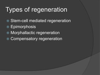
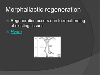
Tuhar Mukherjee
 We have seen that tail of house lizards,
legs of spiders to be of normal size after
they are shed by the organism when
they try to escape from a danger.
 The shedding of own body part is called
autotomy
What is regeneration?
 Regeneration is the reactivation of
development in postembryonic life to
restore missing tissue.
 In animal kingdom regeneration is
present in several animal groups.
Regeneration is marked in lower animal
groups like Porifera, Cnidaria.
 Porifera include those animals that are
called sponges. They multicellular
animals without tissue grade
organization.
 Cnidaria includes animals like jelly fish,
sea anemone and corals. They have
nematocysts lodged in cnidoblast cells.
 Regeneration whether minor wound
healing or regeneration of a leg takes
place in all organisms
Regeneration experiments
 Tremblay 1741 (Hydra)
 Réaumur 1712 (Crustaceans)
 Spallanzani (Salamanders)
 Crustaceans are arthropods that include
crabs, prawns. They predominantly
aquatic except Oniscus (wood louse)
which is a terrestrial crustaceans.
 Salamanders are amphibians that
belong to the Order Caudata. Their body
is lizard-like.
Types of regeneration
 Stem-cell mediated regeneration
 Epimorphosis
 Morphallactic regeneration
 Compensatory regeneration
Epimorphosis
 Adult structure can undergo
dedifferentiation to form a relatively
undifferentiated mass of cells that then
redifferentiates to form the new
structures.
 This type of regeneration is found during
regeneration amphibian limb
Anatomy of the Fore-limbs
Regeneration of Salamander
limbs
1. The skin and muscle retract from the tip
of the humerus after 2 days of
amputation.
2. Thin accumulation of blastema calls is
seen behind a thickened cap-like
epithelial layer or epidermis (Apical
Ectodermal Cap), 5 day stage
3. Large population of mitotically active
blastema cells lie distal to the humerus,
7 day stage
4. Blastema enlarge mitotically,
dedifferentiation occurs, 8 day
5. Early redifferentiation begins with
chondrogenesis begins in the proximal
part of the humerus and radius-ulna, 9
day
6. Pre cartilaginous condensation in the
carpal bones and digits
Molecular mechanism
 Experiments have been done by
transplantation and immuno-blocking.
 The growth of the regeneration blastema
depend on Apical ectodermal cap and
the surrounding nerve tissue.
 AEC secretes Fgf8 which stimulates the
growth of the blastema.
 The presence of nerves are essential for
the development of the regeneration
blastema (Mullen et al 1996).
 The nerve cells secrete release factors like the new
anterior gradient protein (nAG) necessary for the
proliferation of the blastema cells.
 Maintenance of ion current is also important
 The field is maintained by V-ATPase proton pump in
Xenopus laevis.
 V-ATPase also called vacuolar ATPase are
evolutionary conserved ATPases that are found in
eukaryotic cells. They maintain low pH or acidic
environment in a wide variety of intracellular
organelles like lysosomes
 Suppression the field inhibits the development of the
limb (Altizer et al 2002).
Pattern formation
 The dorsal-ventral and anterior-posterior
axes between the stump and the
regenerating tissue are conserved, and
cellular and molecular studies have
confirmed that the patterning
mechanisms of developing and
regenerating limbs are very similar. The
blastema cells can respond to the limb
bud of a developing limb (Muneoka and
Bryant, 1982)
 Sonic Hedgehog is seen in the posterior
region of the blastema and the developing limb
bud.
 Retinoic acid synthesized by the wound
epidermis specifies the proximal distal axis
and also the anterior posterior axis.
 RA activates Hoxa gene differentially in the
that specifies the pattern in the regenerating
Limbs.
 It might also establish the domain of Meis
genes (1 & 2) across the limb bud. Fgfs
suppress the Meis gene activation restricting
them to the proximal side
 Epimorphic regeneration is also seen in
planarian flatworms.
Morphallactic regeneration
 Regeneration occurs due to repatterning
of existing tissues.
 Hydra
Morphollactic regeneration in
Hydra
 Body Hydra has head (hypostome region)
at the distal
 Foot (basal disc) at proximal end
 If the body is cut into several pieces, each
piece will regenerate a head at its apical
end and a foot at its basal end. The result
is a smaller size organism as no growth
takes place.
 The polarity of Hydra is determined by a
series of morphogenetic gradients that
permit the head to form at one region and
basal disc at another.
Morphogen gradients
 Head activation gradient
 Head inhibition gradient
 Basal disc activation gradient
 Basal disc inhibition gradient
Head activation gradient
 Gradient can be measured by implanting
rings of tissue from various levels of a
donor Hydra into the host trunk
 Wilby and Webster 1970
 Herands and Bode 1974
 MacWilliams 1983
 Peptide gradients involved are Heady,
Head Activator and Hym301
 Heady and Head Activator critical for head
formation and initiation of bud
 Hym301 for number of tentacles formed
Head inhibition gradient
 Normal regeneration of hypostome is inhibited when
an intact hypostome is grafted adjacent to the site of
amputation
 If subhypostomal tissue is grafted to the same site of
amputation, no secondary axis forms. Host’s head
inhibits the formation of head and secondary axis
 If subhypostomal tissue is grafted to a decapitated
host, secondary axis forms.
 No head will be produced if the tissue is implanted
into the apical region of intact host
 Head will be produced if implanted below the host.
Hypostome
 Broun and Bode 2002
 Hypostome can induce secondary axis in host tissue
 Produces both head activation and head inhibition signals
 Self-differentiating region
 head inhibition signals inhibit the formation of new
organizing centers
 Signaling through the canonical Wnt pathway form the
head organizer.
 Inhibition of GSK3 leads to ectopic tentacles at all levels
and each piece of the trunk has the ability to stimulate the
outgrowth of new buds.
 Goosecoid (vertebrate organizer molecule) and
Brachyury(induces formation of mesoderm in vertebrates)
are also found
Basal disc
 Source foot activation and foot inhibition
gradients.
 Gradients of head and foot inhibitors
appear to block bud formation
 mannacle might activate shineguard in
the basal disc ectoderm
 shineguard, a tyrosine kinase extends in
a gradient from the ectoderm just above
the basal disc through the lower region.
Bud form where this gradient fades.
 Bud location is a function of both the
head and foot inhibitors.
 When the head is removed the head
inhibitor is not made. The region with the
highest head activator forms the head.
 After the head is formed, head inhibitor
generates.
Compensatory regeneration
 the differentiated cells divide and
maintain their differentiated function.
 Cells do not come from stem cells or
dedifferentiated cells.
 Found in mammalian liver, heart of
zebra fish
Histology of liver
Regeneration of liver cells
 Loss of liver cells is sensed by the absence
of some liver specific factors and increase
of bile salts and gut lipopolysaccharides
 Lipopolysaccharides activate the non-
hepatocyte cells (Kupffer’s cells and
stellate cells) to secrete paracrine factors
like IL6. These factors induce the
hepatocytes to reenter cell cycle.
 Kupffer cells secrete IL6 and TNF alpha
 Stellate cells secrete TGF beta and
hepatocyte growth factor.
 Hepatocytes activate cMet.
 cMet is the receptor for HGF.
 Blocking of cMet blocks liver regeneration
 Trauma of partial hepatectomy releases
metalloproteinases that digests the extracellular
matrix and permit the hepatocyte to separate
and proliferate.
 Enzymes may also activate HGF by proteolysis
cleavage.
 All these factors promote cell division by
activating cyclin D and E, repressing cyclin
inhibitors like p27 and preventing apoptosis
 Fxr transcription factor activated by bile is
necessary for liver regeneration
 Oval cells are small progenitor cells that can
produce hepatocytes and bile duct cells
Regeneration in other animals
 Polypoid stage of cnidarians
 Earthworms can regenerate their lost
body parts if they are cut antero-
posteriorly
 Arthropod limb
 Echinoderms like star fish can
regenerate arms.
 Holothurians can regenerate parts of
alimentary canal and respiration tree.
Regeneration in animals

Regeneration in animals

  • 1.
  • 2.
     We haveseen that tail of house lizards, legs of spiders to be of normal size after they are shed by the organism when they try to escape from a danger.  The shedding of own body part is called autotomy
  • 3.
    What is regeneration? Regeneration is the reactivation of development in postembryonic life to restore missing tissue.  In animal kingdom regeneration is present in several animal groups. Regeneration is marked in lower animal groups like Porifera, Cnidaria.
  • 4.
     Porifera includethose animals that are called sponges. They multicellular animals without tissue grade organization.  Cnidaria includes animals like jelly fish, sea anemone and corals. They have nematocysts lodged in cnidoblast cells.  Regeneration whether minor wound healing or regeneration of a leg takes place in all organisms
  • 5.
    Regeneration experiments  Tremblay1741 (Hydra)  Réaumur 1712 (Crustaceans)  Spallanzani (Salamanders)
  • 6.
     Crustaceans arearthropods that include crabs, prawns. They predominantly aquatic except Oniscus (wood louse) which is a terrestrial crustaceans.  Salamanders are amphibians that belong to the Order Caudata. Their body is lizard-like.
  • 7.
    Types of regeneration Stem-cell mediated regeneration  Epimorphosis  Morphallactic regeneration  Compensatory regeneration
  • 8.
    Epimorphosis  Adult structurecan undergo dedifferentiation to form a relatively undifferentiated mass of cells that then redifferentiates to form the new structures.  This type of regeneration is found during regeneration amphibian limb
  • 9.
    Anatomy of theFore-limbs
  • 10.
    Regeneration of Salamander limbs 1.The skin and muscle retract from the tip of the humerus after 2 days of amputation. 2. Thin accumulation of blastema calls is seen behind a thickened cap-like epithelial layer or epidermis (Apical Ectodermal Cap), 5 day stage 3. Large population of mitotically active blastema cells lie distal to the humerus, 7 day stage
  • 12.
    4. Blastema enlargemitotically, dedifferentiation occurs, 8 day 5. Early redifferentiation begins with chondrogenesis begins in the proximal part of the humerus and radius-ulna, 9 day 6. Pre cartilaginous condensation in the carpal bones and digits
  • 13.
    Molecular mechanism  Experimentshave been done by transplantation and immuno-blocking.  The growth of the regeneration blastema depend on Apical ectodermal cap and the surrounding nerve tissue.  AEC secretes Fgf8 which stimulates the growth of the blastema.  The presence of nerves are essential for the development of the regeneration blastema (Mullen et al 1996).
  • 14.
     The nervecells secrete release factors like the new anterior gradient protein (nAG) necessary for the proliferation of the blastema cells.  Maintenance of ion current is also important  The field is maintained by V-ATPase proton pump in Xenopus laevis.  V-ATPase also called vacuolar ATPase are evolutionary conserved ATPases that are found in eukaryotic cells. They maintain low pH or acidic environment in a wide variety of intracellular organelles like lysosomes  Suppression the field inhibits the development of the limb (Altizer et al 2002).
  • 15.
    Pattern formation  Thedorsal-ventral and anterior-posterior axes between the stump and the regenerating tissue are conserved, and cellular and molecular studies have confirmed that the patterning mechanisms of developing and regenerating limbs are very similar. The blastema cells can respond to the limb bud of a developing limb (Muneoka and Bryant, 1982)
  • 16.
     Sonic Hedgehogis seen in the posterior region of the blastema and the developing limb bud.  Retinoic acid synthesized by the wound epidermis specifies the proximal distal axis and also the anterior posterior axis.  RA activates Hoxa gene differentially in the that specifies the pattern in the regenerating Limbs.  It might also establish the domain of Meis genes (1 & 2) across the limb bud. Fgfs suppress the Meis gene activation restricting them to the proximal side
  • 17.
     Epimorphic regenerationis also seen in planarian flatworms.
  • 18.
    Morphallactic regeneration  Regenerationoccurs due to repatterning of existing tissues.  Hydra
  • 19.
    Morphollactic regeneration in Hydra Body Hydra has head (hypostome region) at the distal  Foot (basal disc) at proximal end  If the body is cut into several pieces, each piece will regenerate a head at its apical end and a foot at its basal end. The result is a smaller size organism as no growth takes place.  The polarity of Hydra is determined by a series of morphogenetic gradients that permit the head to form at one region and basal disc at another.
  • 20.
    Morphogen gradients  Headactivation gradient  Head inhibition gradient  Basal disc activation gradient  Basal disc inhibition gradient
  • 22.
    Head activation gradient Gradient can be measured by implanting rings of tissue from various levels of a donor Hydra into the host trunk  Wilby and Webster 1970  Herands and Bode 1974  MacWilliams 1983  Peptide gradients involved are Heady, Head Activator and Hym301  Heady and Head Activator critical for head formation and initiation of bud  Hym301 for number of tentacles formed
  • 23.
    Head inhibition gradient Normal regeneration of hypostome is inhibited when an intact hypostome is grafted adjacent to the site of amputation  If subhypostomal tissue is grafted to the same site of amputation, no secondary axis forms. Host’s head inhibits the formation of head and secondary axis  If subhypostomal tissue is grafted to a decapitated host, secondary axis forms.  No head will be produced if the tissue is implanted into the apical region of intact host  Head will be produced if implanted below the host.
  • 24.
    Hypostome  Broun andBode 2002  Hypostome can induce secondary axis in host tissue  Produces both head activation and head inhibition signals  Self-differentiating region  head inhibition signals inhibit the formation of new organizing centers  Signaling through the canonical Wnt pathway form the head organizer.  Inhibition of GSK3 leads to ectopic tentacles at all levels and each piece of the trunk has the ability to stimulate the outgrowth of new buds.  Goosecoid (vertebrate organizer molecule) and Brachyury(induces formation of mesoderm in vertebrates) are also found
  • 25.
    Basal disc  Sourcefoot activation and foot inhibition gradients.  Gradients of head and foot inhibitors appear to block bud formation  mannacle might activate shineguard in the basal disc ectoderm  shineguard, a tyrosine kinase extends in a gradient from the ectoderm just above the basal disc through the lower region. Bud form where this gradient fades.
  • 26.
     Bud locationis a function of both the head and foot inhibitors.  When the head is removed the head inhibitor is not made. The region with the highest head activator forms the head.  After the head is formed, head inhibitor generates.
  • 27.
    Compensatory regeneration  thedifferentiated cells divide and maintain their differentiated function.  Cells do not come from stem cells or dedifferentiated cells.  Found in mammalian liver, heart of zebra fish
  • 28.
  • 29.
    Regeneration of livercells  Loss of liver cells is sensed by the absence of some liver specific factors and increase of bile salts and gut lipopolysaccharides  Lipopolysaccharides activate the non- hepatocyte cells (Kupffer’s cells and stellate cells) to secrete paracrine factors like IL6. These factors induce the hepatocytes to reenter cell cycle.  Kupffer cells secrete IL6 and TNF alpha  Stellate cells secrete TGF beta and hepatocyte growth factor.
  • 30.
     Hepatocytes activatecMet.  cMet is the receptor for HGF.  Blocking of cMet blocks liver regeneration  Trauma of partial hepatectomy releases metalloproteinases that digests the extracellular matrix and permit the hepatocyte to separate and proliferate.  Enzymes may also activate HGF by proteolysis cleavage.  All these factors promote cell division by activating cyclin D and E, repressing cyclin inhibitors like p27 and preventing apoptosis  Fxr transcription factor activated by bile is necessary for liver regeneration  Oval cells are small progenitor cells that can produce hepatocytes and bile duct cells
  • 31.
    Regeneration in otheranimals  Polypoid stage of cnidarians  Earthworms can regenerate their lost body parts if they are cut antero- posteriorly  Arthropod limb  Echinoderms like star fish can regenerate arms.  Holothurians can regenerate parts of alimentary canal and respiration tree.