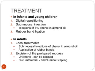
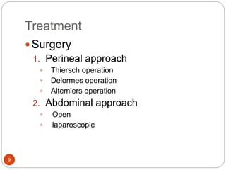
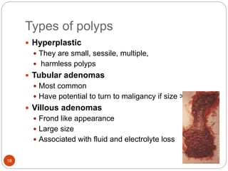
RECTAL DISEASES
1
OVERVIEW
 PROLAPSE
 PROCTITIS
 POLYPS
 BENIGN LESIONS
 CARCINOMA
2
RECTAL PROLAPSE
 Protrusion of rectum through anal canal
 Classified as:
1. Mucosal prolapse
2. Full-thickness prolapse
3
MUCOSAL PROLAPSE
 The mucous membrane and submucosa of the
rectum protrude outside the anus for
approximately 1–4 cm.
 On palpation of prolapsed mucosa between the
finger and thumb it is composed of double layer
of mucous membrane
4
ETIOLOGY
 In infant:
1. Direct downward course of rectum due to
undeveloped sacral curve
 Children:
1. Attack of Diarrhoea
2. Loss of weight leading to consequent loss of fat in
ischiorectal fossa,
3. Others cystic fibrosis, hirschsprung disease,
maldevelopment of pelvis
 Adult:
1. Torn perineum in female
2. Straining from urethral obstruction in males
3. weakness of sphincter mechanism in old age
4. Partial prolapse may follow an operation for
fistula in ano where large portion of muscle has been
divided
5
TREATMENT
 In infants and young children
1. Digital repositioning
2. Submucosal injection
 injections of 5% phenol in almond oil
3. Rubber band ligation
 In Adults
1. Local treatments
 Submucosal injections of phenol in almond oil
 Application of rubber bands
2. Excision of the prolapsed mucosa
 Unilateral - can be excised
 Circumferential - endoluminal stapling
6
FULL THICKNESS
PROLAPSE(PROCIDENTIA)
 All layers of the rectal wall involved
 Usually associated with a weak pelvic floor and
chronic straining
 Less common
 More common in elderly people.
 It begins as a intussusception of rectum usually at
the anterior wall which descends down to
protrude outside the anus.
 It is more than 4cm and may be upto 10-15 cm.
 On palpation between the finger and thumb the
prolapse feels thicker than the mucosal prolapse.
7
Features of full thickness
prolapse
 > 5 cm prolapse contains anteriorly between its
layers a pouch of peritoneum.
 When large, the peritoneal pouch may contains
small Intestine or bladder
 The anal sphincter is patulous and gapes widely
on straining -- to allow the rectum to prolapse.
8
Treatment
 Surgery
1. Perineal approach
 Thiersch operation
 Delormes operation
 Altemiers operation
2. Abdominal approach
 Open
 laparoscopic
9
PERINEAL APPROACH
1. Thiersch operation
 A steel wire or a silastic or nylon suture is
placed around the anal canal
 It has become obsolete
 Suture would often break
 chronic perineal sepsis
 Anal stenosis
 obstructed defaecation.
10
2. Delorme’s operation
 Mucosa of prolapsed rectum is stripped
circumferentially over its length.
 Underlying muscle is plicated with a series of
interupted sutures to the anal canal.
 Excess mucosa is excised.
 Mucosal approximation done.
 Prolapse is reduced, and a ring of
muscle is created above
the anal canal.
 Recurrence rate 30% over 5yrs.
11
3. Altemeier’s procedure
 Full thickness resection of the prolapsed rectum.
 Restoration of colorectal continuity by suturing or
stapled anastomosis.
 Recurrence rate 0-20%.
12
ABDOMINAL APPROACH
Principle is to replace and hold the rectum in its
proper position
 Well’s operation
 Rectum is fixed firmly to the sacrum by inserting a
sheet of polypropylene mesh between them.
 Ripstein’s operation
 Hitching up the rectosigmoid junction by a Teflon
sling to the front of the sacrum
 Sutured rectopexy
 Suture the mobilised rectum to the sacrum using 4-
6 interrupted nonabsorbable sutures
13
 Resection rectopexy
 Sutured rectopexy may lead to constipation so
resection of sigmoid colon is done along with
sutured rectopexy.
 Laparoscopic anterior mesh rectopexy
 Mesh is placed anterior to rectum
 Upper end is sutured to sacral
promontory.
14
PROCTITIS
Inflammation of part of rectum.
 Etiology
• clostridium difficile
• amoebic dysentery
• tuberculous proctitis
• gonococcal proctitis
• Lymphogranuloma venerum
• AIDS – CMV, herpes virus
• radiation proctitis
• Ulcerative proctocolitis
• Proctitis due to crohns disease
15
 Clinical features
 Defaecatory frequency,
 loose stools often mixed with blood
 Malaise, pyrexia
 Investigations
 Proctoscopy sigmoidoscopy
 Colonoscopy with multiple biopsies to know extent of
inflammation
 Stool culture to identify infective organism
 TREATMENT
 Non specific –
 usually self limiting
 topical 5aminosalicylic acid
 oral steroids.
 Specific
 antibiotics
16
POLYPS
 Common sites are rectum and sigmoid colon
 They can be sessile or pedunculated
17
Types of polyps
 Hyperplastic
 They are small, sessile, multiple,
 harmless polyps
 Tubular adenomas
 Most common
 Have potential to turn to maligancy if size >1 cm
 Villous adenomas
 Frond like appearance
 Large size
 Associated with fluid and electrolyte loss
18
 Familial adenomatous polyosis
 Autosomal dominant inherited
 Cause is mutation in APC gene
 This condition is premalignant
 Total colectomy done within 10 years
 Proctocolectomy with permanent ileostomy
 Rectal preservation- colectomy + ileorectal anastomosis
 Juvenile polyp
 Bright red, pedunculated polyp
 Seen in infants and children
 Clinical futures -
 bleeding, pain, if it proplases during
 defeaction
 Histololgy
 Large mucous filled sac lined by cuboidal
epithelium19
MANAGEMENT
 All polyps should be
biopsied/ excised to
rule out cancer
 polyps <1 cm
Endoscopic
polypectomy
Endoscopic mucosal
resection
 Larger > 2 cm
Transanal endoscopic
microsurgery
20
BENIGN RECTAL LESIONS
 Endometrioma
 Hemangioma
 Gastrointestinal stromal tumour
 Neuroendocrine tumours
21
 ENDOMETRIOMA
 Seen in 20 to 40 yrs
 Ectopic endometrial tissue produces either
 Constricting lesions of rectosigmoid
 Tumour invading rectum
 Clinical features - dysmenorrhea rectal bleeding
 Management –
 hormonal manipulation
 total abdominal hysterectomy
 bilateral salpingo-ophorectomy
 Isolated deposits- diathermy ablation.
22
 HEMANGIOMA
 Uncommon cause of hemorrhage
 Mimics ulcerative colitis and diagnosis is delayed
 Treatment
 selective angiography with embolisation
 In severe cases complete excision
 GASTROINTESTINAL STROMAL TUMOUR
 These tumours have high mitotic rate vary in number
size shape and hyperchromasia
 Management radiclal excision
23
 NEUROENDOCRINE TUMOUR
 Grading
 Grade 1-well differentiated
 Grade 2 –moderately differentiated
 Grade 3 – poorly differentiated
 Grade 1 are carcinoid tumours and have good
prognosis
 Grade 3 are aggressive and metastasize early
 Treatment
 Small NETs excised endoscopically or transanally
 Large NETs oncological resection
24
CARCIONOMA
 Epidemiology
 Colorectal carcinoma
 2nd most common cancer in women and 3rd in
males
 4th most common cause of death due to cancer
after lungs, stomach, liver cancer
 More than 1 million cases/year in with 715,000
deaths occur
 Rectosigmoid junction is commonest site involved in
⅓ cases
25
PATHOGENESIS
 AT RISK
 Patient with IBD – 2% cases
 Patient with family history – 20%
 Patient associated with genetic syndrome
o HNPCC/ lynch syndrome – 3%
o Gardern syndrome
o Familial adenomatous
 Other genetic abnormalities
 Over expression of oncogens
o KRAS – kirsten rat sarcoma homologue
o RAF – rapidly accelerated fibro sarcoma
 Inactivation of tumour suppression gene
o PTEN – phosphates and tensive homologue
AT RISK
MUTATION OF APC
GENE
ACCUMULATION OF
β CATENIN
ACTIVATES
TRANSCRIPTION OF
PROTO-ONCOGENS
26
CLINICAL FEATURES
 Early
 Bleeding per rectum; painless and bright red
 Tenesmus
 Early morning diarrhea
 Late
 Pain
 weight loss
27
INVESTIGATIONS
 Clinical examination
 Abdominal examination
 Signs of large bowel obstruction with distention
 Enlarged liver – liver metastasis
 Ascites – peritoneal metastasis
 Rectal examination – elevated irregular hard mass
is felt
 Colonoscopy (best), look for synchronous lesions
- Alternative: air contrast barium enema (“apple
core” lesion)
 sigmoidoscopy
 Metastatic workup: CXR, abdominal
CT/ultrasound
28
TYPES OF SPREAD
 Local spread
 Occurs circumferentially rather than longitudinally
 Anterior penetration -
 Prostate, Seminal vesicles, Bladder in male
 Vagina or uterus in female
 Lateral penetration - Ureter
 Posterior penetration - Sacrum and sacral plexus
 Lymphatic spread
 Exclusively in an upward direction
 Metastatic at higher level than superior rectal artery in
late disease
 Venous spread
 Principal sites are: Liver (34%) , Lungs (22%) , Adrenal
(11%)
29
STAGES OF PROGNOSIS
 DUKE’S STAGING 3 stages
 Stage A Growth limited to the rectal wall (15%):
Prognosis excellent (90% 5 year survival)
 Stage B Growth extended to extrarectal tissue but
no matastasis to regional lymph nodes (35%:
Prognosis reasonable (70% 5 year survival)
 Stage C Secondary deposits in regional lymph
nodes
 C1: Local pararectal lymph node alone involved
 C2: Nodes accompanying supplying blood vessels involved
 Stage D Not described by Duke’s Signifies
presence of widespread metastasis usually hepatic
30
Duke’s staging
31
TNM STAGING
Primary Tumor Regional Lymph Nodes Distant Metastasis
T0 No Primary Tumor N0 No Regional LN M0 No Metastasis
Tis CA in situ N1 Metastasis in 1-3 pericolic
nodes
M1 Distant
Metastasis
T1 Invasion into
submucosa
N2 Metastasis into 4 or more
pericolic nodes
T2 Invasion into
muscularis propria
N3 Metastasis into any nodes
along the course of named
vascular trunks
T3 Invasion into
serosa
T4 Invasion into
adjacent structures
32
TREATMENT
 Radical excision of rectum , together with
mesorectum and associated lymph nodes choice
of treatment in most cases.
 When tumor is locally advanced:
 Course of neoadjuvant (preoperative)
chemoradiotherapy over approx. 6 weeks may
reduce its size and make curative surgery possible.
 When rectal excision possible, aim should be to
restore gastrointestinal continuity and continence
by preserving anal sphincter
33
1. Local operation
 For small low grade T1 tumors
 Done via the anus and TEM (Transanal Endoscopic
Microsurgies) techniuqes
2. Anterior resection
 Anterior proctosigmoidscopy with colorectal
anastomosis
 For removing the portion of bowel containing the
cancer and the mesorectum completely, containing
lymphatic channels draining tumoe bed
 Lower anterior resection: Resection of rectum below
peritoneal reflection
 Intestinal continuity reestablished by anastomosis
between descending colon and rectum
34
35
3. Hartmann’s operation
 Excellent procedure in elderly, not fit for major
surgery
 Through an abdominal incision
 Rectum excised,
 Anorectal stump transected
 End colostomy formed
4. Abdominoperineal excision of rectum
 For the tumors of lower third of rectum (Unsuitable
for sphincter saving procedure)
 Complete excision of rectum and anus by concomitant
dissection through abdomen and perineum
 Suture closure of perineum
 Creation of permanent colostomy
36
5. Endoluminal stenting
 Done endoscopically often with fluoroscopic
guidance
 Used as palliative procedure or to relieve
obstruction
 Only colonic or upper rectal tumors suitable for
stenting
6. Palliative colostomy
 In cases with intestinal obstruction or gross
infiltration of neoplasm
37
 Radiotherapy
 To sterilize the operative field short course
radiotherapy for 5 days and surgery is done after 7
to 10 days .
 Radiotherapy + Chemotherapy:
 shrink extensive tumor prior to surgery
Chemotherapy
 5-Flurouracil alone or in combination with
Oxaliplatin improve survival by 10-15 % in node
positive patient
38
THANK YOU
Reference : Bailey & Love's Short
Practice of Surgery, 27th Edition
39

Rectal diseases

  • 1.
  • 2.
    OVERVIEW  PROLAPSE  PROCTITIS POLYPS  BENIGN LESIONS  CARCINOMA 2
  • 3.
    RECTAL PROLAPSE  Protrusionof rectum through anal canal  Classified as: 1. Mucosal prolapse 2. Full-thickness prolapse 3
  • 4.
    MUCOSAL PROLAPSE  Themucous membrane and submucosa of the rectum protrude outside the anus for approximately 1–4 cm.  On palpation of prolapsed mucosa between the finger and thumb it is composed of double layer of mucous membrane 4
  • 5.
    ETIOLOGY  In infant: 1.Direct downward course of rectum due to undeveloped sacral curve  Children: 1. Attack of Diarrhoea 2. Loss of weight leading to consequent loss of fat in ischiorectal fossa, 3. Others cystic fibrosis, hirschsprung disease, maldevelopment of pelvis  Adult: 1. Torn perineum in female 2. Straining from urethral obstruction in males 3. weakness of sphincter mechanism in old age 4. Partial prolapse may follow an operation for fistula in ano where large portion of muscle has been divided 5
  • 6.
    TREATMENT  In infantsand young children 1. Digital repositioning 2. Submucosal injection  injections of 5% phenol in almond oil 3. Rubber band ligation  In Adults 1. Local treatments  Submucosal injections of phenol in almond oil  Application of rubber bands 2. Excision of the prolapsed mucosa  Unilateral - can be excised  Circumferential - endoluminal stapling 6
  • 7.
    FULL THICKNESS PROLAPSE(PROCIDENTIA)  Alllayers of the rectal wall involved  Usually associated with a weak pelvic floor and chronic straining  Less common  More common in elderly people.  It begins as a intussusception of rectum usually at the anterior wall which descends down to protrude outside the anus.  It is more than 4cm and may be upto 10-15 cm.  On palpation between the finger and thumb the prolapse feels thicker than the mucosal prolapse. 7
  • 8.
    Features of fullthickness prolapse  > 5 cm prolapse contains anteriorly between its layers a pouch of peritoneum.  When large, the peritoneal pouch may contains small Intestine or bladder  The anal sphincter is patulous and gapes widely on straining -- to allow the rectum to prolapse. 8
  • 9.
    Treatment  Surgery 1. Perinealapproach  Thiersch operation  Delormes operation  Altemiers operation 2. Abdominal approach  Open  laparoscopic 9
  • 10.
    PERINEAL APPROACH 1. Thierschoperation  A steel wire or a silastic or nylon suture is placed around the anal canal  It has become obsolete  Suture would often break  chronic perineal sepsis  Anal stenosis  obstructed defaecation. 10
  • 11.
    2. Delorme’s operation Mucosa of prolapsed rectum is stripped circumferentially over its length.  Underlying muscle is plicated with a series of interupted sutures to the anal canal.  Excess mucosa is excised.  Mucosal approximation done.  Prolapse is reduced, and a ring of muscle is created above the anal canal.  Recurrence rate 30% over 5yrs. 11
  • 12.
    3. Altemeier’s procedure Full thickness resection of the prolapsed rectum.  Restoration of colorectal continuity by suturing or stapled anastomosis.  Recurrence rate 0-20%. 12
  • 13.
    ABDOMINAL APPROACH Principle isto replace and hold the rectum in its proper position  Well’s operation  Rectum is fixed firmly to the sacrum by inserting a sheet of polypropylene mesh between them.  Ripstein’s operation  Hitching up the rectosigmoid junction by a Teflon sling to the front of the sacrum  Sutured rectopexy  Suture the mobilised rectum to the sacrum using 4- 6 interrupted nonabsorbable sutures 13
  • 14.
     Resection rectopexy Sutured rectopexy may lead to constipation so resection of sigmoid colon is done along with sutured rectopexy.  Laparoscopic anterior mesh rectopexy  Mesh is placed anterior to rectum  Upper end is sutured to sacral promontory. 14
  • 15.
    PROCTITIS Inflammation of partof rectum.  Etiology • clostridium difficile • amoebic dysentery • tuberculous proctitis • gonococcal proctitis • Lymphogranuloma venerum • AIDS – CMV, herpes virus • radiation proctitis • Ulcerative proctocolitis • Proctitis due to crohns disease 15
  • 16.
     Clinical features Defaecatory frequency,  loose stools often mixed with blood  Malaise, pyrexia  Investigations  Proctoscopy sigmoidoscopy  Colonoscopy with multiple biopsies to know extent of inflammation  Stool culture to identify infective organism  TREATMENT  Non specific –  usually self limiting  topical 5aminosalicylic acid  oral steroids.  Specific  antibiotics 16
  • 17.
    POLYPS  Common sitesare rectum and sigmoid colon  They can be sessile or pedunculated 17
  • 18.
    Types of polyps Hyperplastic  They are small, sessile, multiple,  harmless polyps  Tubular adenomas  Most common  Have potential to turn to maligancy if size >1 cm  Villous adenomas  Frond like appearance  Large size  Associated with fluid and electrolyte loss 18
  • 19.
     Familial adenomatouspolyosis  Autosomal dominant inherited  Cause is mutation in APC gene  This condition is premalignant  Total colectomy done within 10 years  Proctocolectomy with permanent ileostomy  Rectal preservation- colectomy + ileorectal anastomosis  Juvenile polyp  Bright red, pedunculated polyp  Seen in infants and children  Clinical futures -  bleeding, pain, if it proplases during  defeaction  Histololgy  Large mucous filled sac lined by cuboidal epithelium19
  • 20.
    MANAGEMENT  All polypsshould be biopsied/ excised to rule out cancer  polyps <1 cm Endoscopic polypectomy Endoscopic mucosal resection  Larger > 2 cm Transanal endoscopic microsurgery 20
  • 21.
    BENIGN RECTAL LESIONS Endometrioma  Hemangioma  Gastrointestinal stromal tumour  Neuroendocrine tumours 21
  • 22.
     ENDOMETRIOMA  Seenin 20 to 40 yrs  Ectopic endometrial tissue produces either  Constricting lesions of rectosigmoid  Tumour invading rectum  Clinical features - dysmenorrhea rectal bleeding  Management –  hormonal manipulation  total abdominal hysterectomy  bilateral salpingo-ophorectomy  Isolated deposits- diathermy ablation. 22
  • 23.
     HEMANGIOMA  Uncommoncause of hemorrhage  Mimics ulcerative colitis and diagnosis is delayed  Treatment  selective angiography with embolisation  In severe cases complete excision  GASTROINTESTINAL STROMAL TUMOUR  These tumours have high mitotic rate vary in number size shape and hyperchromasia  Management radiclal excision 23
  • 24.
     NEUROENDOCRINE TUMOUR Grading  Grade 1-well differentiated  Grade 2 –moderately differentiated  Grade 3 – poorly differentiated  Grade 1 are carcinoid tumours and have good prognosis  Grade 3 are aggressive and metastasize early  Treatment  Small NETs excised endoscopically or transanally  Large NETs oncological resection 24
  • 25.
    CARCIONOMA  Epidemiology  Colorectalcarcinoma  2nd most common cancer in women and 3rd in males  4th most common cause of death due to cancer after lungs, stomach, liver cancer  More than 1 million cases/year in with 715,000 deaths occur  Rectosigmoid junction is commonest site involved in ⅓ cases 25
  • 26.
    PATHOGENESIS  AT RISK Patient with IBD – 2% cases  Patient with family history – 20%  Patient associated with genetic syndrome o HNPCC/ lynch syndrome – 3% o Gardern syndrome o Familial adenomatous  Other genetic abnormalities  Over expression of oncogens o KRAS – kirsten rat sarcoma homologue o RAF – rapidly accelerated fibro sarcoma  Inactivation of tumour suppression gene o PTEN – phosphates and tensive homologue AT RISK MUTATION OF APC GENE ACCUMULATION OF β CATENIN ACTIVATES TRANSCRIPTION OF PROTO-ONCOGENS 26
  • 27.
    CLINICAL FEATURES  Early Bleeding per rectum; painless and bright red  Tenesmus  Early morning diarrhea  Late  Pain  weight loss 27
  • 28.
    INVESTIGATIONS  Clinical examination Abdominal examination  Signs of large bowel obstruction with distention  Enlarged liver – liver metastasis  Ascites – peritoneal metastasis  Rectal examination – elevated irregular hard mass is felt  Colonoscopy (best), look for synchronous lesions - Alternative: air contrast barium enema (“apple core” lesion)  sigmoidoscopy  Metastatic workup: CXR, abdominal CT/ultrasound 28
  • 29.
    TYPES OF SPREAD Local spread  Occurs circumferentially rather than longitudinally  Anterior penetration -  Prostate, Seminal vesicles, Bladder in male  Vagina or uterus in female  Lateral penetration - Ureter  Posterior penetration - Sacrum and sacral plexus  Lymphatic spread  Exclusively in an upward direction  Metastatic at higher level than superior rectal artery in late disease  Venous spread  Principal sites are: Liver (34%) , Lungs (22%) , Adrenal (11%) 29
  • 30.
    STAGES OF PROGNOSIS DUKE’S STAGING 3 stages  Stage A Growth limited to the rectal wall (15%): Prognosis excellent (90% 5 year survival)  Stage B Growth extended to extrarectal tissue but no matastasis to regional lymph nodes (35%: Prognosis reasonable (70% 5 year survival)  Stage C Secondary deposits in regional lymph nodes  C1: Local pararectal lymph node alone involved  C2: Nodes accompanying supplying blood vessels involved  Stage D Not described by Duke’s Signifies presence of widespread metastasis usually hepatic 30
  • 31.
  • 32.
    TNM STAGING Primary TumorRegional Lymph Nodes Distant Metastasis T0 No Primary Tumor N0 No Regional LN M0 No Metastasis Tis CA in situ N1 Metastasis in 1-3 pericolic nodes M1 Distant Metastasis T1 Invasion into submucosa N2 Metastasis into 4 or more pericolic nodes T2 Invasion into muscularis propria N3 Metastasis into any nodes along the course of named vascular trunks T3 Invasion into serosa T4 Invasion into adjacent structures 32
  • 33.
    TREATMENT  Radical excisionof rectum , together with mesorectum and associated lymph nodes choice of treatment in most cases.  When tumor is locally advanced:  Course of neoadjuvant (preoperative) chemoradiotherapy over approx. 6 weeks may reduce its size and make curative surgery possible.  When rectal excision possible, aim should be to restore gastrointestinal continuity and continence by preserving anal sphincter 33
  • 34.
    1. Local operation For small low grade T1 tumors  Done via the anus and TEM (Transanal Endoscopic Microsurgies) techniuqes 2. Anterior resection  Anterior proctosigmoidscopy with colorectal anastomosis  For removing the portion of bowel containing the cancer and the mesorectum completely, containing lymphatic channels draining tumoe bed  Lower anterior resection: Resection of rectum below peritoneal reflection  Intestinal continuity reestablished by anastomosis between descending colon and rectum 34
  • 35.
  • 36.
    3. Hartmann’s operation Excellent procedure in elderly, not fit for major surgery  Through an abdominal incision  Rectum excised,  Anorectal stump transected  End colostomy formed 4. Abdominoperineal excision of rectum  For the tumors of lower third of rectum (Unsuitable for sphincter saving procedure)  Complete excision of rectum and anus by concomitant dissection through abdomen and perineum  Suture closure of perineum  Creation of permanent colostomy 36
  • 37.
    5. Endoluminal stenting Done endoscopically often with fluoroscopic guidance  Used as palliative procedure or to relieve obstruction  Only colonic or upper rectal tumors suitable for stenting 6. Palliative colostomy  In cases with intestinal obstruction or gross infiltration of neoplasm 37
  • 38.
     Radiotherapy  Tosterilize the operative field short course radiotherapy for 5 days and surgery is done after 7 to 10 days .  Radiotherapy + Chemotherapy:  shrink extensive tumor prior to surgery Chemotherapy  5-Flurouracil alone or in combination with Oxaliplatin improve survival by 10-15 % in node positive patient 38
  • 39.
    THANK YOU Reference :Bailey & Love's Short Practice of Surgery, 27th Edition 39