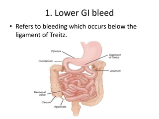
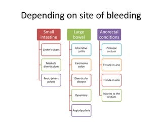
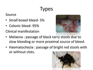
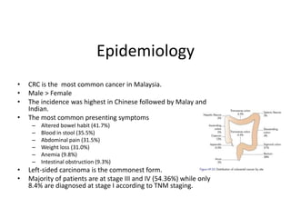
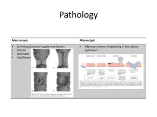
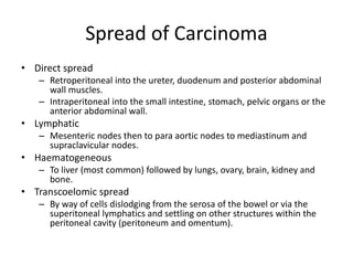
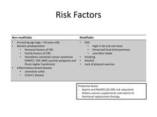
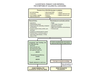
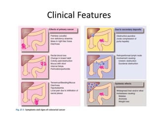
2. OUTLINE
1. Lower GI bleed
2. Colorectal cancer
3. Inflammatory & diverticular disease
4. Perianal disease
3. 1. Lower GI bleed
⢠Refers to bleeding which occurs below the
ligament of Treitz.
4. Depending on site of bleeding
Small
intestine
Crohnâs ulcers
Meckelâs
diverticulum
Peutz-jehers
polyps
Large
bowel
Ulcerative
colitis
Carcinoma
colon
Diverticular
disease
Dysentery
Angiodysplasia
Anorectal
conditions
Prolapse
rectum
Fissure-in-ano
Fistula-in-ano
Injuries to the
rectum
5. Types
Source
⢠Small bowel bleed- 5%
⢠Colonic bleed- 95%
Clinical manifestation
⢠Melaena : passage of black tarry stools due to
slow bleeding or more proximal source of bleed.
⢠Haematochezia : passage of bright red stools with
or without clots.
7. Epidemiology
⢠CRC is the most common cancer in Malaysia.
⢠Male > Female
⢠The incidence was highest in Chinese followed by Malay and
Indian.
⢠The most common presenting symptoms
â Altered bowel habit (41.7%)
â Blood in stool (35.5%)
â Abdominal pain (31.5%)
â Weight loss (31.0%)
â Anemia (9.8%)
â Intestinal obstruction (9.3%)
⢠Left-sided carcinoma is the commonest form.
⢠Majority of patients are at stage III and IV (54.36%) while only
8.4% are diagnosed at stage I according to TNM staging.
9. Spread of Carcinoma
⢠Direct spread
â Retroperitoneal into the ureter, duodenum and posterior abdominal
wall muscles.
â Intraperitoneal into the small intestine, stomach, pelvic organs or the
anterior abdominal wall.
⢠Lymphatic
â Mesenteric nodes then to para aortic nodes to mediastinum and
supraclavicular nodes.
⢠Haematogeneous
â To liver (most common) followed by lungs, ovary, brain, kidney and
bone.
⢠Transcoelomic spread
â By way of cells dislodging from the serosa of the bowel or via the
superitoneal lymphatics and settling on other structures within the
peritoneal cavity (peritoneum and omentum).
10. Risk Factors
Non-modifiable Modifiable
⢠Increasing age (age > 50 years old)
⢠Genetic predisposition
⢠Personal history of CRC
⢠Family history of CRC
⢠Hereditary colorectal cancer syndrome
(HNPCC, FAP, MAP, juvenile polyposis and
Peutz-Jegher Syndrome)
⢠Inflammatory bowel disease
⢠ulcerative colitis
⢠Crohnâs disease
⢠Diet
⢠High in fat and red meat
⢠Preserved food (nitrosamines)
⢠Low fibre intake
⢠Smoking
⢠Alcohol
⢠Lack of physical exercise
Protective factor
- Aspirin and NSAIDS (30-50% risk reduction)
- Dietary calcium supplements and vitamin D.
- Hormonal replacement therapy.
16. Diagnostic and Radiological
Investigations for Staging
⢠Diagnostic investigation
â Colonoscopy ( first line investigation( diagnostic and staging)
â Barium enema
â CT Colonography/ Virtual Colonoscopy
â Carcinoembryonic Antigen
⢠Radiological investigation
â CT TAP â accuracy in identifying CRC and nodal metastases
depends on the stages of tumour
â MRI â accurate assessment of rectal ca local spread
â Abdominal ultrasound - to evaluate abdominal organ especially
liver (hyperechoic liver mass)
â F-fluorodeoxyglucose PET CT â for detection of extrahepatic
metastases and local recurrence of XRX.
18. Histopathology examination
⢠HPE of the resected colorectal specimen is
essential for patientâs management.
⢠Including the estimation of post-operative
outcome and the rationale for adjuvant
therapy.
⢠Adenocarcinoma is the most common type of
CRC.
⢠Signet-ring and mucin-producing carcinoma
have significant poor prognostic features.
25. Standard operation for rectal
carcinoma
Abdominoperineal
resection of
rectum
Anterior resection
of rectum
Construction of J
pouch
Hartmann's
operation
25
27. Management for Rectal Carcinoma
27
Stage of disease Treatment
T1 Involvement of submucosa, but no
penetration through muscularis propria
Local excision (AR or APR)
T2 Invasion into, but not penetration
through, muscularis propria
a) Local excision + adjuvant Chemo/RT OR
b) radical resection
T3 Penetration through muscularis propria
into subserosa (if present), or pericolic fat,
but not into peritoneal cavity or other
organs
Neoadjuvant chemo / RT before radical
resection
T4 Invasion of other organs or involvement
of free peritoneal cavity
28. Total Mesorectal Excision (TME)
⢠Presence of radial spread is
an important prognostic
indicator.
⢠TME is indicated as part of
low anterior resection for
patients with adenocarcinoma
of the middle and lower rectum
(gold standard) - reduce local
recurrence rates
29. Operative complication
Immediate < 24 h Damage to other organs eg. ureters
Early < 30 days Wound infection
Bleeding
Abscess
Anastomosis breakdown/leak â faecal
peritonitis
Early stoma complications
Late>30 days Diarrhoea
Impotence( damage of pelvic nerves)
Adhesions(i/o)
Anastomotic stricture
Late stoma complications
30. Role of adjuvant radiotherapy and chemotherapy :
⢠Adjuvant radiotherapy and chemotherapy ď Duke Câs patient to increase
chance of prolonged survival.
⢠Neoadjuvant chemoradiotherapy is particularly relevant for rectal tumours
tethered in the pelvis by shrinking large tumour in order to make it
operable.
⢠This enables preservation of anal sphincter by downsizing the tumour.
⢠If rectal tumours extend through the bowel wall, particularly anteriorly, a
course of radiotherapy directly before surgery ď reduce local pelvic
recurrence.
⢠Choice of chemotherapy in large bowel cancer ď 5-fluorouracil (5-FU) +
folinic acid
30
31. Follow up and Surveillance
⢠Follow-up strategies in post-surgery and/or
adjuvant treatment are:
⢠History, physical examination and CEA levels
every three to six months for five years.
⢠Surveillance colonoscopy at year one and every
three to five years.
⢠CT scan of thorax, abdomen and pelvis is
performed annually for three year. For high-risk
patients every six to 12 months for the first three
years.
32. DIVERTICULAR DISEASE
Congenital
All three layers of the bowel are
present in the wall of the diverticulum
(e.g. Meckelâs diverticulum).
Acquired
There is no muscularis layer
present in the diverticulum
(e.g. sigmoid diverticular disease)
Diverticula (hollow out-
pouchings)
32
33. ⢠Mainly in Sigmoid but can affect the whole colon
⢠But never the Rectum? Taeni coli has fused. The rectum has a complete
muscular coat and a wider lumen.
⢠Often asymptomatic (diverticulosis) and found incidentally, 10-30% -
symptomatic
⢠Risk factor
â Lack of dietary fiber
PATHOGENESIS
1. â intraluminal diameter ⨠â intraluminal pressure by segmentation
movement ⨠push mucosa through weak point ⨠vasa recta penetrate
muscularis propria between the taenia coli
2. Disorder of motility
3. Degenerative changes in colonic wall - altered collagen structure with ageing
⨠at the point of entry of terminal arteries, where serosa is weakest
33
34. CLINICAL FEATURES
⢠Depends on location of the affected diverticulum, the severity of
inflammatory process, and presence of complications
A. Mild cases : Abdominal distension, flatulence and a sensation of
heaviness in the lower abdomen
B. Diverticulitis:
â Persistent lower abdominal pain, usually in the left iliac fossa
â Pyrexia, tachycardia
â Diarrhoea or constipation
â The sigmoid colon may be tender and thickened on palpation
C. Rectal examination may reveal a tender mass (abscess)
D. Signs of peritonitis â indicate perforation
E. Painless, profuse haemorrhage (sigmoid: bright red with clots)
34
35. COMPLICATIONS
⢠Pain and inflammation
Diverticulitis/ diverticular abscess
⢠most often contained leading to pericolic abscess formation but occasionally
leading to generalised peritonitis.
Perforation
⢠progressive fibrosis â fibrosis sigmoid and large bowel obstruction / loops of
small intestine can adhere to an inflamed sigmoid
Intestinal obstruction
⢠due to erosion of vessels adjacent to a diverticulum.
Haemorrhage
⢠occurs in 5% of cases, colovesical fistulation is most commonly seen.
⢠Recurrent UTI, pneumaturia or even faeces in the urine.
Fistula formation (colovesical, colovaginal, enterocolic,
colocutaneous)
35
37. HINCHEY CLASSIFICATION
for Staging of Complicated Diverticulitis
STAGING INVOLVEMENT
Stage I Pericolonic / mesenteric abscess <4cm
Stage II Large pelvic / retroperitoneal abscess
Stage III Small perforation causing purulent peritonitis
Stage IV Free rupture with faecal peritonitis
* need for surgery is reflected by degree of infective
complications
37
38. INVESTIGATIONS
Imaging
Chest X-ray
⢠tro perforation:
pneumoperitoneum
Abdominal X-ray
⢠Ileus, air fluid level
within an abscess
Spiral CT
⢠Identifying bowel wall
thickening, abscess
formation and
extraluminal disease.
Colonoscopy
Demonstrate the necks of
diverticula within the bowel
lumen
Has significant risk of
endoscopic perforation in
narrowed area
Blood
FBC: Anemia,
leucocytosis
Urea & electrolytes
LFT: TRO other
causes of abdominal
pain
38
39. MANAGEMENT - conservative
⢠Advice for high-fibre diet and bulk-forming laxatives
⢠Acute diverticulitis
â Resuscitation and analgesia
â NBM
â IV broad spectrum antibiotics (augmentin / ciproflaxin / metronidazole) â
Stage 1
â Stage 2 & 3 â drained percutenously
â Stage 4 â urgent laparotomy
⢠After acute phase subsided (4-6weeks)
â Investigated by endoscopy, barium enema or CT virtual colonoscopy to
confirm the diagnosis and assess for complications.
â Colonoscopy and biopsy â exclude colon carcinoma
â Barium enema - only performed if the patient has strictures or an
excessively tortuous sigmoid colon
â The pericolic abscesses can be drained percutaneously. A diameter of 5 cm
is frequently regarded as the cut off between an abscess likely to settle with
antibiotics and one likely to require intervention.
39
40. Operative
procedures for
diverticular disease
The aim of
emergency surgery
is to control
peritoneal infection
Indications for
surgery:
⢠Generalised
peritonitis
⢠Failure to
respond to
optimum medical
management
40
42. INFLAMMATORY
BOWEL DISEASE
Conditions characterized by the presence of idiopathic
intestinal inflammation
Ulcerative colitis and Crohnâs disease
- considered to be separate entities but in 10â15% of cases no
clear distinction can be made â Indeterminate colitis
Bailey & Love's Short Practice of
Surgery, 27th ed.
Essential Surgery Churchill Livingstone,
5th ed.
42
44. ⢠Crude incidence rates in Malaysia of ulcerative colitis 0.46 per
100,000 persons.
⢠Most commonly diagnosed at age between 20-40 years old,
male=female ; older age, male>female
⢠Far more common in the USA and Western Europe
⢠10â20% of patients have a first-degree relative with inflammatory
bowel disease.
⢠Unlike CD, smoking seems to have a protective effect in UC and has
even been the basis of therapeutic trials of nicotine.
⢠Relapses associated with periods of stress
44
45. ⢠Usually begins in the rectum and can extend proximally to affect
entire colon
⢠Longitudinal mucosal continuity â âcontinuous lesionâ
⢠Colonic inflammation:
⢠Diffuse
⢠Confluent and superficial
⢠Affect the mucosa and superficial submucosa
⢠âPseudopolyposisâ occurs in Âź of cases.
⢠Stricturing in UC is very unusual (unlike CD) - should prompt urgent
assessment, possibility of coexisting carcinoma
⢠Location: 40% - Rectum alone
40% - Left colon
20% - Total colitis
10% - Backwash ileitis : 30cm ileum affected,
incompetent ileocecal valve â reflux of noxious inflammatory
mediators
PATHOLOGY
45
46. ⢠Crypt abscess
⢠Depletion if goblet cell mucin
⢠High grade dysplasia
HISTOLOGICAL
⢠Depends in large part on the extent of disease.
⢠Rectal bleed
⢠Tenesmus
⢠Mucous discharge
⢠Bloody diarrhea and urgency (colitis)
⢠In severe cases
⢠Anemia
⢠Hypoproteinaemia
⢠Electrolytes disturbances
⢠Extensive colitis â systemic illness involvement
⢠Malaise, fever, loss of appetite
⢠Children â may impaired the growth
SIGNS & SYMPTOMS
46
50. MANAGEMENT â MEDICAL
⢠For mild attack- more effective than steroids
⢠Given locally- suppositories or enema
⢠Act as inhibitors of the cyclo-oxygenase enzyme system
Local corticosteroid or 5-aminosalicylic acid (5-
ASA) preparations
⢠For moderate to severe acute exacerbation
⢠Widespread anti-inflammatory action â oral or IV
Systemic corticosteroids
⢠Azathioprine and cyclosporin
⢠To maintain remission and as âsteroid-sparingâ agents
Immunosuppressive drugs
⢠Eg: sulfasalazine, mesalazine, olsalazine
⢠Used long term as maintenance therapy.
Oral aminosalicyclate preparations
50
51. INDICATION FOR SURGERY
Severe or fulminating disease failing to respond to medical
therapy
Chronic disease with anaemia, frequent stools, urgency and
tenesmus
Acute case â toxic megacolon/ perforation/ haemorrhage
Inability of the patient to tolerate medical therapy required
to control the disease
Neoplastic change: patients who have severe dysplasia or
carcinoma on review colonoscopy
Fail medical treatment â no symptoms improvement after 1
week intensive treatment
51
52. SURGICAL
⢠Subtotal colectomy and ileostomy : Emergency â 1st line
⢠Proctocolectomy and permanent end ileostomy
â Removes all colon and rectum, removing any risk of colorectal
neoplasia or colitic symptoms + Permanent stoma
â Indicated â for patients, not candidates for restorative surgery
due to sphincter problems or patient preference.
⢠Restorative proctocolectomy with ileoanal pouch
â a pouch is made out of ileum as a substitute for the rectum
and sewn or stapled to the anal canal.
â This avoids a permanent stoma.
â Indicated - patients with adequate anal sphincters
52
55. ⢠Incidence of 0.2 per 100 000 recorded in Malaysia
⢠Rare in Asian population, most common in North
America and Northern Europe
⢠Slightly more common in women than in men
⢠Common in younger age : 60% patients aged below
25 years old.
⢠Smoking increases the relative risk of CD three-fold
⢠10% of patients have a first-degree relative with the
disease
55
56. PATHOLOGY
⢠The terminal ileum is most commonly involved (65%)
⢠Perianal lesions are common, affecting up to 50â75% of
patients.
⢠Fibrotic thickening of the intestinal wall with narrowing of the
lumen and fat wrapping
⢠Dilated bowel just proximal to the stricture & deep mucosal
ulcerations.
⢠Cobblestone appearance- criss-cross of deep fissured ulcers
⢠Transmural inflammation- inflammation involves entire
thickness of bowel wall
â Adhesions
â Perforation
â Fistulae
⢠âSkipâ lesions
56
57. CLINICAL FEATURES
⢠CD more commonly presents with features of chronicity
i. abdominal colicky pain, which may be postprandial
ii. mild diarrhoea extending over many months occurring in
bouts
iii. palpable tender mass in the right iliac fossa- omentum
wrapped around terminal ileum
iv. Intermittent fevers, secondary anaemia and weight loss are
common
v. chronic obstructive symptoms
⢠For Colon: symptoms of colitis and proctitis as described for UC.
â Toxic megacolon is much less common
⢠Perianal problems
i. Bluish perianal skin
ii. Superficial ulcers (painless); Deep cavitating ulcers (painful)
iii. Risk of fistula
57
58. INVESTIGATIONS
LABORATORY
FBC (anemia)
fall in serum
albumin,
magnesium, zinc
and selenium
ESR & CRP (disease
activity)
ENDOSCOPY
Colonoscopic
examination with
biopsies
⢠areas of normal mucosa
in between areas of
inflammation
⢠Irregular and ulcerated,
with a mucopurulent
exudate
⢠Stricturing, biopsy TRO
malignancy
OGDS â upper GI
involvement
IMAGING
High-resolution ultrasound
⢠Demonstrate inflamed and
thickened bowel loops
⢠fluid collections and abscesses
Computed tomography (CT)
scans with oral contrast
⢠can demonstrate fistulae, intra-
abdominal abscesses
⢠bowel thickening or dilatation
Magnetic resonance imaging
⢠assessing complex perianal
disease
⢠investigating the small bowel
58
59. MANAGEMENT â MEDICAL
⢠TPN
Nutritional support
⢠induce remission in 70â80% of cases
⢠used in short courses only and tapered when a response has been achieved
⢠should not be used for maintenance therapy
Steroids
⢠Induce & maintain remission (sulfasalazine, mesalazine)
Aminosalicylates (5-ASA agents)
⢠in perianal disease
Antibiotics (Metronidazole and ciprofloxacin)
⢠Monoclonal antibody therapy
⢠targeting TNF-alpha and other key pro-inflammatory mediators
⢠Infliximab
Immunomodulatory agents (Azathioprine & Cyclosporin)
59
60. SURGICAL TREATMENT
⢠70% of patients with CD will require a bowel
resection within 10 years
⢠40% will require a further resection in the decade
following
⢠Fundamental principle: to preserve healthy gut and
to maintain adequate function
⢠Surgery doesnât cure CD, recurrence is common
60
61. 1. Ileocaecal resection
2. Segmental resection of short segments of small or large
bowel strictures
3. Colectomy and ileorectal anastomosis
ďź for colonic CD with rectal sparing and a normal anus.
4. Subtotal colectomy and ileostomy
5. Temporary loop ileostomy
6. Proctectomy and proctocolectomy
7. Strictureplasty
61
65. HAEMORRHOIDS
⢠Is symptomatic enlargement and distal
displacement of normal anal cushions located
at 3, 7 and 11 oâclock position
⢠Pathophysiology â degeneration of supporting
fibroelastic tissue and smooth muscle
⢠Common when
â increase intraabdominal pressure : eg: pregnancy,
constipation, straining
65
72. PERIANAL FISSURE / FISSURE-IN-
ANO
- Painful linear tear in distal anal canal extended to
but not beyond dentate line
- Long term can involve full thickness of anal
mucosa
- Aetiology
- Stretching of anal mucosa (strained evacuation of
hard stool)
- Birth trauma
- Anal sex
- Inflammatory bowel disease
72
73. Symptoms
⢠Fissure is concealed
by anal spasm
⢠Small skin tag
(sentinel pile)
Signs
⢠Pain on defaecation
⢠Bright-red bleeding
⢠Mucous discharge
⢠Constipation
⢠Itchiness
73
74. Management
⢠Conservative and medical treatment
- normalization bowel habit
⢠high fibre diet
⢠increase water intake
⢠stool softener
- topical application
⢠GTN (relax sphincter spasm)
⢠Injection of botulinum toxin
⢠Surgical approach
â Lateral internal anal sphincterotomy - definitive
management
74
76. AETIOLOGY
⢠Mostly sporadic.
â Most abscesses are cryptoglandular.
â Intersphincter, perianal, ischiorectal and
supralevator abcess
⢠Predisposing factor
â Inflammatory bowel disease (IBD)
â Immunosuppresed patient
⢠Diabetes mellitus
⢠Human immunosuppressive virus (HIV) infection
⢠Chronic corticosteroid treatment
⢠Untreated can progress to sepsis 76
77. CLINICAL FEATURES (depends on space that been infected)
⢠Interspinchter abscess
â Acute anal pain and tenderness
â No suppuration on inspection of perianal region
â Digital rectal examination (DRE) only can be done under general anaesthetic -
localized pea-sized lump in intersphincter space
⢠Perianal abscess
â Acute anal pain
â Pus discharge appeared at perianal margin
⢠Ischiorectal abscess
â Common in poor controlled Diabetes Mellitus (DM)
â Can surround posterior anal and affected contralateral side (the space is
horseshoes-shaped and no fascial barrier)
â Patient come with toxic looking, pyrexia and painful, muscular swelling both
buttocks.
â May have history of perianal pain and difficulty in sitting
⢠High intersphincter abscess or pelvirectal abscess
â may presented with major systemic upset
â Abscess may encircle anorectum above levator muscle
â Needs to consider intraabdominal sepsis (eg: diverticular abscess, crohnâs disease)
77
78. MANAGEMENT
⢠Surgical
â Incision and drainage
â Pus â sent for culture and sensitivity
⢠Antibiotic
â Uncomplicated â not required
â Complicated â extensive cellulitis, diabetic patient
with perianal sepsis
⢠Parenteral broad spectrum antibiotic â cephalosporin and
metronidazole
⢠Sigmoidoscopy and rectal biopsy needed if suspicious of underlying
Inflammatory bowel disease
78
80. AETIOLOGY
⢠Abnormal communication lined with granulation tissue
connecting anal canal to perineal skin.
⢠Obstruction of anal gland duct -> stasis-> cryptoglandular
infection -> abscess -> fistula-in-ano (30 â 50% of
abscesses).
⢠Secondary causes could be
â Crohnâs disease
â Anorectal trauma
â Iatrogenic (surgical)
â Anorectal carcinoma
o Ulcerative colitis
80
81. CLINICAL FEATURES
⢠Chronically discharging opening in perianal skin
⢠Pruritus
⢠Perianal discomfort
⢠May have history of underlying medical condition
or previous surgery
* Recurrent perianal abscess -> suspicious for fistula
tract
81
82. ASSESSMENT
⢠Goodsallâs rule is used to know likely course of fistulous tract
â It is essential to avoid inducing further fistula
during probing
⢠Need to do examination under anesthetic (EUA)
â DRE
â Proctosigmoidoscopic inspection
⢠Classification of low and high fistula is needed as the
management and prognosis are differ.
⢠Colonoscopy â to rule out Crohnâs disease. anorectal cancer
⢠MRI scan can help to identify complex high level fistula.
82
84. MANAGEMENT
LOW FISTULA HIGH FISTULA
Internal opening below the
puborectalis muscle
internal opening above the
puborectalis muscle
- ďŹstulotomy
- Complex fistula â used
tighter seton
- Anorectal advancement flap
- Drainage using a seton may
also be performed.
- High complex perianal
fistula may required
diverting stoma
All fistula tract need to be sent for histopathological
examination and mycobacterial staining and culture (if
clinically suspected) 84
85. RECTAL PROLAPSE
⢠Herniation of rectum into pelvic floor
⢠Encompasses 3 types of abnormal protrusion of rectal wall
- Full thickness rectal prolapse (procidentia)
includes mucosa and muscular layers
- Weak pelvic floor
- Muscular prolapse â mucosal lining of rectum
- Predisposing factor : haemorrhoid, constipation,
straining
- Occult rectal prolapse â intussusception of
rectal wall but without prolapse protruding of
anal canal
⢠Pathological process behind it is incompletely understood
85
86. Clinical Presentation Examination Investigation
-Uncomfortable
sensation of
âsomething coming
downâ the back
passage
-Constipation
-Faecal incontinence
-Mucous discharge
-Blood stained mucus
-Ulcerated and
strangulated
-May associated with
uterine prolapse
DRE
- patulous anus
- Poor sphincter
tone
Rigid
sigmoidoscopy
- Occult rectal
prolapse
86
87. MANAGEMENT
Childhood rectal prolapse Mucosal rectal prolapse Full thickness rectal
prolapse
-Self-limiting and rarely
surgical is needed
-Reassurance to parent
-Maintain regular bowel
habit
-Stool softener
- Applied barronâs bands
- Stapled prolapsectomy
Perineal approaches
- plication of the
prolapsed rectal muscle
(Delormeâs procedure)
Abdominal approaches
- Suture fixation
rectopexy - rectum is
mobilized and the
mesorectum sutured
to the sacral
promontory and
presacral fascia.
- Resection rectopexy â
same but with sigmoid
colectomy. 87
89. References
In Burnand, K. G., & In Browse, N. L. (2015). Browse's
introduction to the symptoms & signs of surgical disease.
In Williams, N. S., In O'Connell, P. R., & In McCaskie, A. W.
(2018). Bailey & Love's short practice of surgery.
Ramachandran, M., & Gladman, M. A. (2010). Clinical cases and
OSCEs in surgery. Edinburgh: Churchill Livingstone.
Essential Surgery: Problems, Diagnosis and Management, 5th
edition.
Principles & Practice of Surgery, 6th Edition.
CPG Management of Colorectal Carcinoma, 2017. MOH.
89
Editor's Notes
Melena is defined as black stools resulting from the
oxidation of hematin in the gut
CTC requires bowel preparation similar to conventional colonoscopy (CC) and during the procedure, air or carbon dioxide is introduced into the rectum via a rubber catheter. No sedation is required and patient is usually able to return to work post procedure.
drawbacks of CTC include radiation exposure and the need for colonoscopy after the identification of polyps for excision and tissue diagnosis, while smaller lesions need to be followed up by surveillance CTC.
MRI - rectal carcinoma requires detailed pre- operative planning that includes the assessment of the relation of tumour to the mesorectal fascia
MRI staging provides an accurate assessment of rectal carcinoma local spread pre-operatively.
MRI is the best modality in assessing the relation of the rectal carcinoma with the potential CRM (circumferential resection margin). MRI predicts whether the surgical resection margins will be clear or affected by the carcinoma
Bowel prep is process removing feces from colon prior to medical/surgical procedure. Start 1 day prior to the procedure. Give laxative (dulcolax)
Bowel prep
ô Modification of diet â 3 days low residue diet (reduce frequence and volume of stools â low fibre, reduce food that increase
bowel activity), NBM day before operation
ô Bowel clearance with polyethylene glycol. CI obstruction?
- Prophylactic antibiotics (max at first incision)
ô ampi/ genta/ metronidazole at induction of anesthesia
For exp, there is ascending colon lesion and are treated by right hemicolectomy,so the right colic artery must be ligated in order to remove the section of right colon.
Epidemiological studies indicate that diverticular disease is a
consequence of a refined Western diet, deficient in dietary
fibre.
In mild cases, symptoms such as distension, flatulence and a
sensation of heaviness in the lower abdomen may be indistinguishable
from those of irritable bowel syndrome. These symptoms
are thought to result from a combination of increased
luminal pressure affecting wall tension and increased visceral
hypersensitivity.
Bleeding from the sigmoid will be bright red with
clots, whereas right-sided bleeding will be darker. Torrential
bleeding is fortunately rare and, in fact, more commonly due
to angiodysplasia, but diverticular bleeding may persist or
recur requiring transfusion and resection.
- Colonoscopy â confirm dx & exclude CA colon and/ or
Barium enema is inferior to colonoscopy in terms of image quality and is usually only performed if the patient has strictures or an excessively tortuous sigmoid colon where colonoscopy is difficult or dangerous â CI in acute as may leaked out
Primary anastomosis - young fit patient without gross contamination or overwhelming sepsis.
Diverticular fistulae can only be cured by resecting the affected bowel, although a defunctioning stoma can worsen the symptoms.
In colovesical fistula the sigmoid can often be pinched off the bladder and the sigmoid resected.
If an anastomosis is performed - place an omental pedicle between the bowel and bladder to prevent recurrent fistulation + ureteric stent
The disease remains confined to the rectum
in 90% of cases but proctitis may extend proximally.
Endoscopy ď Rigid/flexible sigmoidoscopy can detect proctitis in the clinic;
the mucosa is hyperaemic and bleeds on touch, and there
may be a purulent exudate. Where there has been remission
and relapse, there may be regenerative mucosal nodules or
pseudopolyps. Later, tiny ulcers may be seen that appear to
coalesce. Colonoscopy and biopsy has a key role in diagnosis
and management:
Xray
Toxic megacolon should be suspected in patients who develop severe abdominal pain and confirmed by the presence on a plain
abdominal radiograph of a colon with a diameter of more than 6 cm
Initial management
Corticosteroid + 5ASA sulfasalazine
Immunosupressive drug â often effective in bringing steroid resistant disease under control
- tacrolimus, cyclosporine
Tnf alpha inhibitor â prevent endogenous cytokine from binding to the surface of cell receptor
s/e adversely affect normal immune response â allow to reactivation/superinfection such as latent tb
Bouts â short period of intense activity
Rectal bleed
Tenesmus
Mucous discharge
Bloody diarrhea and urgency
Definition : ncbi (varut lohsiriwat)
Anal cushion â contain vascular tissue, smooth muscle (treitz muscle), connective tissue â lined with epithelium of anal canal andre tan
Pathogenesis â straining during constipation -> raised intaabdominal pressure ->obstruct venous rectum causing venous plexuses to engorge
->bulging mucosa is dragged distally by hard stool ->persistent straining causes extruding anal mucosa
To rule out colorectal cancer and indication not to do colonoscopy
Outlet type bleeding â fresh red blood, dripping in pan, on wiping, separate from bowel motion in toilet pan
No alteration of bowel habit
Age less than 50 years old
DRE + proctoscopy and rigid sigmoidoscopy â may secure dx
If one of it not fulfilled â proceed with colonoscopy
Thrombosed prolapsed piles â large , swollen, irreducible haemorrhoid, dark blue/ black due to necrosis and submucosal hâge
Torrential haemorrhage â hypovolaemic, anemia
Venotonic agent - Oral flavonoids, oral calcium dobesilate
Topical treatment
Bulk laxative or stool softener
SCLEROSANT THERAPY
- provoked fibrotric reaction and obliterate the haemorhoidal vessel â causing atrophy of haemorrhoid
May need repetation 2-3 occasion at 4-6 weeks interval
CI â nut allergic
BANDING
Cone of mucosa above Haemorrhoidal neck is tight by elastic band, constricting haemorroidal vessel
Not band at stalk â painful d/t innevation
HALO
- locate artery using ultrasound â encircle with stitch via insensitive lower rectal mucosa
Haemorhoidal shrink leaving the skin tags
SE GTN â headache
Surgical only done if medical failed
Goodsallâs rule for ďŹnding the internal opening of an anal ďŹstula based on the location of the external opening. When an imaginary line is drawn through the center of the anus,
if external openings anterior to the line follow a radial (straight path towards the anal canal).
If the external opening is posterior to the line, the ďŹstulous tract will curve and leave the anal canal in the posterior midline
ďŹstulotomy or laid open of the ďŹstulous tract (following the insertion of a probe into the external opening). Then allowed to heal
Complex fistula â fecal incontinence happened due to significant proportion internal/external sphincter involvement
Probed and seton - Fistula tract is probed and seton passed along the tract to allow drainage. After drainage â tighter seton can be applied that will gradually cut out through the sphincter allowing healing behind the seton -> minimized incontinence ask it maintain end of sphincter together
Anorectal advancement flap - Raising a flap of rectal wall and upper internal sphincter. Flap is advanced distally to close internal opening while external opening can be heal by its own as no fecal stream
May do seton drainage beforehand and may required defunctioning colostomy