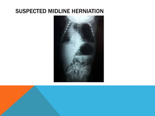
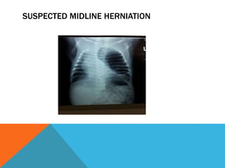
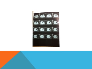
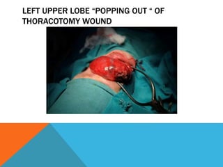
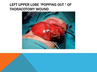
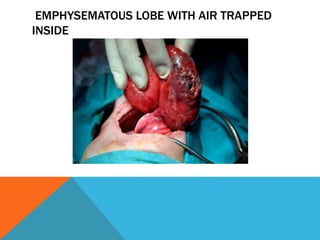
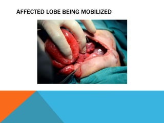
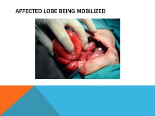
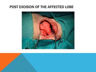
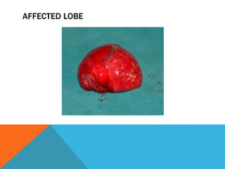
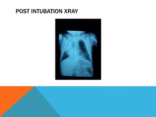
HISTORY
Patient name: XYZ
Father Name : XYZ
Age : 14 days
Sex: Male
Adress: Multan
D.O.B: 14-10-16
D.o.A : 27-10-16
PRESENTING COMPLAINT
Shortness of Breath 2 days post birth
HISTORY OF PRESENT ILLNESS
Patient developed the complaint of shortness of breath 2 days post birth
and presented to a consultant at Multan Hospital where he was
diagnosed as having the pneumothorax secondary to pneumonia and
was intubated. Antibiotics were started for pneumonia treatment
PRE INTUBATION XRAY
POST INTUBATION XRAY
HISTORY OF PRESENT ILLNESS
But it didn’t relieve his shortness of breath
Patient was referred to lahore for the management and presented to paeds
medicine emergency Mayo Hospital Lahore
His Chest tube was taken out and patient was put on ventilator on CMV
mode for 24 hours
Call was sent to the Paeds surgery Department meanwhile
EXAMINATION
On examination patient was tachyponeic
Trachea was shifted to the right
Percussion note was hyper resonant on left side
Air entry was reduced on left side
SUSPECTED DIAGNOSIS
Congenital lobar emphysema involving left upper lobe of lung was
made which is present in 40-50% of congenital lobar emphysema due
to suspected herniation of lung across midline on Xrays
CPAM was other differential diagnosis
SUSPECTED MIDLINE HERNIATION
CT scan was advised
LEFT UPPER LOBE “POPPING OUT “ OF
THORACOTOMY WOUND
LEFT UPPER LOBE “POPPING OUT “ OF
THORACOTOMY WOUND
LEFT UPPER LOBE “POPPING OUT “ OF
THORACOTOMY WOUND
LEFT UPPER LOBE “POPPING OUT “ OF
THORACOTOMY WOUND
EMPHYSEMATOUS LOBE WITH AIR TRAPPED
INSIDE
AFFECTED LOBE BEING MOBILIZED
POST EXCISION OF THE AFFECTED LOBE
AFFECTED LOBE
AFFECTED LOBE
POST OP RECOVERY
Was smooth
Patient discharged on 5th post op day
POST INTUBATION XRAY
DISCUSSION
Causes of CLE:
1. Dysplastic bronchial Cartilage
2. Endo bronchial obstruction from inssipisated mucus or extensive
mucosal bronchial atresia
3. Extrinsic compession of bronchi by abberant vessels
4. Diffuse Bronchial Abnormalities
FREQUENCY OF CLE
Left uppper Lobe (40-50%)
Right middle lobe (30-40%)
Right upper Lobe (20%)
Lower lobes 1%
AGE OF DIAGNOSIS
At birth 25%
In first year of life 50%
Sporadic after 6 monts
TEST TO DIAGNOSE
Pre natal ultrasound
Ultrafast fetal MRI
Chest xray
CT scan
Xenon Radio isoptope scan
PRE OP MANAGEMENT
High frequency ventilation
Selective Bronchial Intubation
Endoscopic Decompression of emphysema
DEFINITE MANGEMENT
Thoracotomy and segmentectomy

Congenital lobar emphysema