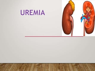
2. UREMIA
Azotemia refers to high levels of urea but is used primarily
when the abnormality can be measured chemically but is not yet
so severe as to produce symptoms.
Uremia is the pathological manifestations of severe azotemia.
There is no specific time for the onset of uremia for people with
progressive loss of kidney function.
Both uremia and the uremic syndrome have been used
interchangeably to define a very high plasma urea concentration
that is the result of renal failure
3. INTRODUCTION
All patients with renal disease should undergo an assessment of
renal function by estimating (GFR) from ser creatinine.
This measurement is used clinically
to evaluate the degree of renal impairment,
to follow the course of the disease, and
to assess the response to therapy.
An attempt must also be made to obtain a specific diagnosis.
careful urinalysis, kidney ultrasound, referral to a nephrologist,
and a kidney biopsy.
4. IDENTIFICATION OF RISK FACTORS AND STAGING OF
CKD
Risk factors:
1. hypertension,
2. diabetes mellitus,
3. autoimmune disease,
4. older age,
5. a family history of renal disease,
6. a previous episode of acute kidney injury,
7. Kidney donors and transplant recipients
8. and the presence of
a. proteinuria,
b. abnormal urinary sediment, or
c. structural abnormalities of the urinary tract
5. CALCULATION OF GFR
Methods of calculation
Cockcroft-Gault formula
MDRD formula/modified MDRD
CKD-EPI when eGFR values above 60 ml/min/1.7 sq meter
is desired
cr
This CKD-EPI equation calculator should be used when Scr reported in mg/dL. This equation is recommended when eGFR values above 60 mL/min/1.73 m2 are desired
This CKD-EPI equation calculator should be used when Scr reported in mg/dL. This equation is recommended when eGFR values above 60 mL/min/1.73 m2 are desired
This CKD-EPI equation calculator should be used when Scr reported in mg/dL. This equation is recommended when eGFR values above 60 mL/min/1.73 m2 are desir
7. 84 F 22 M 66 M 66 F
45.5 104.5 77.2
• Wt (kg)
71.8
1.2 1.2 1.2 1.2
•Screat
• eGFR 26.9 142.7 66.1 52.3
(Calculated with Cockcroft-Gault)
8. MDRD GFR
*From Levey et al, 1999
Ann Intern Med 130: 461-470
• MDRD GFR Formula*
170 x [SCr]-0
.
9
9
9 x [Age]-0
.
1
7
6 x
[0.762 if female] x [1.180 if
black] x [Alb]+0.318
• Modified MDRD
Formula
186.338 x [SCr]-1
.
1
5
4 x [Age]-0
.
2
0
3 x
[1.212 if black] x [0.742 if
female]
(A calculator may be found at
www.hdcn.org)
10. HOW THE KIDNEY RESPONDS TO INJURY?
The kidney is able to adapt to damage by increasing the filtration
rate in the remaining normal nephrons, a process called adaptive
hyperfiltration.
As a result, the patient with mild RI often has a normal or near-
normal ser creatinine.
Additional homeostatic mechanisms (most frequently occurring
within the renal tubules) permit the serum concentrations of
sodium, potassium, calcium, and phosphorous and the total body
water to also remain within the normal range.
11. PHYSIOLOGIC CHANGES IN CHRONIC
KIDNEY DISEASE
Increased single nephron GFR
Afferent arteriolar vasodilation
Intraglomerular hypertension
Loss of glomerular permselectivity
Inabilty to appropriately dilute or concentrate the urine
in the face of volume challenge
12. PHYSIOLOGIC CHANGES IN CHRONIC
KIDNEY DISEASE
Intraglomerular hypertension and glomerular hypertrophy
leading to glomerular scarring (glomerulosclerosis).
Additional causes may include systemic hypertension,
hyperlipidemia, metabolic acidosis, and tubulointerstitial disease.
Thus, proteinuria typically is present in patients with progressive
CKD, even in primary tubulointerstitial diseases such as reflux
nephropathy.
Principal targets for renal protection —the blood pressure goal
and, the proteinuria goal
13. PHYSIOLOGIC CHANGES IN CKD
- ACE inhibitors or ARBs agents in patients with
proteinuric CKD if begun before irreversible scarring
- ARBs do not appear to be more beneficial than other
antihypertensive agents in patients with nonproteinuric
CKD.
- When used in patients with CKD, common side
effects of ARBs include a mild to moderate reduction in
GFR and hyperkalemia, which occurs soon after the
initiation of therapy or an increase in dose
15. LEFT:SCHEMA OF THE NORMAL GLOMERULAR
ARCHITECTURE.
RIGHT: SECONDARY GLOMERULAR CHANGES
16. PHYSIOLOGIC CHANGES IN CKD
The gradual decline in function in patients with (CKD)
is initially asymptomatic.
However, different signs and symptoms may be
observed with advanced RF, including volume overload,
hyperkalemia, metabolic acidosis, hypertension, anemia,
and (MBDs).
The onset of (ESRD) results in a constellation of signs
and symptoms referred to as uremia.
17. WHAT IS CKD?
Chronic kidney disease is defined based on the presence of either
kidney damage or decreased kidney function for three or more
months,irrespective of cause.
• Criteria:
Duration ≥3 months, based on documentation or inference
Glomerular filtration rate (GFR) <60 mL/min/1.73 m2
Kidney damage, as defined by structural abnormalities or functional abnormalities
other than decreased GFR
18. CHRONIC KIDNEY DISEASE
Kidney damage, as defined by structural abnormalities or functional abnormalities other than
decreased GFR
A) Pathologic abnormalities (examples). Cause is based on underlying illness and
pathology. Markers of kidney damage may reflect pathology.
1.Glomerular diseases (diabetes, autoimmune diseases, systemic infections, drugs,
neoplasia)
2.Vascular diseases (atherosclerosis, hypertension, ischemia, vasculitis, thrombotic
microangiopathy)
3.Tubulointerstitial diseases (urinary tract infections, stones, obstruction, drug
toxicity)
4.Cystic disease (polycystic kidney disease)
19. CHRONIC KIDNEY DISEASE
Kidney damage, as defined by structural abnormalities or functional abnormalities other
than decreased GFR
B) History of kidney transplantation. In addition to pathologic
abnormalities observed in native kidneys, common pathologic
abnormalities include the following:
1.Chronic allograft nephropathy (non-specific findings of tubular
atrophy, interstitial fibrosis, vascular and glomerular sclerosis)
2.Rejection
3.Drug toxicity (calcineurin inhibitors)
4.BK virus nephropathy
5.Recurrent disease (glomerular disease, oxalosis, Fabry disease)
20. CHRONIC KIDNEY DISEASE
C) Albuminuria as a marker of kidney damage (increased glomerular
permeability, urine albumin-to-creatinine ratio [ACR] >30 mg/g).*
1.The normal urine ACR in young adults is <10 mg/g. Urine ACR
categories 10-29, 30-300 and >300 mg are termed "high normal, high,
and very high" respectively. Urine ACR >2200 mg/g is accompanied by
signs and symptoms of nephrotic syndrome
2.Threshold value corresponds approximately to urine dipstick values of
trace or 1+
3.High urine ACR can be confirmed by urine albumin excretion in a timed
urine collection
Kidney damage, as defined by structural abnormalities or functional abnormalities other
than decreased GFR
21. CHRONIC KIDNEY DISEASE
Kidney damage, as defined by structural abnormalities or functional abnormalities other
than decreased GFR
D) Urinary sediment abnormalities as markers of kidney damage
1.RBC casts in proliferative glomerulonephritis
2 . W B C casts in pyelonephritis or interstitial nephritis
3.Oval fat bodies or fatty casts in diseases with proteinuria
4.Granular casts and renal tubular epithelial cells in many
parenchymal diseases (non-specific)
22. CHRONIC KIDNEY DISEASE
Kidney damage, as defined by structural abnormalities or functional
abnormalities other than decreased GFR
E) Imaging abnormalities as markers of kidney damage (ultrasound,
computed tomography and magnetic resonance imaging with or without
contrast, isotope scans, angiography).
1.Polycystic kidneys
2.Hydronephrosis due to obstruction
3.Cortical scarring due to infarcts, pyelonephritis or vesicoureteral reflux
4.Renal masses or enlarged kidneys due to infiltrative diseases
5.Renal artery stenosis
6.Small and echogenic kidneys (common in later stages of CKD due to
many parenchymal diseases)
23.
24. ETIOLOGY OF CHRONIC KIDNEY DISEASE
Diabetes
43%
HTN
25%
GN
12%
Other
20%
Diabetes
HTN
GN
Other
25. CHRONIC KIDNEY DISEASE
No direct correlation exists between the absolute serum levels
of (BUN) or creatinine and the development of uremic
symptoms.
Some patients have relatively low levels (eg, a BUN of 60 mg/dL
in an older patient) but are markedly symptomatic, while others
have marked elevations (eg, a BUN of 140 mg/dL]) but remain
asymptomatic.
26. CHRONIC KIDNEY DISEASE
Certain drugs also interfere with either creatinine
secretion or the assay used to measure the serum
creatinine. These include cimetidine, trimethoprim,
cefoxitin, and flucytosine.
In these settings,
There will be no change in the true GFR;
Absence of a concurrent elevation in the (BUN)
29. CLINICAL ABNORMALITIES IN UREMIA
1. Fluid and electrolyte disturbances
2. Endocrine-metabolic disturbances
3. Neuromuscular disturbances
4. Cardiovascular and pulmonary disturbances
5. Dermatologic disturbances
6. Gastrointestinal disturbances
7. Hematologic and immunologic disturbances
(I) improves with an optimal program of dialysis and related
therapy;
(P) persist or even progress, despite an optimal program; (D)
develops only after initiation of dialysis therapy.
30. CLINICAL ABNORMALITIES IN UREMIA
1. Fluid and electrolyte disturbances
a. Volume expansion (I)
b. Hyponatremia (I)
c. Hyperkalemia (I)
d. Hyperphosphatemia (I)
(I) improves with an optimal program of dialysis and related therapy;
(P) persist or even progress, despite an optimal program;
(D) develops only after initiation of dialysis therapy.
31. FLUID, ELECTROLYTE,AND ACID-BASE DISORDERS
Hyponatremia – water restriction
ECFV expansion – salt restriction
Thiazides – limited utility in stages 3-5 CKD
- loop diuretics needed
Loop Diuretics resistance – Higher doses
Metolazone – combined with loop diuretics, which inhibits the
sodium chloride co-transporter of the distal convoluted tubule, can help
effect renal salt excretion
32. FLUID, ELECTROLYTE,AND ACID-BASE DISORDERS
• HYPERKALEMIA
• Precipitated by
• increased dietary potassium intake,
• protein catabolism,
• hemolysis,
• hemorrhage,
• transfusion of stored red blood cells,
• and metabolic acidosis
• Medications .
33. FLUID, ELECTROLYTE,AND ACID-BASE DISORDERS
Hyperkalemia
A common reason for initiation of RRT
There is limited K excretion as GFR falls
Diabetics may have a type IV RTA (hyporeninemic
hypoaldosteronism)
Use of ACE-I can exacerbate hyperkalemia
34. FLUID, ELECTROLYTE,AND ACID-BASE DISORDERS
Hyperkalemia
- Potassium balance usually remains intact
until GFR < 10-20 mL/min
- Tx of acute hyperkalemia involves cardiac
monitoring, IV calcium chloride or
gluconate, insulin with glucose, bicarbonate,
and potassium-binding resins
- Chronic hyperkalemia tx’d with dietary k
restriction, and Ca resonium PRN
35. FLUID, ELECTROLYTE,AND ACID-BASE DISORDERS
Hypokalemia:
• Not common in CKD
• reduced dietary potassium intake
• GI losses
• Diuretic therapy
• Fanconi’s syndrome
• RTA
• Hereditary or acquired Tubulointerstitial disease
36. FLUID, ELECTROLYTE,AND ACID-BASE DISORDERS
Metabolic acidosis
•
•
•
•
•
common disturbance in advanced CKD
combination of hyperkalemia and hyperchloremic
metabolic acidosis is often present, even at earlier
stages of CKD (stages 1–3)
Treat hyperkalemia
the pH is rarely <7.35
usually be corrected with oral sodium bicarbonate
supplementation
37. CLINICAL ABNORMALITIES IN UREMIA
(I) improves with an optimal program of dialysis and related therapy;
(P) persist or even progress, despite an optimal program;
(D) develops only after initiation of dialysis therapy.
2. Endocrine-metabolic disturbances
38. CLINICAL ABNORMALITIES IN UREMIA
(I) improves with an optimal program of dialysis and related therapy;
(P) persist or even progress, despite an optimal program;
(D) develops only after initiation of dialysis therapy.
2. Endocrine-metabolic disturbances
39. PTH
Pi Ca2+
Renal Mass
25(OH)D3 1,25(OH)2D3
11
-a
-a
lp
lp
h
ha
a-
-h
hy
y
d
d
rr
oo
xx
yl
y
al
sa
ese
+
Acidosis
+
Hyperparathyroid Related Bone Disease
Impaired
Absorption
Osteitis Fibrosa
Cystica
41. DISORDERS OF CALCIUM A N D PHOSPHATE
METABOLISM
Other complications of abnormal mineral
metabolism:
• Calciphylaxis (calcific uremic arteriolopathy)
• Other etiologies
•
•
use of oral calcium as a phosphate binder
Warfarin
42. CLINICAL ABNORMALITIES IN UREMIA
(I) improves with an optimal program of dialysis and related therapy;
(P) persist or even progress, despite an optimal program;
(D) develops only after initiation of dialysis therapy.
2. Endocrine-metabolic disturbances
43. CKD patients have a nutritional deficiency of 25-OH Vitamin D
which itself leads to an increase in PTH secretion
Levels of 25-OH D should be measured when PTH-Intact
>70pg/ml
Treatment
<5ng/ml 50,000U Ergocalciferol/wk x12, then q mo x6
5-15ng/ml 50,000/wk x 4, then q mo x 6
16-30ng/ml 50,000/month x 6
Measure 25(OH)-D at 6 months
Maintenance 800-1200 IU qd
Calcium and Phosphorus Balance:
Recommendations (KDOQI)
44. DISORDERS OF CALCIUM A N D PHOSPHATE
METABOLISM
The principal complications of abnormalities of calcium and
phosphate metabolism in CKD
1. occur in the skeleton and
2. the vascular bed,
3. with occasional severe involvement of extraosseous soft
tissues
Bone manifestations of CKD, classified as:
•
• associated with high bone turnover with increased PTH
levels
low bone turnover with low or normal PTH levels
45. DISORDERS OF CALCIUM A N D PHOSPHATE
METABOLISM
The principal complications of abnormalities of calcium and
phosphate metabolism in CKD
1. occur in the skeleton and
2. the vascular bed,
3. with occasional severe involvement of extraosseous soft
tissues
Bone manifestations of CKD, classified as:
•
• associated with high bone turnover with increased PTH
levels
low bone turnover with low or normal PTH levels
46. COMPLICATIONS OF LONG TERM CALCIUM
A N D PHOSPHORUS IMBALANCE
Tertiary hyperparathyroidism
Renal osteodystrophy
Demineralization
Bone pain
Fractures
Systemic toxicity
Cutaneous - Calciphylaxis
Cardiovascular, accelerated vascular
calcification
47. How are these goals achieved ?
Control of dietary phosphorus intake to 0.8-1g/d
May need initiation of “Phosphate binders” with
meals
When 25(OH)-D < 30pg/ml and PTH-I > target,
initiate treatment with exogenous “Active Vitamin
D”
A few patients with very elevated PTH-I values
may benefit from Calcimimetics
Calcium and Phosphorus Balance
KDOQI Recommendations
48. The use of calcium based binders is now falling out of
favor because of the recognition of accelerated
vascular calcification proposed to be associated with
them.
Although controversial, this is thought by some to be
associated with the development of coronary
atherosclerosis and is related to the presence and/or
consequences of elevated serum phosphorus, calcium,
and (PTH)
Calcium and Phosphorus Balance
51. 13C.1 Adynamic bone disease in stage 5 CKD (as
determined either by bone biopsy or intact PTH <100
pg/ml [11.0 pmol/L]) should be treated by allowing
plasma levels of intact PTH to rise in order to increase
bone turnover. (OPINION)
13C.1a This can be accomplished by decreasing doses
of calcium-based phosphate binders and vitamin D or
eliminating such therapy. (OPINION)
Adynamic bone disease
52. Indication
Bio-Intact PTH > 800 pg/mL refractory to medical
therapy
Severe hypercalcemia
Progressive high turnover bone disease
Complications
May result in excessive low PTH levels
Symptomatic hypocalcemia
Risk for injury to recurrent laryngeal nerve
Parathyroidectomy
53. CLINICAL ABNORMALITIES IN UREMIA
(I) improves with an optimal program of dialysis and related therapy;
(P) persist or even progress, despite an optimal program;
(D) develops only after initiation of dialysis therapy.
3. Neuromuscular disturbances
54. CLINICAL ABNORMALITIES IN UREMIA
(I) improves with an optimal program of dialysis and related therapy;
(P) persist or even progress, despite an optimal program;
(D) develops only after initiation of dialysis therapy.
4. Cardiovascular and pulmonary disturbances
55. CLINICAL ABNORMALITIES IN UREMIA
(I) improves with an optimal program of dialysis and related therapy;
(P) persist or even progress, despite an optimal program;
(D) develops only after initiation of dialysis therapy.
4. Cardiovascular and pulmonary disturbances
56. CLINICAL ABNORMALITIES IN UREMIA
(I) improves with an optimal program of dialysis and related therapy;
(P) persist or even progress, despite an optimal program;
(D) develops only after initiation of dialysis therapy.
4. Cardiovascular and pulmonary disturbances
57. CLINICAL ABNORMALITIES IN UREMIA
(I) improves with an optimal program of dialysis and related therapy;
(P) persist or even progress, despite an optimal program;
(D) develops only after initiation of dialysis therapy.
4. Cardiovascular and pulmonary disturbances
58. CLINICAL ABNORMALITIES IN UREMIA
(I) improves with an optimal program of dialysis and related therapy;
(P) persist or even progress, despite an optimal program;
(D) develops only after initiation of dialysis therapy.
4. Cardiovascular and pulmonary disturbances
59. CLINICAL ABNORMALITIES IN UREMIA
(I) improves with an optimal program of dialysis and related therapy;
(P) persist or even progress, despite an optimal program;
(D) develops only after initiation of dialysis therapy.
4. Cardiovascular and pulmonary disturbances
60. CARDIOVASCULAR ABNORMALITIES
Ischemic vascular disease
The CKD-related risk factors comprise
1. anemia,
2. hyperphosphatemia,
3. hyperparathyroidism,
4. sleep apnea, and
5. generalized inflammation
Cardiac troponin levels are frequently elevated in
CKD without evidence of acute ischemia.
61. CLINICAL ABNORMALITIES IN UREMIA
1.Pallor (I)b
2.Hyperpigmentation (I, P, or D)
3.Pruritus (P)
4.Ecchymoses (I)
5.Nephrogenic fibrosing dermopathy (D)
6.Uremic frost (I)
(I) improves with an optimal program of dialysis and related therapy;
(P) persist or even progress, despite an optimal program;
(D) develops only after initiation of dialysis therapy.
5. Dermatologic disturbances
62. CLINICAL ABNORMALITIES IN UREMIA
1.Anorexia (I)
2.Nausea and vomiting (I)
3.Gastroenteritis (I)
4.Peptic ulcer (I or P)
5.Gastrointestinal bleeding (I, P, or D)
6.Idiopathic ascites (D)
7.Peritonitis (D)
(I) improves with an optimal program of dialysis and related therapy;
(P) persist or even progress, despite an optimal program;
(D) develops only after initiation of dialysis therapy.
6. Gastrointestinal disturbances
63. IDIOPATHIC ASCITES
Ascites in ESKD patients is predominantly of the low SAAG and
high protein variety which is a manifestation of the combined
effect of altered peritoneal membrane permeability, fluid
overload and under-dialysis.
The severity of ascites is affected by the presence of concomitant
cardiac failure and hypoalbuminemia.
Investigations to rule out tuberculosis, if the clinical and
laboratory based suspicion is strongly in favour of this diagnosis.
65. CLINICAL ABNORMALITIES IN UREMIA
1.Anemia (I)b
2.Lymphocytopenia (P)
3.Bleeding diathesis (I or D)b
4.Increased susceptibility to infection
5.(I or P)
6.Leukopenia (D)
7.Thrombocytopenia (D)
(I) improves with an optimal program of dialysis and related therapy;
(P) persist or even progress, despite an optimal program;
(D) develops only after initiation of dialysis therapy.
7. Hematologic and immunologic disturbances
66. HEMATOLOGIC ABNORMALITIES
Anemia
A normocytic, normochromic anemia develops when
the GFR decreases to < 30-35 ml/min :
decreasing production of erythropoietin
Reduced renal mass
Uremic inhibition of bone marrow
Decreased RBC life-span
PTH induced marrow fibrosis
Iron deficiency
Folate or vitamin B12 deficiency
Aluminum related bone disease
67. The 2012 KDIGO guidelines,
Patients who do not have anemia, Hgb should be
checked when it is clinically indicated and at least
yearly
Patients with stage 3 CKD at least every six months
Patients with stage 4 to 5 CKD, at least every three
months
Patients with stage 5D should be monitored monthly.
68. Investigations
Nonrenal causes of anemia.
Red blood cell indices,
Absolute reticulocyte count,
Serum iron, total iron-binding capacity, percent
Transferrin saturation, serum ferritin,
White blood cell count and differential, platelet count,
B12 and folate if (MCV) is increased,
Ocult blood in stool.
This work-up should be performed prior to
administering ESA therapy.
69. HEMATOLOGIC ABNORMALITIES
Abnormal hemostasis
1. prolonged bleeding time,
2. decreased activity of platelet factor III,
3. abnormal platelet aggregation and adhesiveness,
4. and impaired prothrombin consumption.
Clinical manifestations include
1. an increased tendency to bleeding and bruising,
2. prolonged bleeding from surgical incisions,
3. menorrhagia,
4. and spontaneous GI bleeding
70. Balancing the impact of decreased protein intake on
the rate of progression of renal disease, against
hypoalbuminemia and malnutrition
Can we restrict protein intake sufficiently, without
leading to malnutrition, especially important in patients
with eGFR < 25 ml/min
Nutrition
71. common in patients with advanced CKD because of
a lower food intake (principally due to anorexia),
decreased intestinal absorption and digestion, and
metabolic acidosis
Many additional studies have shown a strong
correlation between malnutrition and death in
maintenance dialysis patients.
To best assess nutritional status, the serum albumin
and BW should be measured serially; these should be
measured approximately every one to three months
and more frequently, if necessary
Malnutrition
72. Patients with CKD are at increased risk for infection
The risk of bacterial infection (particularly pulmonary
and genitourinary) increases with the decline in kidney
function .
Preventive measures such as influenza and
pneumococcal immunization
Infection and vaccination
73. 2012 KDIGO guidelines :
●Adults with all stages of CKD should be offered annual vaccination with
influenza virus unless contraindicated.
●Adults with stage 4 and 5 CKD who are at high risk of progression of
CKD should be immunized against hepatitis B and the response
confirmed by immunologic testing.
●Adults with CKD stages 4 and 5 should be vaccinated with polyvalent
pneumococcal vaccine unless contraindicated.
Patients who have received pneumococcal vaccination should be offered
revaccination within five years.
US has recommended two forms of pneumococcal vaccine, including
(PPSV23 [Pneumovax or Pnu-Immune]) and the (PCV13 [Prevnar for
individuals aged ≥19 years with an immunocompromising condition,
including CKD
Vaccination
74. The primary finding in CKD is hypertriglyceridemia, with the total
cholesterol concentration usually being normal (perhaps due in part to
malnutrition in some patients).
2013 KDIGO guidelines that recommend an initial evaluation with lipid
profile, including total cholesterol, (LDL-C), (HDL-C), and triglycerides
As per the KDIGO guidelines, follow-up evaluation of lipid profiles is
generally not necessary for patients age ≥50 years since statin therapy is
not titrated to the lipid profile.
Dyslipidemia
75. Follow-up testing may be performed among patients who are age
<50 years who are not already on a statin
Follow-up evaluation may also be performed
to assess adherence to statin treatment,
if there is a change in the modality of renal replacement therapy,
or
if there is concern about new secondary causes of dyslipidemia.
Dyslipidemia
77. Patients with (CKD) should be referred to a nephrologist when (eGFR)
is <30 mL/min/1.73 m2 in order to discuss and potentially plan for renal
replacement therapy.
There is less consensus about referral for patients with higher eGFRs.
Lower costs and/or decreased morbidity and mortality may be
associated with early referral and care by nephrologists
REFERRAL TO NEPHROLOGISTS
78. ■
■
Poorly controlled HTN
Diabetes mellitus with atypical renal manifestations
■
■
■
Proteinuria or nephrotic syndrome without retinopathy
Renal insufficiency without proteinuria or retinopathy
Sudden onset of nephrotic syndrome or rapidly changing serum
creatinine
■
■
■
■
Systemic disease associated with renal involvement
Heavy proteinuria
Urine-sediment abnormalities
Prior to onset of uremic symptoms
REFERRAL TO NEPHROLOGISTS
79. In most studies, referral to the nephrologist is considered late if it is
within one to six months of the requirement for renal replacement
therapy .
25 to 50 percent of patients beginning chronic renal replacement
therapy in US required dialysis within one month of their first
nephrology visit ,
22 to 49 percent of patients were first seen by a nephrologist less than
four months prior to the initiation of dialysis
REFERRAL TO NEPHROLOGISTS
80. One retrospective study has suggested that a multidisciplinary
approach may improve survival .
The 2012 (KDIGO) CKD guidelines suggest management of
CKD patients in a multidisciplinary setting, with access to dietary
counseling, renal replacement therapies, transplant options,
vascular access surgery, and ethical, psychological and social
care .
REFERRAL TO NEPHROLOGISTS
81. ●Pericarditis or pleuritis (urgent indication).
●Progressive uremic encephalopathy or neuropathy, (urgent indication).
●A clinically significant bleeding diathesis attributable to uremia (urgent
indication).
●Fluid overload refractory to diuretics.
●Hypertension poorly responsive to antihypertensive medications.
●Persistent metabolic disturbances that are refractory to medical
therapy.
●Persistent nausea and vomiting.
●Evidence of malnutrition.
Indications for renal replacement
therapy
82. The timing of initiation of dialysis is unclear
To help avoid the onset of possible life-threatening
complications of uremia, the initiation of dialysis
should be considered in the asymptomatic patient with
an extremely low eGFR.
However, some clinicians may choose to closely
monitor (weekly) even when the eGFR is less than 8
to 10 mL/min/1.73 m2, with the initiation of dialysis
upon the onset of uremic signs/symptoms.
Asymptomatic patients with progressive
CKD
83. Dialysis provided evidence for the validity of the
intoxication :
Visual Evidence: uremic frost disappeared
Comatose patients were waking up
Survival was increased
What we see today is a different life-threatening
condition that is known as the “residual uremic
syndrome”
The “Residual” Syndrome
84. A. Accumulation of:
1. large molecular weight solutes that are difficult to
remove bydialysis
2. protein-bound small molecular weight solutes that are
difficult to remove bydialysis
3. dialyzable solutes that are incompletely removed
B. Indirect phenomena:
1. Accelerated protein “aging”
2. Inflammation
3. Tissue calcification
4. Toxic effect of hormone imbalance
C.A toxic effect of the dialysisitself
Possible Causes of the “Residual”
Syndrome
85. Subtle signs of malnutrition
Increased susceptibility to infection
Increased susceptibility to cardiovascular complications
Low-grade serositis
Impaired vascular reactivity
Hypothermia
Reduced exercise capacity and O2 utilization
Fatigue
Subtle psychological disturbances such as loss of focus and
ambition (or is it depression?)
Sleep disturbances
Restless Legs
Clinical Manifestations of the “Residual”
Syndrome
86. Uremia is the pathological manifestations of severe azotemia.
All patients with renal disease should undergo an assessment of
renal function
Chronic kidney disease is defined based on the presence of either
kidney damage or decreased kidney function for three or more
months, irrespective of cause.
No direct correlation exists between the absolute serum levels
of (BUN) or creatinine and the development of uremic
symptoms.
Lower costs and/or decreased morbidity and mortality may be
associated with early referral and care by nephrologists
Conclusion