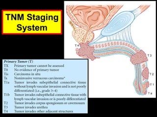
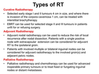
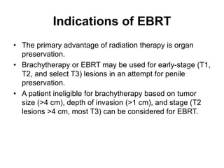
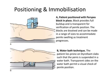
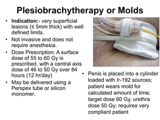
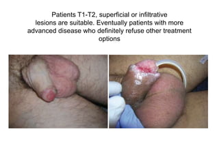
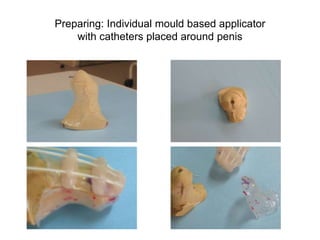
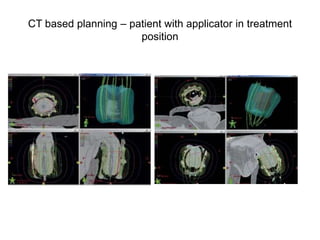
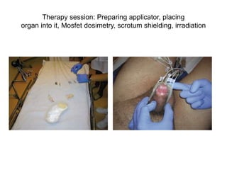
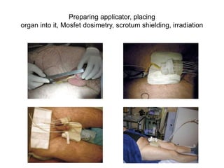
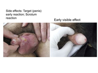
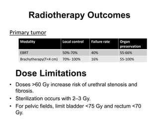
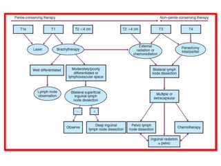
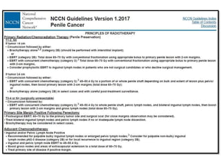
Radiotherapy in Penile
Carcinoma
Dr. Ayush Garg
ANATOMY
TNM Staging
System
Types of RT
Curative Radiotherapy
• Selected early stage I and II tumours 4 cm in size, and where there
is invasion of the corpora cavernosa 1 cm, can be treated with
interstitial brachytherapy.
• EBRT can be used for selected stage II and III tumours in patients
unfit for or refusing surgery.
Adjuvant Radiotherapy
• Adjuvant nodal radiotherapy can be used to reduce the risk of local
recurrence after nodal dissection. Patients with a single positive
node with extracapsular extension can be considered for adjuvant
RT to the ipsilateral groin.
• Patients with involved multiple or bilateral inguinal nodes can be
considered for adjuvant radiotherapy to the involved groin(s) and
adjacent pelvic nodes.
Palliative Radiotherapy
• Palliative radiotherapy and chemotherapy can be used for advanced
inoperable primary tumours or to treat fixed or fungating inguinal
nodes or distant metastases.
Indications of EBRT
• The primary advantage of radiation therapy is organ
preservation.
• Brachytherapy or EBRT may be used for early-stage (T1,
T2, and select T3) lesions in an attempt for penile
preservation.
• A patient ineligible for brachytherapy based on tumor
size (>4 cm), depth of invasion (>1 cm), and stage (T2
lesions >4 cm, most T3) can be considered for EBRT.
RADIATION THERAPY TECHNIQUES
• If indicated, circumcision must be performed before radiation
therapy is initiated.
• The purpose of this procedure is to minimize radiation
therapy–associated morbidity: swelling, irritation of the skin,
moist desquamation, and secondary infection.
• Frequently, a plastic box with a central circular opening that
can be fitted over the penis is used.
• The space between the skin and the box must be filled with
tissue-equivalent material.
• This box can then be treated with parallel opposed beams.
• An alternative to the box technique is the use of a water-filled
container to envelop the penis while the patient is in a prone
position
Positioning & Immobilisation
B, Water bath technique. The
patient lies prone on Styrofoam slabs
such that the penis is suspended in a
water bath. Transparent sides on the
water bath permit a visual check of
penile position.
A, Patient positioned with Perspex
block in place. Block provides full
buildup and is transparent for
verification of penile position. The
blocks are bivalved and can be made
in a range of sizes to accommodate
penile swelling as treatment
progresses.
NODAL
IRRADIATION
• Inguinal lymph node
irradiation for clinically
negative nodes results in
regional control in about
95% of cases.
• In a study it was seen that
without irradiation to the
inguinal lymph nodes, as
many as 20% of patients
can be expected to
develop positive nodes
later.
• Both groins should be
irradiated. The fields
should include inguinal
and pelvic (external iliac
and hypogastric) lymph
nodes
RT Doses
• Use parallel-opposed lateral photon beams to 45 to 50.4
Gy to the whole penile shaft, followed by a cone down to
60 to 70 Gy to 2 cm margin.
• For in situ tumor (Tis), consider 125 to 250 kV
orthovoltage or 13 MeV electrons to 35 Gy in 10 fx of 3
to 5 Gy/fx.
• Use AP/PA to cover inguinal LN. Negative nodes should
receive 50 Gy. Palpable/unresectable nodes should be
treated to 70 to 75 Gy.
Simulation and Field Design
• Simulate patient supine; apply foley catheter and suspend
penis; surround penis by tissue bolus for MV RT.
• If treating inguinal nodes, patient is treated in the frog-leg
position. If treating pelvic nodes, may secure penis cranially
into pelvic field.
Volumes
• GTV: palpable/visible disease (physical exam, CT, MRI)
• CTV:
• (a) GTV + whole shaft of penis
• (b) ±superficial and deep inguinal nodes
• (c) ±pelvic nodes (internal + external iliacs, obturator nodes)
• PTV: 1 cm appropriate(Institutional Protocol)
Brachytherapy
The ideal tumor for brachytherapy should be less than 4 cm
in its maximum diameter, with less than 1 cm of invasion.
Types:
1. Interstitial BT
2. Mold BT
Iridium-192 (192Ir) mold plesiotherapy, and interstitial
implant using 192Ir wires are used.
Indications of Brachytherapy
• Can be considered for lesions less than 4 cm with
invasion of corpora cavernosa less than 1 cm.
• Typically reserved for T1, T2, and select T3 lesions.
• Consider limiting brachytherapy for volume size less
than 8 cc and use of less than six needles to minimize
adverse effects.
Brachytherapy
• A mold is usually built in the form of a box or cylinder
with a central opening and channels for placement of
radioactive sources (needles or wires) in the periphery of
the device.
• The cylinder and sources should be long enough to
prevent underdosage at the tip of the penis. A dose of 60
to 65 Gy at the surface and approximately 50 Gy at the
center of the organ is delivered over 6 to 7 days.
• Alternatively, single- or double-plane implants can be
used to deliver 60 to 70 Gy in 5 to 7 days
Interstitial Brachytherapy
• Implant requires general or spinal anesthesia, takes 30–
45 min
• Catheterize to assist urethra identification to avoid
transfixing with needles/catheters; patients remain
catheterized for duration of treatment.
• May use rigid steel needles held in predrilled parallel
acryli templates or parallel flexible nylon catheters,
placed 1–1.5 cm apart.
• HDR with afterloaded Ir-192 is used.
Interstitial Brachytherapy
• LDR‐Brachytherapy: 0.4–
0.5 Gy/h ~ 60–65 Gy over
100-120 hrs (4-5 days)
• HDR‐Brachytherapy: twice
daily 18 x 3.0 Gy ~ 54.0 Gy
or 12 x 3.2 ~ 38.4 Gy
• Needle placement should
be planned such that the
prescription isodose will
cover 10 mm beyond
visible or palpable tumor.
Plesiobrachytherapy or Molds
• Indication:- very superficial
lesions (≤ 5mm thick) with well
defined limits.
• Not invasive and does not
require anesthesia.
• Dose Prescription: A surface
dose of 55 to 60 Gy is
prescribed, with a central axis
dose of 46 to 50 Gy over 84
hours (12 hr/day)
• May be delivered using a
Perspex tube or silicon
monomer.
• Penis is placed into a cylinder
loaded with Ir-192 sources;
patient wears mold for
calculated amount of time;
target dose 60 Gy, urethra
dose 50 Gy; requires very
compliant patient
Patients T1-T2, superficial or infiltrative
lesions are suitable. Eventually patients with more
advanced disease who definitely refuse other treatment
options
Preparing: Individual mould based applicator
with catheters placed around penis
CT based planning – patient with applicator in treatment
position
Therapy session: Preparing applicator, placing
organ into it, Mosfet dosimetry, scrotum shielding, irradiation
Preparing applicator, placing
organ into it, Mosfet dosimetry, scrotum shielding, irradiation
Side effects: Target (penis)
early reaction; Scrotum
reaction
Early visible effect
Radiotherapy Outcomes
Primary tumor
Modality Local control Failure rate Organ
preservation
EBRT 50%-70% 40% 55-66%
Brachytherapy(T<4 cm) 70%- 100% 16% 55-100%
Dose Limitations
• Doses >60 Gy increase risk of urethral stenosis and
fibrosis.
• Sterilization occurs with 2–3 Gy.
• For pelvic fields, limit bladder <75 Gy and rectum <70
Gy.
COMPLICATIONS
Telengiectasia >90%
Meatal stenosis 30%
Dermatitis, Dysuria, Urethral strictures / fistula 10-40%
Impotence (10–20%),
Penile fibrosis, penile necrosis (3–15%, higher with IS)
Small bowel obstruction (rare).
RESULTS: The specific survival and the disease-free survival at 5
years was
87.6% and 84% respectively. The rate of preservation of the penis was
66% (n531). Sexual data: Among the 17 patients sexually active before
brachytherapy, 10 patients remained sexually active after treatment
(58.8%). Of the 18 patients who had erections before PB, 17 still had
them after treatment (94.4%).
CONCLUSION: The PB seems to have a moderated impact on the
sexual functions and the sexual behavior of the patients.
According to the 2013 ABS-GEC-ESTRO consensus statement,
the good tumor control rates, acceptable morbidity, and functional
organ preservation warrant recommendation of brachytherapy as
the initial treatment for invasive T1, T2, and selected T3 penile
cancers.
• Adequate surgical salvage possibilities with a success rate
between 70% and 100% are observed and reported penis
conservation rate is between 52% and 86%.
• The most important predictors for successful brachytherapy
seem to be tumour size (less or more than 4 cm) and tumour
location limited to the glans or the prepuce without corpus
cavernosum involvement.
• For patients meeting these criteria, different studies report
local recurrence rates of about 20% after 5–10 years with a
secondary control of about 85% of the recurrences by salvage
surgery.
THANK YOU

Radiotherapy in CA Penis

  • 1.
  • 2.
  • 3.
  • 5.
    Types of RT CurativeRadiotherapy • Selected early stage I and II tumours 4 cm in size, and where there is invasion of the corpora cavernosa 1 cm, can be treated with interstitial brachytherapy. • EBRT can be used for selected stage II and III tumours in patients unfit for or refusing surgery. Adjuvant Radiotherapy • Adjuvant nodal radiotherapy can be used to reduce the risk of local recurrence after nodal dissection. Patients with a single positive node with extracapsular extension can be considered for adjuvant RT to the ipsilateral groin. • Patients with involved multiple or bilateral inguinal nodes can be considered for adjuvant radiotherapy to the involved groin(s) and adjacent pelvic nodes. Palliative Radiotherapy • Palliative radiotherapy and chemotherapy can be used for advanced inoperable primary tumours or to treat fixed or fungating inguinal nodes or distant metastases.
  • 6.
    Indications of EBRT •The primary advantage of radiation therapy is organ preservation. • Brachytherapy or EBRT may be used for early-stage (T1, T2, and select T3) lesions in an attempt for penile preservation. • A patient ineligible for brachytherapy based on tumor size (>4 cm), depth of invasion (>1 cm), and stage (T2 lesions >4 cm, most T3) can be considered for EBRT.
  • 7.
    RADIATION THERAPY TECHNIQUES •If indicated, circumcision must be performed before radiation therapy is initiated. • The purpose of this procedure is to minimize radiation therapy–associated morbidity: swelling, irritation of the skin, moist desquamation, and secondary infection. • Frequently, a plastic box with a central circular opening that can be fitted over the penis is used. • The space between the skin and the box must be filled with tissue-equivalent material. • This box can then be treated with parallel opposed beams. • An alternative to the box technique is the use of a water-filled container to envelop the penis while the patient is in a prone position
  • 8.
    Positioning & Immobilisation B,Water bath technique. The patient lies prone on Styrofoam slabs such that the penis is suspended in a water bath. Transparent sides on the water bath permit a visual check of penile position. A, Patient positioned with Perspex block in place. Block provides full buildup and is transparent for verification of penile position. The blocks are bivalved and can be made in a range of sizes to accommodate penile swelling as treatment progresses.
  • 9.
    NODAL IRRADIATION • Inguinal lymphnode irradiation for clinically negative nodes results in regional control in about 95% of cases. • In a study it was seen that without irradiation to the inguinal lymph nodes, as many as 20% of patients can be expected to develop positive nodes later. • Both groins should be irradiated. The fields should include inguinal and pelvic (external iliac and hypogastric) lymph nodes
  • 10.
    RT Doses • Useparallel-opposed lateral photon beams to 45 to 50.4 Gy to the whole penile shaft, followed by a cone down to 60 to 70 Gy to 2 cm margin. • For in situ tumor (Tis), consider 125 to 250 kV orthovoltage or 13 MeV electrons to 35 Gy in 10 fx of 3 to 5 Gy/fx. • Use AP/PA to cover inguinal LN. Negative nodes should receive 50 Gy. Palpable/unresectable nodes should be treated to 70 to 75 Gy.
  • 11.
    Simulation and FieldDesign • Simulate patient supine; apply foley catheter and suspend penis; surround penis by tissue bolus for MV RT. • If treating inguinal nodes, patient is treated in the frog-leg position. If treating pelvic nodes, may secure penis cranially into pelvic field. Volumes • GTV: palpable/visible disease (physical exam, CT, MRI) • CTV: • (a) GTV + whole shaft of penis • (b) ±superficial and deep inguinal nodes • (c) ±pelvic nodes (internal + external iliacs, obturator nodes) • PTV: 1 cm appropriate(Institutional Protocol)
  • 12.
    Brachytherapy The ideal tumorfor brachytherapy should be less than 4 cm in its maximum diameter, with less than 1 cm of invasion. Types: 1. Interstitial BT 2. Mold BT Iridium-192 (192Ir) mold plesiotherapy, and interstitial implant using 192Ir wires are used.
  • 13.
    Indications of Brachytherapy •Can be considered for lesions less than 4 cm with invasion of corpora cavernosa less than 1 cm. • Typically reserved for T1, T2, and select T3 lesions. • Consider limiting brachytherapy for volume size less than 8 cc and use of less than six needles to minimize adverse effects.
  • 14.
    Brachytherapy • A moldis usually built in the form of a box or cylinder with a central opening and channels for placement of radioactive sources (needles or wires) in the periphery of the device. • The cylinder and sources should be long enough to prevent underdosage at the tip of the penis. A dose of 60 to 65 Gy at the surface and approximately 50 Gy at the center of the organ is delivered over 6 to 7 days. • Alternatively, single- or double-plane implants can be used to deliver 60 to 70 Gy in 5 to 7 days
  • 15.
    Interstitial Brachytherapy • Implantrequires general or spinal anesthesia, takes 30– 45 min • Catheterize to assist urethra identification to avoid transfixing with needles/catheters; patients remain catheterized for duration of treatment. • May use rigid steel needles held in predrilled parallel acryli templates or parallel flexible nylon catheters, placed 1–1.5 cm apart. • HDR with afterloaded Ir-192 is used.
  • 16.
    Interstitial Brachytherapy • LDR‐Brachytherapy:0.4– 0.5 Gy/h ~ 60–65 Gy over 100-120 hrs (4-5 days) • HDR‐Brachytherapy: twice daily 18 x 3.0 Gy ~ 54.0 Gy or 12 x 3.2 ~ 38.4 Gy
  • 17.
    • Needle placementshould be planned such that the prescription isodose will cover 10 mm beyond visible or palpable tumor.
  • 18.
    Plesiobrachytherapy or Molds •Indication:- very superficial lesions (≤ 5mm thick) with well defined limits. • Not invasive and does not require anesthesia. • Dose Prescription: A surface dose of 55 to 60 Gy is prescribed, with a central axis dose of 46 to 50 Gy over 84 hours (12 hr/day) • May be delivered using a Perspex tube or silicon monomer. • Penis is placed into a cylinder loaded with Ir-192 sources; patient wears mold for calculated amount of time; target dose 60 Gy, urethra dose 50 Gy; requires very compliant patient
  • 19.
    Patients T1-T2, superficialor infiltrative lesions are suitable. Eventually patients with more advanced disease who definitely refuse other treatment options
  • 20.
    Preparing: Individual mouldbased applicator with catheters placed around penis
  • 21.
    CT based planning– patient with applicator in treatment position
  • 22.
    Therapy session: Preparingapplicator, placing organ into it, Mosfet dosimetry, scrotum shielding, irradiation
  • 23.
    Preparing applicator, placing organinto it, Mosfet dosimetry, scrotum shielding, irradiation
  • 24.
    Side effects: Target(penis) early reaction; Scrotum reaction Early visible effect
  • 25.
    Radiotherapy Outcomes Primary tumor ModalityLocal control Failure rate Organ preservation EBRT 50%-70% 40% 55-66% Brachytherapy(T<4 cm) 70%- 100% 16% 55-100% Dose Limitations • Doses >60 Gy increase risk of urethral stenosis and fibrosis. • Sterilization occurs with 2–3 Gy. • For pelvic fields, limit bladder <75 Gy and rectum <70 Gy.
  • 26.
    COMPLICATIONS Telengiectasia >90% Meatal stenosis30% Dermatitis, Dysuria, Urethral strictures / fistula 10-40% Impotence (10–20%), Penile fibrosis, penile necrosis (3–15%, higher with IS) Small bowel obstruction (rare).
  • 27.
    RESULTS: The specificsurvival and the disease-free survival at 5 years was 87.6% and 84% respectively. The rate of preservation of the penis was 66% (n531). Sexual data: Among the 17 patients sexually active before brachytherapy, 10 patients remained sexually active after treatment (58.8%). Of the 18 patients who had erections before PB, 17 still had them after treatment (94.4%). CONCLUSION: The PB seems to have a moderated impact on the sexual functions and the sexual behavior of the patients.
  • 28.
    According to the2013 ABS-GEC-ESTRO consensus statement, the good tumor control rates, acceptable morbidity, and functional organ preservation warrant recommendation of brachytherapy as the initial treatment for invasive T1, T2, and selected T3 penile cancers.
  • 29.
    • Adequate surgicalsalvage possibilities with a success rate between 70% and 100% are observed and reported penis conservation rate is between 52% and 86%. • The most important predictors for successful brachytherapy seem to be tumour size (less or more than 4 cm) and tumour location limited to the glans or the prepuce without corpus cavernosum involvement. • For patients meeting these criteria, different studies report local recurrence rates of about 20% after 5–10 years with a secondary control of about 85% of the recurrences by salvage surgery.
  • 32.

Editor's Notes

  • #3 Corpus cavernosum: Two columns of tissue running along the sides of the penis. Blood fills this tissue to cause an erection. • Corpus spongiosum: A column of sponge-like tissue running along the front of the penis and ending at the glans penis; it fills with blood during an erection, keeping the urethra -- which runs through it -- open.
  • #6 Curative Radiotherapy Electron beam radiotherapy can be used for very small stage I superficial tumours. Selected early stage I and II tumours 4 cm in size, and where there is invasion of the corpora cavernosa 1 cm, can be treated with interstitial brachytherapy. EBRT can be used for selected stage II and III tumours in patients unfit for or refusing surgery. Adjuvant radiotherapy Adjuvant nodal radiotherapy can be used to reduce the risk of local recurrence after nodal dissection. Patients with a single positive node with extracapsular extension can be considered for adjuvant radiotherapy to the ipsilateral groin. Patients with involved multiple or bilateral inguinal nodes can be considered for adjuvant radiotherapy to the involved groin(s) and adjacent pelvic nodes.
  • #9 A, Patient positioned with Perspex block in place. Block provides full buildup and is transparent for verification of penile position. The blocks are bivalved and can be made in a range of sizes to accommodate penile swelling as treatment progresses. B, Water bath technique. The patient lies prone on Styrofoam slabs such that the penis is suspended in a water bath. Transparent sides on the water bath permit a visual check of penile position.