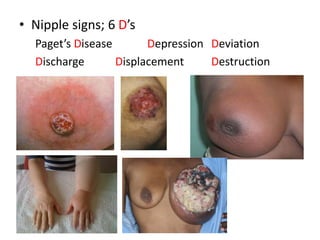
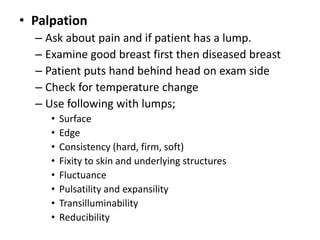
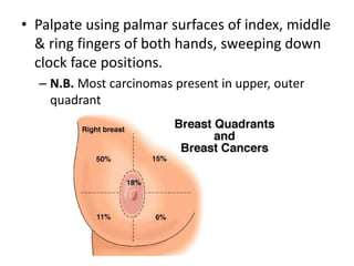
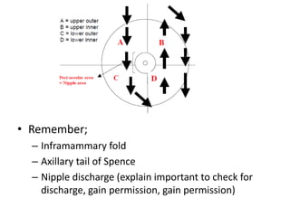
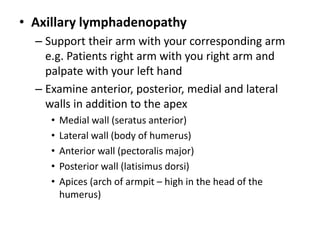
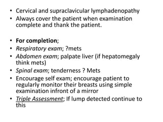
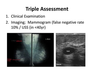
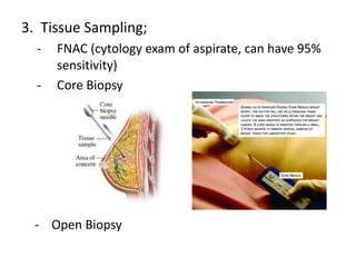
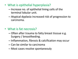
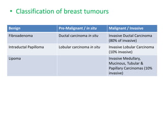
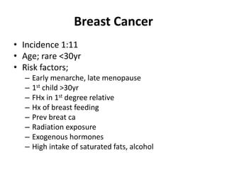
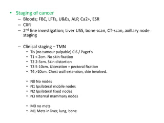
DEPARTMENT OF MIDWIFERY
GROUP ASSIGNMENT
TITTLE:- BRAST EXAMINATION
GROUP MEMBERS
1.ABDULHAKIM MOHAMMED
2.BELAYNEW ASHAGIRE
3.WORKU BELAYNEW
4.
MINILIK II MEDICAL AND
🚑 HEALTH SCIENCE 🚑
🎓 COLLEGE 🎓
Breast Examination
Topics
• Breast history
• Examination
• Investigations
• Breast conditions
– Benign / Malignant
• Treatment
History
• Presenting complaint is important
• Lump;
– always ask how long been present
– Relation to menstrual cycle
– Does its size vary? Is it getting larger?
• Pain;
– Is it cyclical? Is the lump painful?
• Nipple discharge; ascertain
– Colour, Quantity, pattern, frequency
• Age of patient; cancers are uncommon
<30yrs, but fibroadenomas are
• Ask if noticed any;
– Nipple retraction
– Breast distortion
– Metastatic related symptoms
• Previous breast disease
– Was it investigated / treated
• Family history
– Genetics; 5-10% are inherited dominantly
• They have early onset & associated with other tumours
e.g. Bowel, ovarian.
• BRCA1 (chromosome 17q21)
• BRCA2 (chromosome 13q24)
• P53 gene chromosome 17
• Medications; HRT, pill
• Gynae / Obstetric Hx;
– Menarche, menses
– Parity? When? After 30 increases risk
Examination
• Introduce yourself to patient
• Undress to waist, sit on couch at 45 degrees
• Maintain patient dignity e.g. Bed sheet
• Assess in following positions
– Patient’s hands behind their head (accentuate lumps,
asymmetry, tethering)
– Pushing against their hips (accentuate lumps attached
to pectoralis muscle)
– Patient leaning over side of bed (accentuate
abnormalities in large breasts)
• Exam good breast first, then the ‘diseased’ breast
• Inspection
– 6 S’s
• Site
• Size
• Shape
• Symmetry
• overlying Skin
• associated Scars
• Fungation; comment on presence of fungating carcinoma
(check inframammory fold)
• Asymmetry; carcinoma may be present in higher breast
• Tethering; due to infiltration of ligaments of Astley-Cooper
• Peau d’orange; micro-oedema
• Lymphoedema; may indicate lymphatic infiltration by
carcinoma or previous surgery with LN removal
• Erythema
• Nipple signs; 6 D’s
Paget’s Disease Depression Deviation
Discharge Displacement Destruction
• Palpation
– Ask about pain and if patient has a lump.
– Examine good breast first then diseased breast
– Patient puts hand behind head on exam side
– Check for temperature change
– Use following with lumps;
• Surface
• Edge
• Consistency (hard, firm, soft)
• Fixity to skin and underlying structures
• Fluctuance
• Pulsatility and expansility
• Transilluminability
• Reducibility
• Palpate using palmar surfaces of index, middle
& ring fingers of both hands, sweeping down
clock face positions.
– N.B. Most carcinomas present in upper, outer
quadrant
• Remember;
– Inframammary fold
– Axillary tail of Spence
– Nipple discharge (explain important to check for
discharge, gain permission, gain permission)
• Axillary lymphadenopathy
– Support their arm with your corresponding arm
e.g. Patients right arm with you right arm and
palpate with your left hand
– Examine anterior, posterior, medial and lateral
walls in addition to the apex
• Medial wall (seratus anterior)
• Lateral wall (body of humerus)
• Anterior wall (pectoralis major)
• Posterior wall (latisimus dorsi)
• Apices (arch of armpit – high in the head of the
humerus)
• Cervical and supraclavicular lymphadenopathy
• Always cover the patient when examination
complete and thank the patient.
• For completion;
• Respiratory exam; ?mets
• Abdomen exam; palpate liver (if hepatomegaly
think mets)
• Spinal exam; tenderness ? Mets
• Encourage self exam; encourage patient to
regularly monitor their breasts using simple
examination infront of a mirror
• Triple Assessment; If lump detected continue to
this
Triple Assessment
1. Clinical Examination
2. Imaging; Mammogram (false negative rate
10% / USS (in <40yr)
3. Tissue Sampling;
- FNAC (cytology exam of aspirate, can have 95%
sensitivity)
- Core Biopsy
- Open Biopsy
Breast Disease
• Classify as benign or malignant
– Benign aetiology classified as Aberrations of
normal development and involution (ANDI)
Peak Age (years)
15-25 Development Fibroadenoma & excessive Breast
development
25-40 Cyclical Hormonal Cyclical nodularity & mastalgia
35-55 Involution Lobular:
Ductal:
Epithelial:
Cyst
Duct ectasia & periductal mastitis
Hyperplasia & fibrosis
• What is a fibroadenoma?
– Most common benign neoplasm. Fibroepithelial
tumour, composed of glandular tissue & stroma.
– Peak onset 15-25yrs.
– Painless, smooth, firm, rubbery lump, highly mobile.
– Approx 10% resolve spontaneously within 1yr
• What are breast cysts?
– Fluid-filled, distended & involuted lobules.
– Present as smooth lumps. Maybe painful
– Peak age onset 35-55yr.
– FNA may relieve symptoms and can be analysed
• What are cyclical nodularity & mastalgia?
– Affect pre-menopausal females & are hormonal
dependent.
– Cyclical breast changes occur, result lumps
(nodularity) & pain (mastalgia) related to
menstrual cycle.
– Treatment options classified as;
Conservative Medical Surgical
Reassurance Evening primrose oil Mastectomy (for
treatment resistant
severe mastalgia)
Firm supporting bra Analgesia
Evening primrose oil OCP
Danazol
Bromocriptin
Tamoxifen
• What is duct ectasia?
– Involution & dilatation of subareolar ducts
– Clinical features; nipple inversion, nipple discharge
(may be cheese / blood stained), subareolar mass,
mastalgia.
• What is periductal mastitis?
– Inflammation, often due to infection of subareolar
ducts.
– May present like duct ectasia
– Pus discharge from nipple & mastalgia
• What is epithelial hyperplasia?
– Increase no. of epithelial lining cells of the
terminal lobular unit.
– Atypical dyplasia increased risk of progression to
carcinoma.
• What is fat necrosis?
– Often after trauma to fatty breast tisssue e.g.
Surgery / breastfeeding.
– Inflammation, fibrosis & calcification may occur
– Can be similar to carcinoma
– Most cases resolve spontaneouly
• Classification of breast tumours
Benign Pre-Malignant / in situ Malignant / Invasive
Fibroadenoma Ductal carcinoma in situ Invasive Ductal Carcinoma
(80% of invasive)
Intraductal Papilloma Lobular carcinoma in situ Invasive Lobular Carcinoma
(10% invasive)
Lipoma Invasive Medullary,
Mucinous, Tubular &
Papillary Carcinomas (10%
invasive)
Breast Cancer
• Incidence 1:11
• Age; rare <30yr
• Risk factors;
– Early menarche, late menopause
– 1st child >30yr
– FHx in 1st degree relative
– Hx of breast feeding
– Prev breat ca
– Radiation exposure
– Exogenous hormones
– High intake of saturated fats, alcohol
• Staging of cancer
– Bloods; FBC, LFTs, U&Es, ALP, Ca2+, ESR
– CXR
– 2nd line investigation; Liver USS, bone scan, CT-scan, axillary node
staging
– Clinical staging – TMN
• Tis (no tumour palpable) CIS / Paget’s
• T1 < 2cm. No skin fixation
• T2 2-5cm. Skin distortion
• T3 5-10cm. Ulceration + pectoral fixation
• T4 >10cm. Chest wall extension, skin involved.
• N0 No nodes
• N1 Ipsilateral mobile nodes
• N2 Ipsilateral fixed nodes
• N3 Internal mammary nodes
• M0 no mets
• M1 Mets in liver, lung, bone
Treatment
• Surgical;
– WLE plus DXT (need 1cm excision margin)
– Mastectomy
– Axillary sampling (removal of lower axillary nodes)
– Axillary clearance (removal of contents below the
level of the axillary vein)
• Level 1 = below pec minor
• Level 2 = behind pec minor
• Level 3 = above pec minor (full clearance)
– SLNB
• Systemic treatment
– Can be adjuvant or neo-adjuvant
1. Radiotherapy
- Breast and chest wall
- Axilla
- Palliation (e.g. For bony tenderness)
2. Chemotherapy
- Recurrent disease
- <70yr with > 1 +ive axillary node
- Very large tumours
• 3. Endocrine therapy and Tamoxifen
– Tamoxifen in ER + ive females
– Up to 15% of ER –ive females also respond
– Beneficial in pre- and postmenopausal women,
not effective in ER –ive premenopausals
– Increased risk of endometrial carcinoma
– Aromatase enzyme inhibitor = Anastrazole
(Arimidex)
• For post-menopausal women ER +ive

breast_surgery.pptx

  • 1.
    DEPARTMENT OF MIDWIFERY GROUPASSIGNMENT TITTLE:- BRAST EXAMINATION GROUP MEMBERS 1.ABDULHAKIM MOHAMMED 2.BELAYNEW ASHAGIRE 3.WORKU BELAYNEW 4. MINILIK II MEDICAL AND 🚑 HEALTH SCIENCE 🚑 🎓 COLLEGE 🎓
  • 2.
  • 3.
    Topics • Breast history •Examination • Investigations • Breast conditions – Benign / Malignant • Treatment
  • 4.
    History • Presenting complaintis important • Lump; – always ask how long been present – Relation to menstrual cycle – Does its size vary? Is it getting larger? • Pain; – Is it cyclical? Is the lump painful?
  • 5.
    • Nipple discharge;ascertain – Colour, Quantity, pattern, frequency • Age of patient; cancers are uncommon <30yrs, but fibroadenomas are • Ask if noticed any; – Nipple retraction – Breast distortion – Metastatic related symptoms • Previous breast disease – Was it investigated / treated
  • 6.
    • Family history –Genetics; 5-10% are inherited dominantly • They have early onset & associated with other tumours e.g. Bowel, ovarian. • BRCA1 (chromosome 17q21) • BRCA2 (chromosome 13q24) • P53 gene chromosome 17 • Medications; HRT, pill • Gynae / Obstetric Hx; – Menarche, menses – Parity? When? After 30 increases risk
  • 7.
    Examination • Introduce yourselfto patient • Undress to waist, sit on couch at 45 degrees • Maintain patient dignity e.g. Bed sheet • Assess in following positions – Patient’s hands behind their head (accentuate lumps, asymmetry, tethering) – Pushing against their hips (accentuate lumps attached to pectoralis muscle) – Patient leaning over side of bed (accentuate abnormalities in large breasts) • Exam good breast first, then the ‘diseased’ breast
  • 8.
    • Inspection – 6S’s • Site • Size • Shape • Symmetry • overlying Skin • associated Scars • Fungation; comment on presence of fungating carcinoma (check inframammory fold) • Asymmetry; carcinoma may be present in higher breast • Tethering; due to infiltration of ligaments of Astley-Cooper • Peau d’orange; micro-oedema • Lymphoedema; may indicate lymphatic infiltration by carcinoma or previous surgery with LN removal • Erythema
  • 9.
    • Nipple signs;6 D’s Paget’s Disease Depression Deviation Discharge Displacement Destruction
  • 10.
    • Palpation – Askabout pain and if patient has a lump. – Examine good breast first then diseased breast – Patient puts hand behind head on exam side – Check for temperature change – Use following with lumps; • Surface • Edge • Consistency (hard, firm, soft) • Fixity to skin and underlying structures • Fluctuance • Pulsatility and expansility • Transilluminability • Reducibility
  • 11.
    • Palpate usingpalmar surfaces of index, middle & ring fingers of both hands, sweeping down clock face positions. – N.B. Most carcinomas present in upper, outer quadrant
  • 12.
    • Remember; – Inframammaryfold – Axillary tail of Spence – Nipple discharge (explain important to check for discharge, gain permission, gain permission)
  • 13.
    • Axillary lymphadenopathy –Support their arm with your corresponding arm e.g. Patients right arm with you right arm and palpate with your left hand – Examine anterior, posterior, medial and lateral walls in addition to the apex • Medial wall (seratus anterior) • Lateral wall (body of humerus) • Anterior wall (pectoralis major) • Posterior wall (latisimus dorsi) • Apices (arch of armpit – high in the head of the humerus)
  • 14.
    • Cervical andsupraclavicular lymphadenopathy • Always cover the patient when examination complete and thank the patient. • For completion; • Respiratory exam; ?mets • Abdomen exam; palpate liver (if hepatomegaly think mets) • Spinal exam; tenderness ? Mets • Encourage self exam; encourage patient to regularly monitor their breasts using simple examination infront of a mirror • Triple Assessment; If lump detected continue to this
  • 15.
    Triple Assessment 1. ClinicalExamination 2. Imaging; Mammogram (false negative rate 10% / USS (in <40yr)
  • 16.
    3. Tissue Sampling; -FNAC (cytology exam of aspirate, can have 95% sensitivity) - Core Biopsy - Open Biopsy
  • 17.
    Breast Disease • Classifyas benign or malignant – Benign aetiology classified as Aberrations of normal development and involution (ANDI) Peak Age (years) 15-25 Development Fibroadenoma & excessive Breast development 25-40 Cyclical Hormonal Cyclical nodularity & mastalgia 35-55 Involution Lobular: Ductal: Epithelial: Cyst Duct ectasia & periductal mastitis Hyperplasia & fibrosis
  • 18.
    • What isa fibroadenoma? – Most common benign neoplasm. Fibroepithelial tumour, composed of glandular tissue & stroma. – Peak onset 15-25yrs. – Painless, smooth, firm, rubbery lump, highly mobile. – Approx 10% resolve spontaneously within 1yr • What are breast cysts? – Fluid-filled, distended & involuted lobules. – Present as smooth lumps. Maybe painful – Peak age onset 35-55yr. – FNA may relieve symptoms and can be analysed
  • 19.
    • What arecyclical nodularity & mastalgia? – Affect pre-menopausal females & are hormonal dependent. – Cyclical breast changes occur, result lumps (nodularity) & pain (mastalgia) related to menstrual cycle. – Treatment options classified as; Conservative Medical Surgical Reassurance Evening primrose oil Mastectomy (for treatment resistant severe mastalgia) Firm supporting bra Analgesia Evening primrose oil OCP Danazol Bromocriptin Tamoxifen
  • 20.
    • What isduct ectasia? – Involution & dilatation of subareolar ducts – Clinical features; nipple inversion, nipple discharge (may be cheese / blood stained), subareolar mass, mastalgia. • What is periductal mastitis? – Inflammation, often due to infection of subareolar ducts. – May present like duct ectasia – Pus discharge from nipple & mastalgia
  • 21.
    • What isepithelial hyperplasia? – Increase no. of epithelial lining cells of the terminal lobular unit. – Atypical dyplasia increased risk of progression to carcinoma. • What is fat necrosis? – Often after trauma to fatty breast tisssue e.g. Surgery / breastfeeding. – Inflammation, fibrosis & calcification may occur – Can be similar to carcinoma – Most cases resolve spontaneouly
  • 22.
    • Classification ofbreast tumours Benign Pre-Malignant / in situ Malignant / Invasive Fibroadenoma Ductal carcinoma in situ Invasive Ductal Carcinoma (80% of invasive) Intraductal Papilloma Lobular carcinoma in situ Invasive Lobular Carcinoma (10% invasive) Lipoma Invasive Medullary, Mucinous, Tubular & Papillary Carcinomas (10% invasive)
  • 23.
    Breast Cancer • Incidence1:11 • Age; rare <30yr • Risk factors; – Early menarche, late menopause – 1st child >30yr – FHx in 1st degree relative – Hx of breast feeding – Prev breat ca – Radiation exposure – Exogenous hormones – High intake of saturated fats, alcohol
  • 24.
    • Staging ofcancer – Bloods; FBC, LFTs, U&Es, ALP, Ca2+, ESR – CXR – 2nd line investigation; Liver USS, bone scan, CT-scan, axillary node staging – Clinical staging – TMN • Tis (no tumour palpable) CIS / Paget’s • T1 < 2cm. No skin fixation • T2 2-5cm. Skin distortion • T3 5-10cm. Ulceration + pectoral fixation • T4 >10cm. Chest wall extension, skin involved. • N0 No nodes • N1 Ipsilateral mobile nodes • N2 Ipsilateral fixed nodes • N3 Internal mammary nodes • M0 no mets • M1 Mets in liver, lung, bone
  • 25.
    Treatment • Surgical; – WLEplus DXT (need 1cm excision margin) – Mastectomy – Axillary sampling (removal of lower axillary nodes) – Axillary clearance (removal of contents below the level of the axillary vein) • Level 1 = below pec minor • Level 2 = behind pec minor • Level 3 = above pec minor (full clearance) – SLNB
  • 26.
    • Systemic treatment –Can be adjuvant or neo-adjuvant 1. Radiotherapy - Breast and chest wall - Axilla - Palliation (e.g. For bony tenderness) 2. Chemotherapy - Recurrent disease - <70yr with > 1 +ive axillary node - Very large tumours
  • 27.
    • 3. Endocrinetherapy and Tamoxifen – Tamoxifen in ER + ive females – Up to 15% of ER –ive females also respond – Beneficial in pre- and postmenopausal women, not effective in ER –ive premenopausals – Increased risk of endometrial carcinoma – Aromatase enzyme inhibitor = Anastrazole (Arimidex) • For post-menopausal women ER +ive

Editor's Notes

  • #20 Danazol (Gonadotrophin inhibitor), Bromocriptine (Dopamine receptor stimulant – reduces level prolactin), tamoxifen (An anti-oestrogen hormone antagonist)