
Recommended
Recommended
More Related Content
What's hot
What's hot (20)
Similar to Shoulder Joint Anatomy and Radiography Guide
Similar to Shoulder Joint Anatomy and Radiography Guide (20)
Recently uploaded
Recently uploaded (20)
Shoulder Joint Anatomy and Radiography Guide
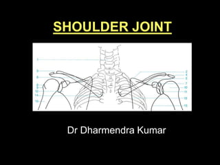
- 1. SHOULDER JOINT Dr Dharmendra Kumar
- 2. CLAVICLE GREATER TUBERCLE HUMERAL HEAD GLENOID ACROMION CORACOID ACROMIOCLAVICULAR JOINT LESSER TUBERCLE VERTEBRAL BORDER OF SCAPULA BICIPITAL GROOVE RADIOLOGICAL ANATOMY
- 3. OSSIFICATION CENTRES 3 CLAVICLE – 1. Lateral head – 5th weeks in utero 2. Medial head – 15th years SCAPULA – 1. Body – 8th weeks in utero 2. coracoid process ( two centres ) – 12 – 18th months 3. Glenoid – 10 – 11 years 4. inferior angle – 14 – 20 years 5. acromian ( three centres) – 14 – 20 years 6. medial border – 14 – 20 years PROXIMAL HUMERUS – 1. Diaphysis – 8th weeks in utero 2. Head – 1-6th months 3. Greater tubercle – 1 year 4. Lesser tubercle – 3-5 years
- 4. Recommended Projections for shoulder joint • 1) Basic Projections i) Antero-posterior (15 Degree) erect ii) Supero-inferior (Axial) iii) Infero- superior (Reverse axial) 2) Alternate Projections for Trauma i) Anterior Oblique (Y-projection) ii) Apical Oblique (Garth Projection) iii) Supero-inferior modified/ Modified axial (Wallace projection) Cntd…….
- 5. .. 3) Additional projections i) Infero-superior modified (West point projection) ii) Antero-posterior modified (stryker projection)
- 6. TECHNICAL FACTORS 6 kVp mAs FFD Grid Collimatio n Film size 40-50 10 100cm- 120 cm No Yes 10” x 8”
- 7. Patient position:- The patient stand with the affected shoulder against the cassette and is rotated 15 degrees . Part position:- The arm is supinated with slightly abducted away from the body. The cassette is positioned 5 cm above the shoulder. Centre:- The horizontal central ray is directed to the palpable coracoid process of the scapula. Antero-posterior erect
- 8. Essential image characteristics :- The head and proximal end of the humerus, the inferior angle of the scapula and the whole of the clavicle. The apex of lung should be included Arrested respiration - good rib detail in acute trauma. The head of humerus slightly overlapping the glenoid cavity. 8
- 9. Patient position:- The patient seated with their affected side adjacent to the table. Part position:- Arm abducted over the cassette. The cassette is placed on the table top , and arm under examination is abducted over the table Centre:- Mid gleno-humeral joint. (SUPERO-INFERIOR (AXIAL)
- 10. Essential image characteristics :- The image must demonstrate the humeral head in relation to the glenoid cavity. The acromion process should be visible over the neck of humerus and ideally the coracoid process will be visible. Radiological considerations :- The lesser tuberosity will be seen in profile while the greater tuberosity is superimposed over the humeral head. 10
- 11. Patient position:- The patient lies supine on the table. Part position:- The arm of the affected side abducted and supinated. A cassette is supported vertically against the shoulder. (Infero-superior (reverse axial) Centre:- The horizontal central ray is directed towards the axilla with minimum angulation towards the trunk.
- 12. Essential Image characteristics :- > Image must demonstrate the humeral head in relation to the glenoid cavity > The acromion process should be visible over the neck of the humerus . 12
- 13. Patient position:- The patient stands or sits with the lateral aspect of the injury arm against the cassette. Part position:- The unaffected shoulder is raised to make the angle between the trunk and cassette approximately 60 degrees. The cassette is positioned to include the superior border of the scapula. Centre: The horizontal ray is centred towards the head of the humerus. Anterior Oblique (‘Y’ projection )
- 14. Radiographic criteria: The exposure should demonstrate the position of the head of the humeus in relation to the glenoid cavity between coracoid and acromion processes 14
- 15. . Patient position:- The patient lies supine on the X-ray table. Part position:- The arm of the affected side is extended fully and the elbow then flexed to allow the hand to rest on the patient’s head. The centre of the cassette is positioned 2.5 cm superior to the head of the humerus. Centre:- The central ray is directed through the centre of the axilla to the head of the humerus. (AP modified) – Stryker’s view
- 16. Radiographic criteria:- The projection demonstrates the postero- lateral aspect of the humeral head. 16
- 19. Centre:- The horizontal central ray is directed to the palpable lateral end of the clavicle at the acromio-clavicular joint. Patient position:- The patient stands facing the Xray tube. Part position:- The shoulder being examined is placed in contact with the cassette, The patient is then rotated approximately 15 degrees towards the side being examined to bring the acromio-clavicular joint space at right-angles to the film Radiography of Acromio-clavicular joint (AP projection)
- 20. • Radiographic criteria:- Acromio-clavicular joint and the clavicle projected above the acromion process. 20
- 21. The AC joint has a week joint capsule and is vulnerable to trauma. Subluxation may be difficult to diagnose in the standard AP image. To prove subluxation, it is necessary to do weight-bearing comparison projections of both AC joints The position of the patient and cassette are same as normal AP view. The normal joint is between 3-8 mm in width. Normal variation between two sides should be less than 2-3mm. Radiography of Acromio-clavicular joint (Weight-bearing AP projection))
- 23. THANK YOU