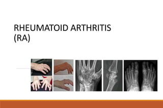
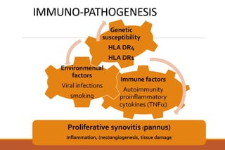
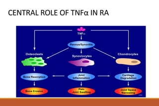
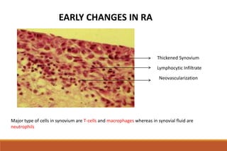
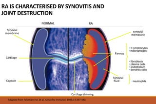
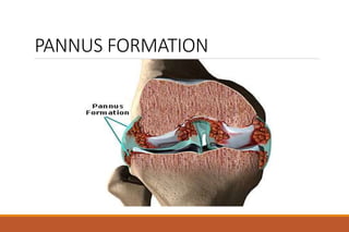
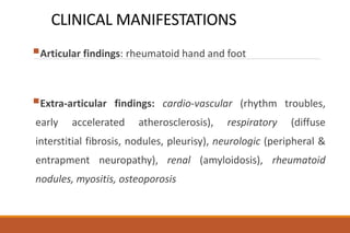
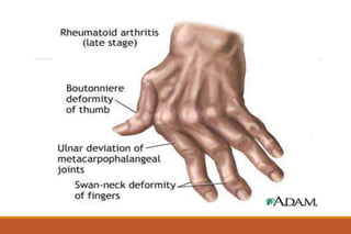
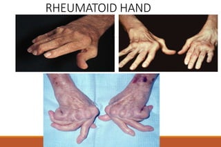
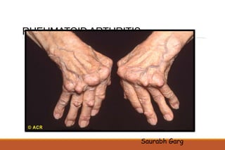
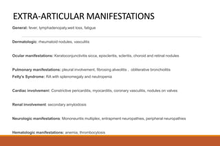
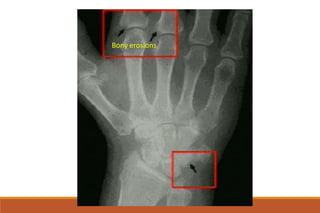
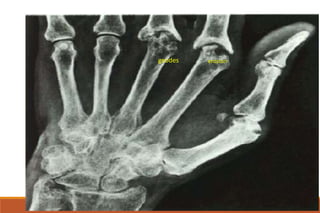
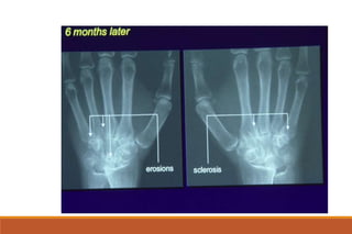
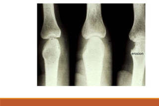
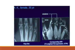
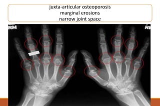
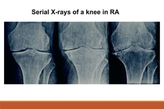
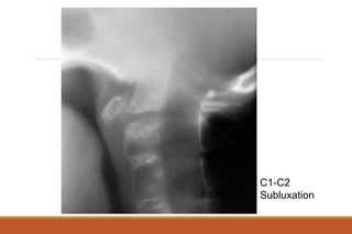
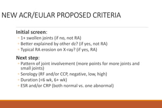
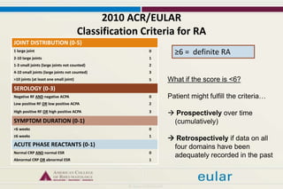
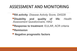
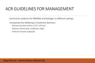
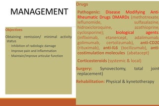
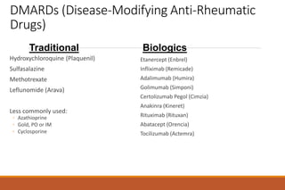
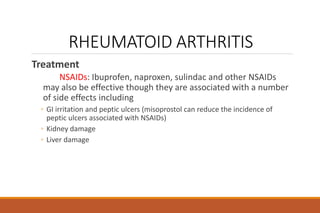
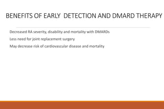
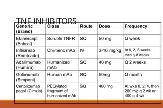
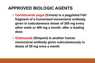
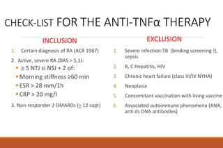
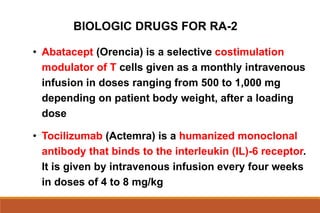
RHEUMATOID ARTHRITIS
(RA)
OVERVIEW
Definition
Immuno-pathogenesis
Clinical findings (articular and systemic manifestations)
Investigations
Assessment & monitoring
Management
RHEUMATOID ARTHRITIS
Chronic multisystemic inflammatory disease of unknown
etiology
Affects the synovial membranes of multiple joints (diarthrodial
joints)
Female : Male ratio 3:1
Most frequent during 4th and 5th decade
Affects approximately 1% of the adult population worldwide
DEFINITION
chronic immune inflammatory disorder, with still
unknown aetiology, characterized by:
Articular manifestations: chronic destructive and
deforming arthritis affecting small joints in a bilateral and
symmetrical pattern;
Systemic manifestations: cardio-vascular, respiratory,
renal, neurologic, ocular;
Progressive irreversible articular damage;
Significant functional disability and impaired quality of life
IMMUNO-PATHOGENESIS
Immune factors
Autoimmunity
proinflammatory
cytokines (TNF)
Environmental
factors
Viral infections
smoking
Genetic
susceptibility
HLA DR4
HLA DR1
Proliferative synovitis (pannus)
Inflammation, (neo)angiogenesis, tissue damage
NORMAL SYNOVIAL JOINT
Articular cartilage
Synovium Membrane
Lamellar bone
Subchondral bone
Type A cell: mphage like,
protective role
Type B cell:
fibroblast
like, produce
matrix and
synovial fluid
Vessel
Synovial Membrane:
only 1-2 cells thick
DESTRUCTION OF JOINT CARTILAGE AND BONE
Proliferating synovial lining => Pannus
Proliferating synovial lining comes in contact with
the cartilage matrix and bone there is
degradation of the cartilage and erosion of the
bone surface (by matrix metalloproteinases and
other proteases produced by synovial cells)
Chondrocytes themselves
IL-1 and TNF-alpha also stimulate production of
metalloproteinase by Chondrocytes of the articular
cartilage. In response to these cytokines,
chondrocytes decrease type II collagen and
proteoglycan synthesis and increase synthesis of
metalloproteinases that contribute to the
degradation of collagen and proteoglycans.
Neutrophils in Synovial Fluid
The main inflammatory cells of the synovial
fluid are neutrophils.
Cytokines such as transforming growth factor
beta (TGF-beta) and interleukin 8 (IL-8) attract
neutrophils.
Neutrophils may undergo degranulation and
cause some damage to surrounding tissues.
Osteoclasts
Osteoclasts may be activated by
inflammatory mediators including IL-
1, TNF and PGE2
bone
cartilage
CENTRAL ROLE OF TNFα IN RA
Thickened Synovium
Lymphocytic Infiltrate
Neovascularization
EARLY CHANGES IN RA
Major type of cells in synovium are T-cells and macrophages whereas in synovial fluid are
neutrophils
RA IS CHARACTERISED BY SYNOVITIS AND
JOINT DESTRUCTION
NORMAL RA
Synovial
membrane
Cartilage
Capsule
Synovial
fluid
Inflamed
synovial
membrane
Pannus
Major cell types:
• T lymphocytes
• macrophages
Minor cell types:
• fibroblasts
• plasma cells
• endothelium
• dendritic cells
Major cell type:
• neutrophils
Adapted from Feldmann M, et al. Annu Rev Immunol. 1996;14:397-440.
Cartilage thinning
PANNUS FORMATION
CLINICAL MANIFESTATIONS
Articular findings: rheumatoid hand and foot
Extra-articular findings: cardio-vascular (rhythm troubles,
early accelerated atherosclerosis), respiratory (diffuse
interstitial fibrosis, nodules, pleurisy), neurologic (peripheral &
entrapment neuropathy), renal (amyloidosis), rheumatoid
nodules, myositis, osteoporosis
RHEUMATOID ARTHRITIS
Clinical Features
◦ the stiffness is characteristically worse in the morning and
improves during the day; its duration is a useful indicator of the
activity of the disease.
◦ the usual joints affected by rheumatoid arthritis are the
metacarpophalangeal joints, the PIP joints, the wrists, knees,
ankles and toes.
◦ Entrapment syndromes may occur especially carpal tunnel
syndrome
RHEUMATOID HAND
DEVIERE CUBITALA DEGETE
POLICE “IN Z”
1. Swelling of the RC,
MCF, PIP joints
2. Fusiform swelling
fingers: synovitis of PIP
joints, causing them to
appear spindle-shaped.
RHEUMATOID ARTHRITIS
After months to years, deformities can occur; the most
common are
◦ ulnar deviation of the fingers
◦ swan neck deformity, which is hyperextension of the distal
interphalangeal joint and flexion of the proximal interphalangeal
joint
◦ boutonniere deformity, which is flexion of the distal
interphalangeal joint and extension of the proximal
interphalangeal joint
◦ valgus deformity of the knee
RHEUMATOID HAND
RHEUMATOID HAND
Boutonniere deformity
Rheumatoid nodules
Artritis mutilans
DEGETE “IN GAT DE LEBADA”
Swan-neck deformity”
Fingers in “swan-neck”
Fingers in “ boutonniere”
artritis mutilans
RHEUMATOID ARTHRITIS
Saurabh Garg
RHEUMATOID FOOT
Hammer toes Complex deformity of the foot
Subluxation of the metatarsal heads
Triangular foot
EXTRA-ARTICULAR MANIFESTATIONS
General: fever, lymphadenopaty,weit loss, fatigue
Dermatologic: rheumatoid nodules, vasculitis
Ocular manifestations: Keratoconjunctivitis sicca, episcleritis, scleritis, choroid and retinal nodules
Pulmonary manifestations: pleural involvement, fibrosing alveolitis , obliterative bronchiolitis
Felty’s Syndrome: RA with splenomegaly and neutropenia
Cardiac involvement: Constrictive pericarditis, myocarditis, coronary vasculitis, nodules on valves
Renal involvement: secondary amyloidosis
Neurologic manifestations: Mononeuritis multiplex, entrapment neuropathies, peripheral neuropathies
Hematologic manifestations: anemia, thrombocytosis
RHEUMATOID NODULES
Subcutaneus nodules
Occur 20-35% of RA patients in
severe, active disease with RF positive
localisation: on the extensor
surface of the forearms, in the olecranon
bursa, over joints, and over pressure
points, like sacrum and occiput
characteristic histology: central
area of fibrinoid necrosis surrounded by a
zone of palisades of elongated histiocytes
and a peripheral layer of cellular
connective tissue
VASCULITIS
In RA patients with long
standing disease, significant joint
involvement, high titers RF, and
nodules
Clinical aspects: palpable
purpura, small infarcts of digital
pulp, visceral arteritis
OCULAR MANIFESTATIONS
keratoconjunctivitis sicca
(30%)
iritis/iridociclitis
episcleritis/scleritis
scleromalacia perforans
retinian vasculitis
EPISCLERITIS SCLERITIS
SCLEROMALACIA PERFORANS
INVESTIGATIONS
RHEUMATOID ARTHRITIS
Labs: inflammatory syndrome:
◦ The ESR is elevated both in the acute and chronic phases of the disease
◦ C reactive protein
◦ a moderate anemia is often present which is usually hypochromic
normocytic
◦ the white count is normal or slightly increased but leukopenia may
occur, often in presence of splenomegaly (e.g., Felty’s syndrome)
◦ the platelet count is often elevated in proportion to the degree of joint
inflammation
RHEUMATOID ARTHRITIS
Labs: immune syndrome:
◦ Rheumatoid factor is an autoantibody directed against the constant region (Fc) of
IgG. High titers of rheumatoid factor are associated with severe disease.
Rheumatoid factor is also found in other diseases like syphilis, sarcoidosis,
infective endocarditis, TB, leprosy, parasitic,SLE, hepatitis, infections; in advanced
age and in asymptomatic relatives of patients with rheumatoid disease.
◦ Antinuclear antibody are seen in 20% of patients with rheumatoid arthritis, though
their titer is lower than in SLE
◦ Anticyclic citrullinated peptide antibody are the most specific for RA; they are
correlate strongly with erosive disease.
RHEUMATOID ARTHRITIS
Labs
◦ joint fluid examination is valuable. The fluid is translucent to
opaque and has between 3000 and 50,000 WBCs /microL. There
are 50% or more polymorphonuclear leukocytes. The culture is
negative.
X-ray
◦ of all the laboratory tests, x-ray changes are most specific for
rheumatoid arthritis. However, they are not sensitive and usually
are negative during the first 6 months of the disease
RHEUMATOID ARTHRITIS
X-rays
◦ The earliest changes occur in the wrist or feet and consist of soft
tissue swelling and juxta-articular demineralization.
◦ Later, diagnostic changes consisting of joint space narrowing and
erosions develop. The erosions are first seen at the ulnar styloid
and at the juxta-articular margin, where the bony surface is not
protected by cartilage.
◦ Diagnostic changes also occur in the cervical spine with C1-2
subluxation, but this can take several years to develop.
Juxta-articular osteoporosis
Bony erosions
erosion
geodes
erosion
juxta-articular osteoporosis
marginal erosions
narrow joint space
Serial X-rays of a knee in RA
C1-C2
Subluxation
Pulmonary rheumatoid nodules
Caplan syndrome
RHEUMATOID ARTHRITIS
1987 American College of Rheumatology Revised criteria for the diagnosis
of Rheumatoid Arthritis:
◦ At least four of the following
◦ 1. Morning stiffness > 1hour
◦ 2. Synovitis in three joints simultaneously
◦ 3. Synovitis in wrist or hand, MCP or PIP joints
◦ 4. Symmetrical arthritis (some joint areas on both sides of the body)
◦ 5. Rheumatoid nodules
◦ 6. Serum rheumatoid factor
◦ 7. Radiographic changes typical of Rheumatoid Arthritis (erosions are
the patognomonical signes)
To be classified as having RA a patient must meet 4 or more criteria
PROBLEMS WITH OLD ACR CRITERIA
Work best in longstanding RA
◦ But DMARDs work best in early RA and the goal is to prevent
development of damage
Need criteria addressing earlier diagnosis given the benefits of
early treatment
Need to include ACPA (CCP)
◦ Balance with need for use in low resource settings where CCP not
available
Goal of new criteria: predict who should be treated with
DMARDs
NEW ACR/EULAR PROPOSED CRITERIA
Initial screen:
◦ 1+ swollen joints (if no, not RA)
◦ Better explained by other dz? (if yes, not RA)
◦ Typical RA erosion on X-ray? (if yes, RA)
Next step:
◦ Pattern of joint involvement (more points for more joints and
small joints)
◦ Serology (RF and/or CCP, negative, low, high)
◦ Duration (<6 wk, 6+ wk)
◦ ESR and/or CRP (both normal vs. one abnormal)
2010 ACR/EULAR
Classification Criteria for RA
JOINT DISTRIBUTION (0-5)
1 large joint 0
2-10 large joints 1
1-3 small joints (large joints not counted) 2
4-10 small joints (large joints not counted) 3
>10 joints (at least one small joint) 5
SEROLOGY (0-3)
Negative RF AND negative ACPA 0
Low positive RF OR low positive ACPA 2
High positive RF OR high positive ACPA 3
SYMPTOM DURATION (0-1)
<6 weeks 0
≥6 weeks 1
ACUTE PHASE REACTANTS (0-1)
Normal CRP AND normal ESR 0
Abnormal CRP OR abnormal ESR 1
≥6 = definite RA
What if the score is <6?
Patient might fulfill the criteria…
 Prospectively over time
(cumulatively)
 Retrospectively if data on all
four domains have been
adequately recorded in the past
ASSESSMENT AND MONITORING
RA activity: Disease Activity Score, DAS28
Disability and quality of life: Health
Assessment Questionnaire, HAQ
Response to treatment: EULAR, ACR criteria
Remission
 Negative prognostic factors
ACR GUIDELINES FOR MANAGEMENT
Summarize evidence for DMARDs and biologics in different settings
Incorporate the following in treatment decisions
◦ Disease duration (<6mo, 6-24, >24 mo)
◦ Disease activity (low, moderate, high)
◦ Features of poor prognosis
Saag KG et al. Arthritis Rheum 2008;59:762
MEASUREMENT OF DISEASE ACTIVITY: DAS28 AS
EXAMPLE
DAS28 = 0.56 * sqrt(tender28) + 0.28 * sqrt(swollen28) + 0.70
* ln(ESR) + 0.014 * GH
Includes:
◦ Tender joint count
◦ Swollen joint count
◦ ESR (or CRP in different version)
◦ GH: Patient global disease activity assessment
Categorized: as low (<3.2), moderate (3.2-5.1), or high (>5.1)
DAS 28
28 joints
counted
POOR PROGNOSTIC FACTORS IN RA
Presence of RF and/or CCP antibodies
Radiographic erosions
Functional limitation
Extraarticular disease
REMISSION CRITERIA
5 or more must be fulfilled for at least 2 consecutive months:
◦ Morning stiffness not exceeding 15 minutes
◦ No fatigue
◦ No joint pain (by history)
◦ No joint tenderness or pain on motion
◦ No soft tissue swelling in joints or tendon sheaths
◦ ESR (W) < 30 mm/h (f); < 20 mm/h (m)
DIFFERENTIAL DIAGNOSIS
◦ Rheumatic fever: migratory arthritis, elevated ASLO and
dramatic response to Aspirin
◦ Systemic Lupus Erythematosus: Butterfly rash, discoid lupus
erythematous, photosensitivity, alopecia, high titers of Anti
Ds-DNA, renal and CNS disease
◦ Osteoarthritis: no constitutional manifestations and no
evidence of joint inflammation
◦ Gouty Arthritis: usually monoarticular initially but can
become polyarticular in the later years
DIFFERENTIAL DIAGNOSIS
◦ Pyogenic arthritis: usually monoarticular, fever and chills, abnormal
joint fluid
◦ Chronic Lyme disease: commonly monoarticular and associated with
positive titers of anti Borrelia antibodies
◦ Human Parvovirus infection: arthralgia more common than arthritis,
rash may be present, serologic evidence of parvovirus B19 infection
◦ Polymyalgia rheumatica is associated with proximal muscle weakness
and stiffness
◦ several cancers produce paraneoplastic syndromes including
polyarthritis; e.g., hypertrophic pulmonary osteoarthropathy produced
by lung and gastrointestinal cancers. Diffuse swelling of the palmar
fascia has been associated with several cancers including ovarian
cancer.
MANAGEMENT
Objectives
Obtaining remission/ minimal activity
status
◦ Inhibition of radiologic damage
◦ Improve pain and inflammation
◦ Maintain/improve articular function
Drugs
Pathogenic: Disease Modifying Anti-
Rheumatic Drugs DMARDs (methotrexate,
leflunomide, sulfasalazine,
hidroxicloroquine, azathioprine,
cyclosporine); biological agents:
(infliximab, etanercept, adalimumab,
golimumab, certolizumab), anti-CD20
(rituximab), anti-IL6 (tocilizumab), anti-
costimulation molecules (abatacept)
Corticosteroids (systemic & local)
Surgery: Synovectomy, total joint
replacement)
Rehabilitation: Physical & kynetotherapy
DMARDs (Disease-Modifying Anti-Rheumatic
Drugs)
Hydroxychloroquine (Plaquenil)
Sulfasalazine
Methotrexate
Leflunomide (Arava)
Less commonly used:
◦ Azathioprine
◦ Gold, PO or IM
◦ Cyclosporine
Etanercept (Enbrel)
Infliximab (Remicade)
Adalimumab (Humira)
Golimumab (Simponi)
Certolizumab Pegol (Cimzia)
Anakinra (Kineret)
Rituximab (Rituxan)
Abatacept (Orencia)
Tocilizumab (Actemra)
Traditional Biologics
RHEUMATOID ARTHRITIS
Treatment
◦ goal of treatment
◦ reduce inflammation and pain,
◦ preservation of function, and
◦ prevention of deformity.
RHEUMATOID ARTHRITIS
Treatment
NSAIDs: Ibuprofen, naproxen, sulindac and other NSAIDs
may also be effective though they are associated with a number
of side effects including
◦ GI irritation and peptic ulcers (misoprostol can reduce the incidence of
peptic ulcers associated with NSAIDs)
◦ Kidney damage
◦ Liver damage
BENEFITS OF EARLY DETECTION AND DMARD THERAPY
Decreased RA severity, disability and mortality with DMARDs
Less need for joint replacement surgery
May decrease risk of cardiovascular disease and mortality
RHEUMATOID ARTHRITIS
Treatment (Disease Modifying Agents (DMARDs)
◦ Methotrexate: the gold standard for RA.
◦ It produces a beneficial effect in 2-6 weeks and is given once weekly.
◦ The usual dose is 10-25 mg once a week. The most common side
effect is gastric irritation and stomatitis. Other side effects are
hepatotoxicity, pancytopenia and interstitial pneumonitis
.
• Combine with folic acid (at least 1 mg per day)
RHEUMATOID ARTHRITIS
Treatment
◦ Antimalarials such as hydroxychloroquine sulfate is effective in
25-50% of patients and in most cases after 3-6 months of
therapy. It is reserved for mild disease. Doses: 200-400 mg/day.
◦ Sulfasalazine: 2-3 g daily
◦ Leflunomide: 20 mg daily; side effects: diarrhea, hepatotoxicity,
hypertension.
RHEUMATOID ARTHRITIS
Treatment.
◦ Corticosteroids produce immediate and dramatic anti-
inflammatory benefit but are limited by their many side effects
◦ “bridge therapy” to shut off the inflammation rapidly
◦ Prednison 10mg daily.
• Consider using low-dose corticosteroids, if necessary
LIMITATIONS OF TRADITIONAL DMARDS
Lack of efficacy in some patients
May not slow radiographic progression
Toxicity (less common reason for discontinuation)
BIOLOGICS IN RA
All recommended with methotrexate
Biologic agents target:
◦ Tumor necrosis factor- (TNF-)
◦ Co-stimulation between B and T cells
◦ B cell surface proteins
◦ Interleukin-6 (IL-6)
◦ More likely to come soon….
TNFα INHIBITORS
Generally accepted as first-line biologics
Add to methotrexate when disease activity remains moderate to high (after
adequate trial)
Five different agents available
TNF INHIBITORS
Generic
(Brand)
Class Route Dose Frequency
Etanercept
(Enbrel)
Soluble TNFR SQ 50 mg Q week
Infliximab
(Remicade)
Chimeric mAb IV 3-10 mg/kg At 0, 2, 6 weeks,
then q 8 weeks
Adalimumab
(Humira)
Humanized
mAb
SQ 40 mg Q 2 weeks
Golimumab
(Simponi)
Human mAb SQ 50mg Q month
Certolizumab
pegol (Cimzia)
PEGylated
fragment of
humanized mAb
SQ 400 mg At wks 0, 2, 4, then
200 mg q 2 wk or
400 q 4 wk
APPROVED BIOLOGIC AGENTS
• Etanercept (Enbrel) is a fusion protein given as a
weekly, 50-mg subcutaneous injection
• Infliximab (Remicade) is a chimeric monoclonal
antibody given in doses of 3 mg/kg to 10 mg/kg every
four to eight weeks
• Adalimumab (Humira) is a fully human monoclonal
antibody given as a subcutaneous injection of 40 mg
every 2 weeks
APPROVED BIOLOGIC AGENTS
• Certolizumab pegol (Cimzia) is a pegylated Fab’
fragment of a humanized monoclonal antibody,
given in subcutaneous doses of 200 mg every
other week or 400 mg a month, after a loading
dose
• Golimumab (Simponi) is another human
monoclonal antibody given subcutaneously in
doses of 50 mg once a month
CHECK-LIST FOR THE ANTI-TNFα THERAPY
INCLUSION
1. Certain diagnosis of RA (ACR 1987)
2. Active, severe RA (DAS > 5,1):
 ≥ 5 NTJ si NSJ + 2 of:
Morning stiffness ≥60 min
ESR > 28 mm/1h
CRP > 20 mg/l
3. Non-responder 2 DMARDs (> 12 sapt)
EXCLUSION
1. Severe infection:TB (binding screening !),
sepsis
2. B, C Hepatitis, HIV
3. Chronic heart failure (class III/IV NYHA)
4. Neoplasia
5. Concomitant vaccination with living vaccine
6. Associated autoimmune phenomena (ANA,
anti ds DNA antibodies)
HOW TO APPROACH FAILURE OF TNFα
TNF inhibitors are not universally efficacious
Options after TNF inhibitor failure:
◦ Within-class switching
◦ TNF to non-TNF class biologic switch
◦ Rituximab
◦ Abatacept
◦ Tocilizumab
Future direction: biomarkers to direct choice
Buch MH. Curr Opin Rheumatol 2010;22:321
BIOLOGIC DRUGS FOR RA-2
• Abatacept (Orencia) is a selective costimulation
modulator of T cells given as a monthly intravenous
infusion in doses ranging from 500 to 1,000 mg
depending on patient body weight, after a loading
dose
• Tocilizumab (Actemra) is a humanized monoclonal
antibody that binds to the interleukin (IL)-6 receptor.
It is given by intravenous infusion every four weeks
in doses of 4 to 8 mg/kg
Biologic Drugs for RA-2
• Rituximab (Mabthera) is a chimeric monoclonal
antibody against the CD20 protein on the surface
of B cells. This drug is given as two 1,000 mg
infusions; the frequency of repeat therapy is at 24
weeks after the last infusion
Common Adverse Effects of the Biologic Drugs
• Etanercept: Infections and injection site
reactions
• Infliximab: Upper respiratory tract infections,
sinusitis, pharyngitis, infusion reactions,
headache, and abdominal pain
• Adalimumab: Upper respiratory tract
infections, sinusitis, injection site reactions,
headache, and rash
Common Adverse Effects of the Biologic Drugs
• Certolizumab pegol: Upper respiratory tract
infections, rash, and urinary tract infections
• Golimumab: Upper respiratory tract infections,
nasopharyngitis
• Abatacept: Headache, upper respiratory tract
infections, nasopharyngitis, and nausea
• Tocilizumab: Upper respiratory tract infections,
nasopharyngitis, headache, hypertension,
increased alanine transaminase levels
• Rituximab: Upper respiratory tract infection,
nasopharyngitis, urinary tract infection, bronchitis.
Other potentially important events include infusion
reactions, serious infections, cardiovascular events
and, progressive multifocal leukoencephalopathy
Common Adverse Effects of the Biologic Drugs

ra-2013-final.pptx

  • 1.
  • 2.
    OVERVIEW Definition Immuno-pathogenesis Clinical findings (articularand systemic manifestations) Investigations Assessment & monitoring Management
  • 3.
    RHEUMATOID ARTHRITIS Chronic multisystemicinflammatory disease of unknown etiology Affects the synovial membranes of multiple joints (diarthrodial joints) Female : Male ratio 3:1 Most frequent during 4th and 5th decade Affects approximately 1% of the adult population worldwide
  • 4.
    DEFINITION chronic immune inflammatorydisorder, with still unknown aetiology, characterized by: Articular manifestations: chronic destructive and deforming arthritis affecting small joints in a bilateral and symmetrical pattern; Systemic manifestations: cardio-vascular, respiratory, renal, neurologic, ocular; Progressive irreversible articular damage; Significant functional disability and impaired quality of life
  • 5.
    IMMUNO-PATHOGENESIS Immune factors Autoimmunity proinflammatory cytokines (TNF) Environmental factors Viralinfections smoking Genetic susceptibility HLA DR4 HLA DR1 Proliferative synovitis (pannus) Inflammation, (neo)angiogenesis, tissue damage
  • 6.
    NORMAL SYNOVIAL JOINT Articularcartilage Synovium Membrane Lamellar bone Subchondral bone Type A cell: mphage like, protective role Type B cell: fibroblast like, produce matrix and synovial fluid Vessel Synovial Membrane: only 1-2 cells thick
  • 7.
    DESTRUCTION OF JOINTCARTILAGE AND BONE Proliferating synovial lining => Pannus Proliferating synovial lining comes in contact with the cartilage matrix and bone there is degradation of the cartilage and erosion of the bone surface (by matrix metalloproteinases and other proteases produced by synovial cells) Chondrocytes themselves IL-1 and TNF-alpha also stimulate production of metalloproteinase by Chondrocytes of the articular cartilage. In response to these cytokines, chondrocytes decrease type II collagen and proteoglycan synthesis and increase synthesis of metalloproteinases that contribute to the degradation of collagen and proteoglycans. Neutrophils in Synovial Fluid The main inflammatory cells of the synovial fluid are neutrophils. Cytokines such as transforming growth factor beta (TGF-beta) and interleukin 8 (IL-8) attract neutrophils. Neutrophils may undergo degranulation and cause some damage to surrounding tissues. Osteoclasts Osteoclasts may be activated by inflammatory mediators including IL- 1, TNF and PGE2 bone cartilage
  • 8.
    CENTRAL ROLE OFTNFα IN RA
  • 9.
    Thickened Synovium Lymphocytic Infiltrate Neovascularization EARLYCHANGES IN RA Major type of cells in synovium are T-cells and macrophages whereas in synovial fluid are neutrophils
  • 10.
    RA IS CHARACTERISEDBY SYNOVITIS AND JOINT DESTRUCTION NORMAL RA Synovial membrane Cartilage Capsule Synovial fluid Inflamed synovial membrane Pannus Major cell types: • T lymphocytes • macrophages Minor cell types: • fibroblasts • plasma cells • endothelium • dendritic cells Major cell type: • neutrophils Adapted from Feldmann M, et al. Annu Rev Immunol. 1996;14:397-440. Cartilage thinning
  • 11.
  • 12.
    CLINICAL MANIFESTATIONS Articular findings:rheumatoid hand and foot Extra-articular findings: cardio-vascular (rhythm troubles, early accelerated atherosclerosis), respiratory (diffuse interstitial fibrosis, nodules, pleurisy), neurologic (peripheral & entrapment neuropathy), renal (amyloidosis), rheumatoid nodules, myositis, osteoporosis
  • 13.
    RHEUMATOID ARTHRITIS Clinical Features ◦the stiffness is characteristically worse in the morning and improves during the day; its duration is a useful indicator of the activity of the disease. ◦ the usual joints affected by rheumatoid arthritis are the metacarpophalangeal joints, the PIP joints, the wrists, knees, ankles and toes. ◦ Entrapment syndromes may occur especially carpal tunnel syndrome
  • 15.
    RHEUMATOID HAND DEVIERE CUBITALADEGETE POLICE “IN Z” 1. Swelling of the RC, MCF, PIP joints 2. Fusiform swelling fingers: synovitis of PIP joints, causing them to appear spindle-shaped.
  • 16.
    RHEUMATOID ARTHRITIS After monthsto years, deformities can occur; the most common are ◦ ulnar deviation of the fingers ◦ swan neck deformity, which is hyperextension of the distal interphalangeal joint and flexion of the proximal interphalangeal joint ◦ boutonniere deformity, which is flexion of the distal interphalangeal joint and extension of the proximal interphalangeal joint ◦ valgus deformity of the knee
  • 18.
  • 19.
    RHEUMATOID HAND Boutonniere deformity Rheumatoidnodules Artritis mutilans DEGETE “IN GAT DE LEBADA” Swan-neck deformity” Fingers in “swan-neck” Fingers in “ boutonniere” artritis mutilans
  • 20.
  • 21.
    RHEUMATOID FOOT Hammer toesComplex deformity of the foot Subluxation of the metatarsal heads Triangular foot
  • 22.
    EXTRA-ARTICULAR MANIFESTATIONS General: fever,lymphadenopaty,weit loss, fatigue Dermatologic: rheumatoid nodules, vasculitis Ocular manifestations: Keratoconjunctivitis sicca, episcleritis, scleritis, choroid and retinal nodules Pulmonary manifestations: pleural involvement, fibrosing alveolitis , obliterative bronchiolitis Felty’s Syndrome: RA with splenomegaly and neutropenia Cardiac involvement: Constrictive pericarditis, myocarditis, coronary vasculitis, nodules on valves Renal involvement: secondary amyloidosis Neurologic manifestations: Mononeuritis multiplex, entrapment neuropathies, peripheral neuropathies Hematologic manifestations: anemia, thrombocytosis
  • 23.
    RHEUMATOID NODULES Subcutaneus nodules Occur20-35% of RA patients in severe, active disease with RF positive localisation: on the extensor surface of the forearms, in the olecranon bursa, over joints, and over pressure points, like sacrum and occiput characteristic histology: central area of fibrinoid necrosis surrounded by a zone of palisades of elongated histiocytes and a peripheral layer of cellular connective tissue
  • 24.
    VASCULITIS In RA patientswith long standing disease, significant joint involvement, high titers RF, and nodules Clinical aspects: palpable purpura, small infarcts of digital pulp, visceral arteritis
  • 25.
    OCULAR MANIFESTATIONS keratoconjunctivitis sicca (30%) iritis/iridociclitis episcleritis/scleritis scleromalaciaperforans retinian vasculitis EPISCLERITIS SCLERITIS SCLEROMALACIA PERFORANS
  • 26.
  • 27.
    RHEUMATOID ARTHRITIS Labs: inflammatorysyndrome: ◦ The ESR is elevated both in the acute and chronic phases of the disease ◦ C reactive protein ◦ a moderate anemia is often present which is usually hypochromic normocytic ◦ the white count is normal or slightly increased but leukopenia may occur, often in presence of splenomegaly (e.g., Felty’s syndrome) ◦ the platelet count is often elevated in proportion to the degree of joint inflammation
  • 28.
    RHEUMATOID ARTHRITIS Labs: immunesyndrome: ◦ Rheumatoid factor is an autoantibody directed against the constant region (Fc) of IgG. High titers of rheumatoid factor are associated with severe disease. Rheumatoid factor is also found in other diseases like syphilis, sarcoidosis, infective endocarditis, TB, leprosy, parasitic,SLE, hepatitis, infections; in advanced age and in asymptomatic relatives of patients with rheumatoid disease. ◦ Antinuclear antibody are seen in 20% of patients with rheumatoid arthritis, though their titer is lower than in SLE ◦ Anticyclic citrullinated peptide antibody are the most specific for RA; they are correlate strongly with erosive disease.
  • 29.
    RHEUMATOID ARTHRITIS Labs ◦ jointfluid examination is valuable. The fluid is translucent to opaque and has between 3000 and 50,000 WBCs /microL. There are 50% or more polymorphonuclear leukocytes. The culture is negative. X-ray ◦ of all the laboratory tests, x-ray changes are most specific for rheumatoid arthritis. However, they are not sensitive and usually are negative during the first 6 months of the disease
  • 30.
    RHEUMATOID ARTHRITIS X-rays ◦ Theearliest changes occur in the wrist or feet and consist of soft tissue swelling and juxta-articular demineralization. ◦ Later, diagnostic changes consisting of joint space narrowing and erosions develop. The erosions are first seen at the ulnar styloid and at the juxta-articular margin, where the bony surface is not protected by cartilage. ◦ Diagnostic changes also occur in the cervical spine with C1-2 subluxation, but this can take several years to develop.
  • 31.
  • 32.
  • 33.
  • 35.
  • 37.
  • 38.
    Serial X-rays ofa knee in RA
  • 39.
  • 40.
  • 41.
  • 42.
    RHEUMATOID ARTHRITIS 1987 AmericanCollege of Rheumatology Revised criteria for the diagnosis of Rheumatoid Arthritis: ◦ At least four of the following ◦ 1. Morning stiffness > 1hour ◦ 2. Synovitis in three joints simultaneously ◦ 3. Synovitis in wrist or hand, MCP or PIP joints ◦ 4. Symmetrical arthritis (some joint areas on both sides of the body) ◦ 5. Rheumatoid nodules ◦ 6. Serum rheumatoid factor ◦ 7. Radiographic changes typical of Rheumatoid Arthritis (erosions are the patognomonical signes) To be classified as having RA a patient must meet 4 or more criteria
  • 43.
    PROBLEMS WITH OLDACR CRITERIA Work best in longstanding RA ◦ But DMARDs work best in early RA and the goal is to prevent development of damage Need criteria addressing earlier diagnosis given the benefits of early treatment Need to include ACPA (CCP) ◦ Balance with need for use in low resource settings where CCP not available Goal of new criteria: predict who should be treated with DMARDs
  • 44.
    NEW ACR/EULAR PROPOSEDCRITERIA Initial screen: ◦ 1+ swollen joints (if no, not RA) ◦ Better explained by other dz? (if yes, not RA) ◦ Typical RA erosion on X-ray? (if yes, RA) Next step: ◦ Pattern of joint involvement (more points for more joints and small joints) ◦ Serology (RF and/or CCP, negative, low, high) ◦ Duration (<6 wk, 6+ wk) ◦ ESR and/or CRP (both normal vs. one abnormal)
  • 45.
    2010 ACR/EULAR Classification Criteriafor RA JOINT DISTRIBUTION (0-5) 1 large joint 0 2-10 large joints 1 1-3 small joints (large joints not counted) 2 4-10 small joints (large joints not counted) 3 >10 joints (at least one small joint) 5 SEROLOGY (0-3) Negative RF AND negative ACPA 0 Low positive RF OR low positive ACPA 2 High positive RF OR high positive ACPA 3 SYMPTOM DURATION (0-1) <6 weeks 0 ≥6 weeks 1 ACUTE PHASE REACTANTS (0-1) Normal CRP AND normal ESR 0 Abnormal CRP OR abnormal ESR 1 ≥6 = definite RA What if the score is <6? Patient might fulfill the criteria…  Prospectively over time (cumulatively)  Retrospectively if data on all four domains have been adequately recorded in the past
  • 46.
    ASSESSMENT AND MONITORING RAactivity: Disease Activity Score, DAS28 Disability and quality of life: Health Assessment Questionnaire, HAQ Response to treatment: EULAR, ACR criteria Remission  Negative prognostic factors
  • 47.
    ACR GUIDELINES FORMANAGEMENT Summarize evidence for DMARDs and biologics in different settings Incorporate the following in treatment decisions ◦ Disease duration (<6mo, 6-24, >24 mo) ◦ Disease activity (low, moderate, high) ◦ Features of poor prognosis Saag KG et al. Arthritis Rheum 2008;59:762
  • 48.
    MEASUREMENT OF DISEASEACTIVITY: DAS28 AS EXAMPLE DAS28 = 0.56 * sqrt(tender28) + 0.28 * sqrt(swollen28) + 0.70 * ln(ESR) + 0.014 * GH Includes: ◦ Tender joint count ◦ Swollen joint count ◦ ESR (or CRP in different version) ◦ GH: Patient global disease activity assessment Categorized: as low (<3.2), moderate (3.2-5.1), or high (>5.1)
  • 49.
  • 51.
    POOR PROGNOSTIC FACTORSIN RA Presence of RF and/or CCP antibodies Radiographic erosions Functional limitation Extraarticular disease
  • 52.
    REMISSION CRITERIA 5 ormore must be fulfilled for at least 2 consecutive months: ◦ Morning stiffness not exceeding 15 minutes ◦ No fatigue ◦ No joint pain (by history) ◦ No joint tenderness or pain on motion ◦ No soft tissue swelling in joints or tendon sheaths ◦ ESR (W) < 30 mm/h (f); < 20 mm/h (m)
  • 53.
    DIFFERENTIAL DIAGNOSIS ◦ Rheumaticfever: migratory arthritis, elevated ASLO and dramatic response to Aspirin ◦ Systemic Lupus Erythematosus: Butterfly rash, discoid lupus erythematous, photosensitivity, alopecia, high titers of Anti Ds-DNA, renal and CNS disease ◦ Osteoarthritis: no constitutional manifestations and no evidence of joint inflammation ◦ Gouty Arthritis: usually monoarticular initially but can become polyarticular in the later years
  • 54.
    DIFFERENTIAL DIAGNOSIS ◦ Pyogenicarthritis: usually monoarticular, fever and chills, abnormal joint fluid ◦ Chronic Lyme disease: commonly monoarticular and associated with positive titers of anti Borrelia antibodies ◦ Human Parvovirus infection: arthralgia more common than arthritis, rash may be present, serologic evidence of parvovirus B19 infection ◦ Polymyalgia rheumatica is associated with proximal muscle weakness and stiffness ◦ several cancers produce paraneoplastic syndromes including polyarthritis; e.g., hypertrophic pulmonary osteoarthropathy produced by lung and gastrointestinal cancers. Diffuse swelling of the palmar fascia has been associated with several cancers including ovarian cancer.
  • 55.
    MANAGEMENT Objectives Obtaining remission/ minimalactivity status ◦ Inhibition of radiologic damage ◦ Improve pain and inflammation ◦ Maintain/improve articular function Drugs Pathogenic: Disease Modifying Anti- Rheumatic Drugs DMARDs (methotrexate, leflunomide, sulfasalazine, hidroxicloroquine, azathioprine, cyclosporine); biological agents: (infliximab, etanercept, adalimumab, golimumab, certolizumab), anti-CD20 (rituximab), anti-IL6 (tocilizumab), anti- costimulation molecules (abatacept) Corticosteroids (systemic & local) Surgery: Synovectomy, total joint replacement) Rehabilitation: Physical & kynetotherapy
  • 56.
    DMARDs (Disease-Modifying Anti-Rheumatic Drugs) Hydroxychloroquine(Plaquenil) Sulfasalazine Methotrexate Leflunomide (Arava) Less commonly used: ◦ Azathioprine ◦ Gold, PO or IM ◦ Cyclosporine Etanercept (Enbrel) Infliximab (Remicade) Adalimumab (Humira) Golimumab (Simponi) Certolizumab Pegol (Cimzia) Anakinra (Kineret) Rituximab (Rituxan) Abatacept (Orencia) Tocilizumab (Actemra) Traditional Biologics
  • 57.
    RHEUMATOID ARTHRITIS Treatment ◦ goalof treatment ◦ reduce inflammation and pain, ◦ preservation of function, and ◦ prevention of deformity.
  • 58.
    RHEUMATOID ARTHRITIS Treatment NSAIDs: Ibuprofen,naproxen, sulindac and other NSAIDs may also be effective though they are associated with a number of side effects including ◦ GI irritation and peptic ulcers (misoprostol can reduce the incidence of peptic ulcers associated with NSAIDs) ◦ Kidney damage ◦ Liver damage
  • 59.
    BENEFITS OF EARLYDETECTION AND DMARD THERAPY Decreased RA severity, disability and mortality with DMARDs Less need for joint replacement surgery May decrease risk of cardiovascular disease and mortality
  • 60.
    RHEUMATOID ARTHRITIS Treatment (DiseaseModifying Agents (DMARDs) ◦ Methotrexate: the gold standard for RA. ◦ It produces a beneficial effect in 2-6 weeks and is given once weekly. ◦ The usual dose is 10-25 mg once a week. The most common side effect is gastric irritation and stomatitis. Other side effects are hepatotoxicity, pancytopenia and interstitial pneumonitis . • Combine with folic acid (at least 1 mg per day)
  • 61.
    RHEUMATOID ARTHRITIS Treatment ◦ Antimalarialssuch as hydroxychloroquine sulfate is effective in 25-50% of patients and in most cases after 3-6 months of therapy. It is reserved for mild disease. Doses: 200-400 mg/day. ◦ Sulfasalazine: 2-3 g daily ◦ Leflunomide: 20 mg daily; side effects: diarrhea, hepatotoxicity, hypertension.
  • 62.
    RHEUMATOID ARTHRITIS Treatment. ◦ Corticosteroidsproduce immediate and dramatic anti- inflammatory benefit but are limited by their many side effects ◦ “bridge therapy” to shut off the inflammation rapidly ◦ Prednison 10mg daily. • Consider using low-dose corticosteroids, if necessary
  • 63.
    LIMITATIONS OF TRADITIONALDMARDS Lack of efficacy in some patients May not slow radiographic progression Toxicity (less common reason for discontinuation)
  • 64.
    BIOLOGICS IN RA Allrecommended with methotrexate Biologic agents target: ◦ Tumor necrosis factor- (TNF-) ◦ Co-stimulation between B and T cells ◦ B cell surface proteins ◦ Interleukin-6 (IL-6) ◦ More likely to come soon….
  • 65.
    TNFα INHIBITORS Generally acceptedas first-line biologics Add to methotrexate when disease activity remains moderate to high (after adequate trial) Five different agents available
  • 66.
    TNF INHIBITORS Generic (Brand) Class RouteDose Frequency Etanercept (Enbrel) Soluble TNFR SQ 50 mg Q week Infliximab (Remicade) Chimeric mAb IV 3-10 mg/kg At 0, 2, 6 weeks, then q 8 weeks Adalimumab (Humira) Humanized mAb SQ 40 mg Q 2 weeks Golimumab (Simponi) Human mAb SQ 50mg Q month Certolizumab pegol (Cimzia) PEGylated fragment of humanized mAb SQ 400 mg At wks 0, 2, 4, then 200 mg q 2 wk or 400 q 4 wk
  • 67.
    APPROVED BIOLOGIC AGENTS •Etanercept (Enbrel) is a fusion protein given as a weekly, 50-mg subcutaneous injection • Infliximab (Remicade) is a chimeric monoclonal antibody given in doses of 3 mg/kg to 10 mg/kg every four to eight weeks • Adalimumab (Humira) is a fully human monoclonal antibody given as a subcutaneous injection of 40 mg every 2 weeks
  • 68.
    APPROVED BIOLOGIC AGENTS •Certolizumab pegol (Cimzia) is a pegylated Fab’ fragment of a humanized monoclonal antibody, given in subcutaneous doses of 200 mg every other week or 400 mg a month, after a loading dose • Golimumab (Simponi) is another human monoclonal antibody given subcutaneously in doses of 50 mg once a month
  • 69.
    CHECK-LIST FOR THEANTI-TNFα THERAPY INCLUSION 1. Certain diagnosis of RA (ACR 1987) 2. Active, severe RA (DAS > 5,1):  ≥ 5 NTJ si NSJ + 2 of: Morning stiffness ≥60 min ESR > 28 mm/1h CRP > 20 mg/l 3. Non-responder 2 DMARDs (> 12 sapt) EXCLUSION 1. Severe infection:TB (binding screening !), sepsis 2. B, C Hepatitis, HIV 3. Chronic heart failure (class III/IV NYHA) 4. Neoplasia 5. Concomitant vaccination with living vaccine 6. Associated autoimmune phenomena (ANA, anti ds DNA antibodies)
  • 70.
    HOW TO APPROACHFAILURE OF TNFα TNF inhibitors are not universally efficacious Options after TNF inhibitor failure: ◦ Within-class switching ◦ TNF to non-TNF class biologic switch ◦ Rituximab ◦ Abatacept ◦ Tocilizumab Future direction: biomarkers to direct choice Buch MH. Curr Opin Rheumatol 2010;22:321
  • 71.
    BIOLOGIC DRUGS FORRA-2 • Abatacept (Orencia) is a selective costimulation modulator of T cells given as a monthly intravenous infusion in doses ranging from 500 to 1,000 mg depending on patient body weight, after a loading dose • Tocilizumab (Actemra) is a humanized monoclonal antibody that binds to the interleukin (IL)-6 receptor. It is given by intravenous infusion every four weeks in doses of 4 to 8 mg/kg
  • 72.
    Biologic Drugs forRA-2 • Rituximab (Mabthera) is a chimeric monoclonal antibody against the CD20 protein on the surface of B cells. This drug is given as two 1,000 mg infusions; the frequency of repeat therapy is at 24 weeks after the last infusion
  • 73.
    Common Adverse Effectsof the Biologic Drugs • Etanercept: Infections and injection site reactions • Infliximab: Upper respiratory tract infections, sinusitis, pharyngitis, infusion reactions, headache, and abdominal pain • Adalimumab: Upper respiratory tract infections, sinusitis, injection site reactions, headache, and rash
  • 74.
    Common Adverse Effectsof the Biologic Drugs • Certolizumab pegol: Upper respiratory tract infections, rash, and urinary tract infections • Golimumab: Upper respiratory tract infections, nasopharyngitis • Abatacept: Headache, upper respiratory tract infections, nasopharyngitis, and nausea
  • 75.
    • Tocilizumab: Upperrespiratory tract infections, nasopharyngitis, headache, hypertension, increased alanine transaminase levels • Rituximab: Upper respiratory tract infection, nasopharyngitis, urinary tract infection, bronchitis. Other potentially important events include infusion reactions, serious infections, cardiovascular events and, progressive multifocal leukoencephalopathy Common Adverse Effects of the Biologic Drugs