RHEUMATOID ARTHRITIS
PREPARED
BY
MARRAH
2
RHEUMATOID ARTHRITIS
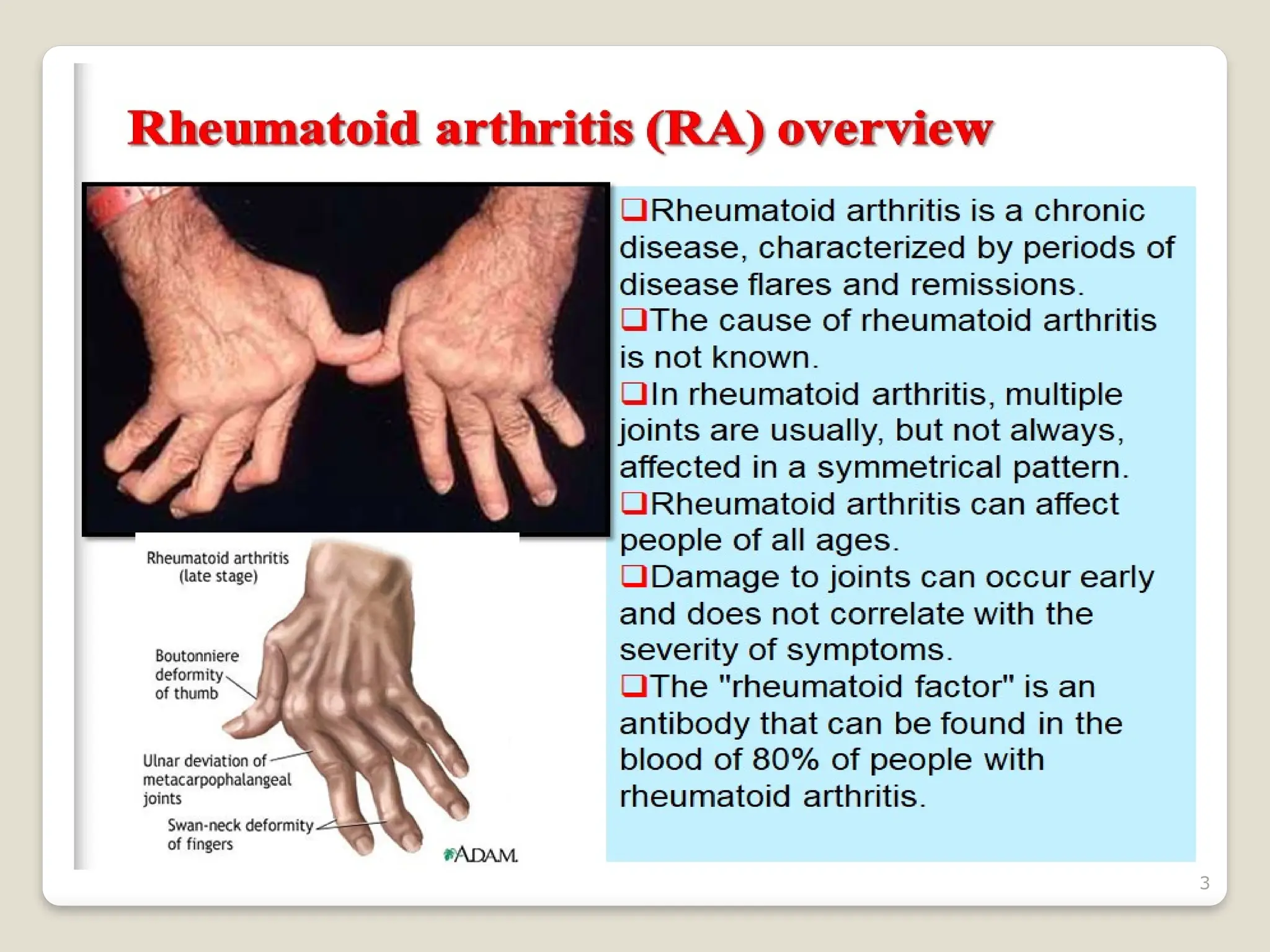
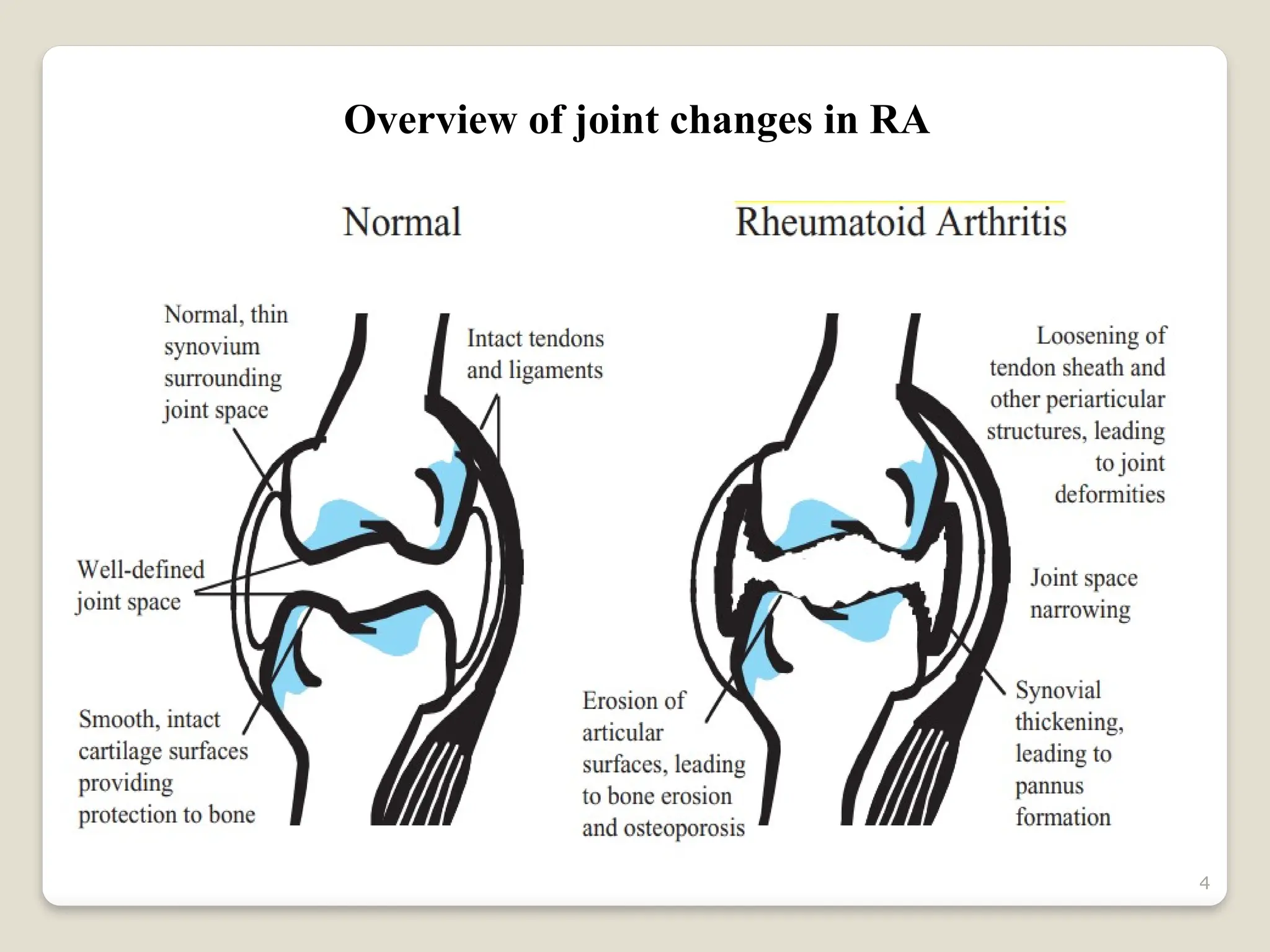
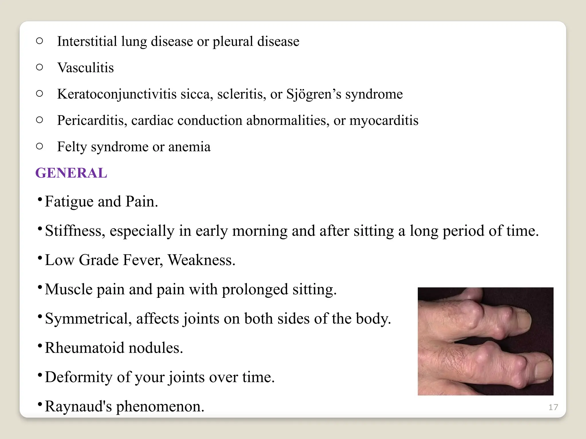
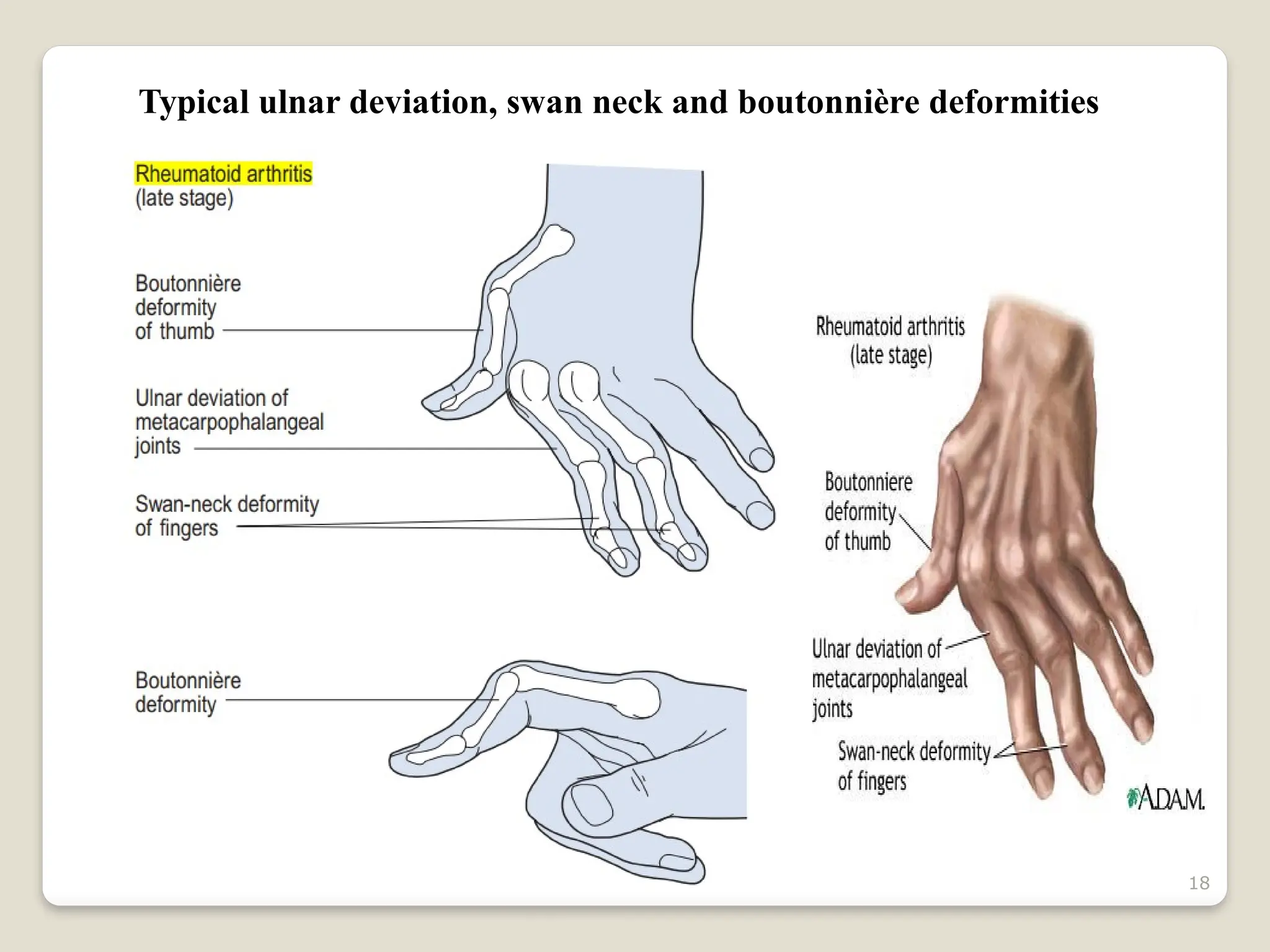
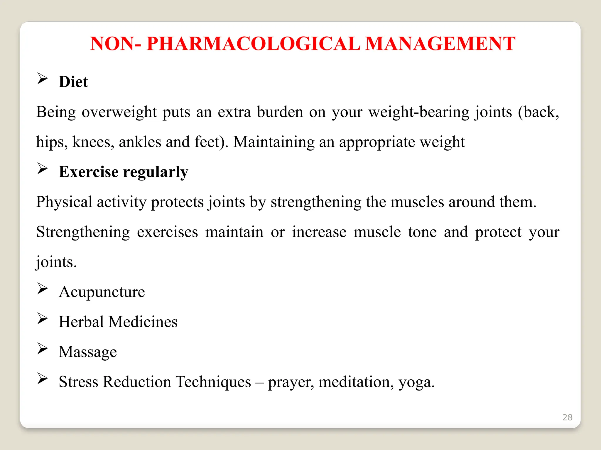
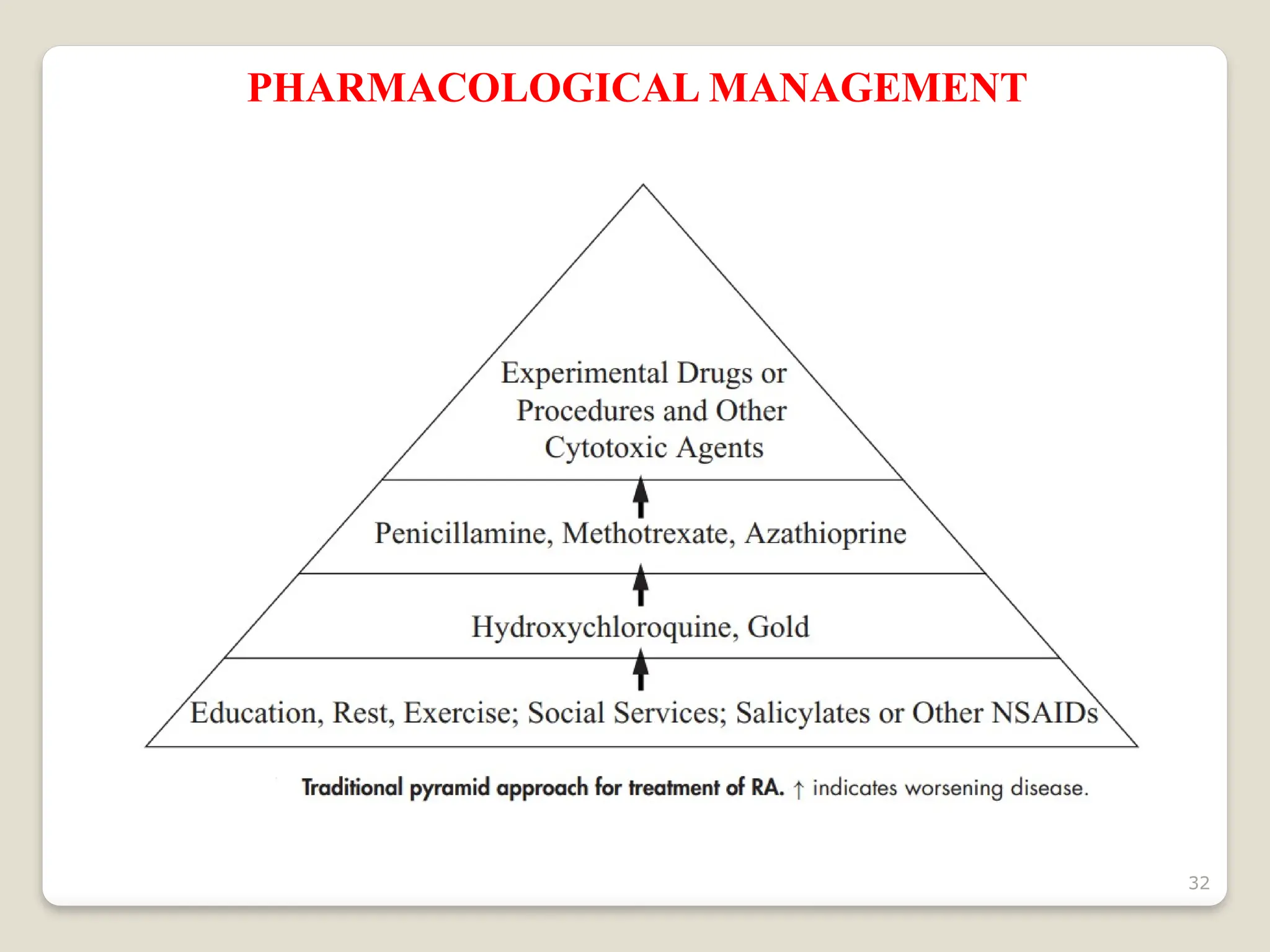
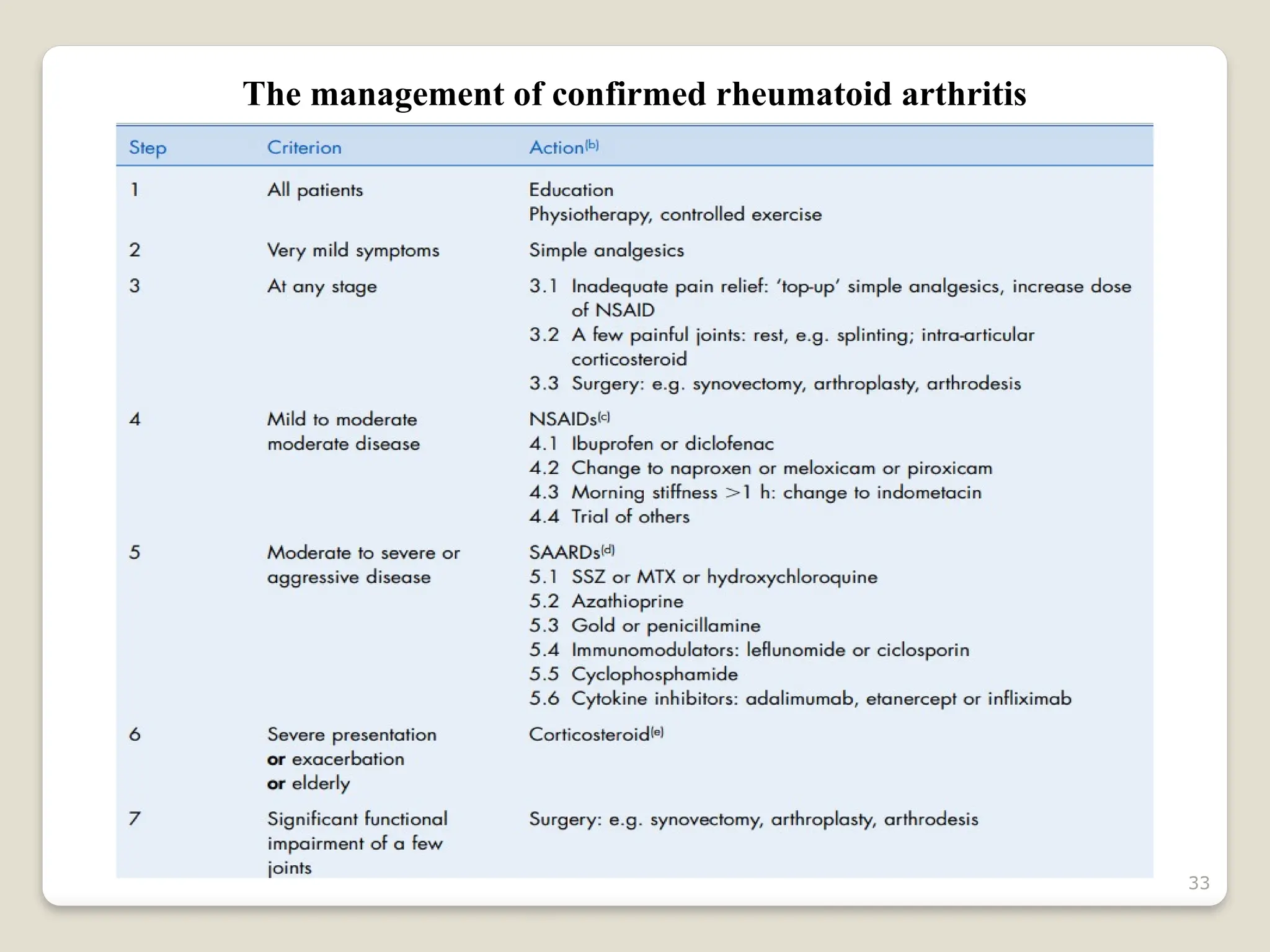
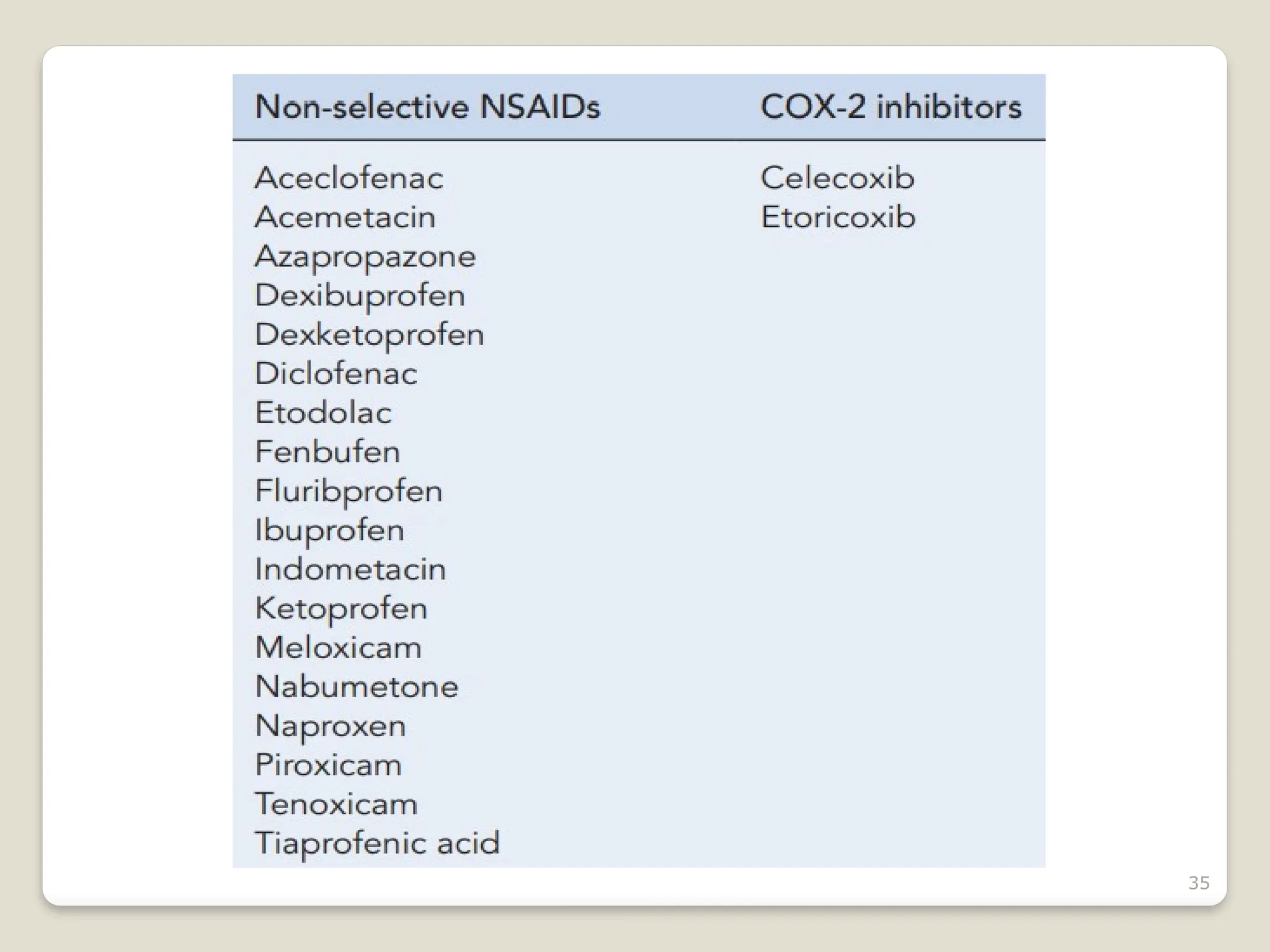
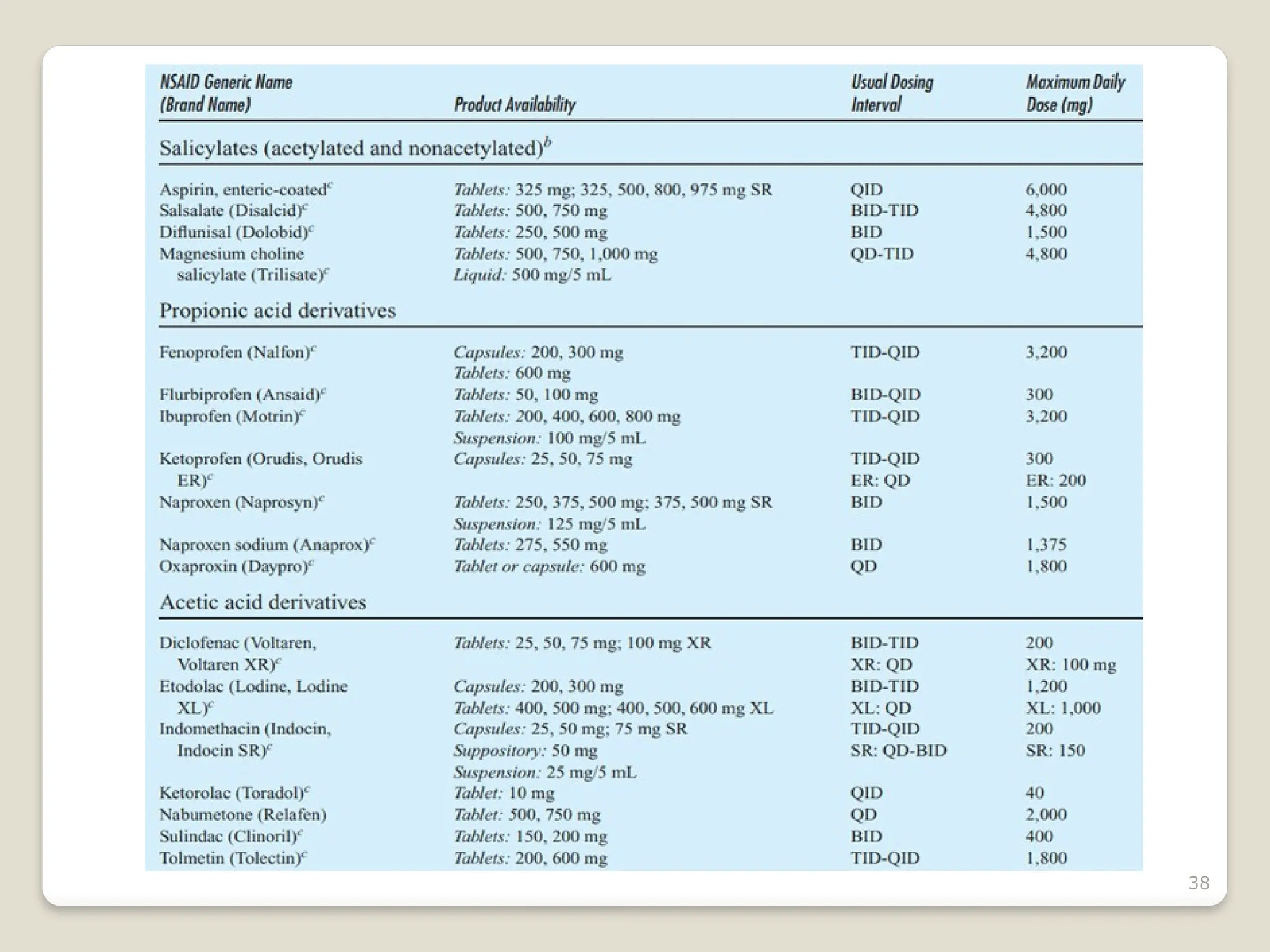
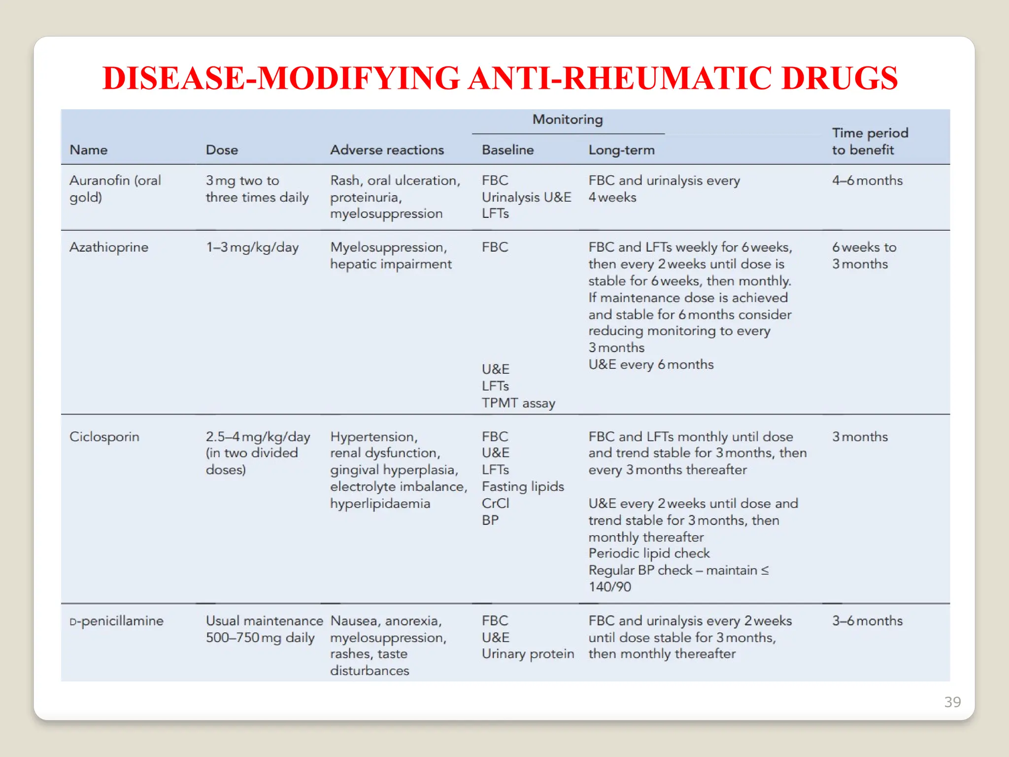
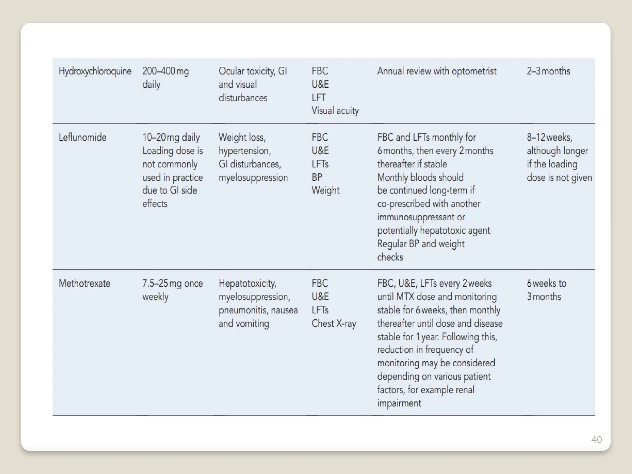
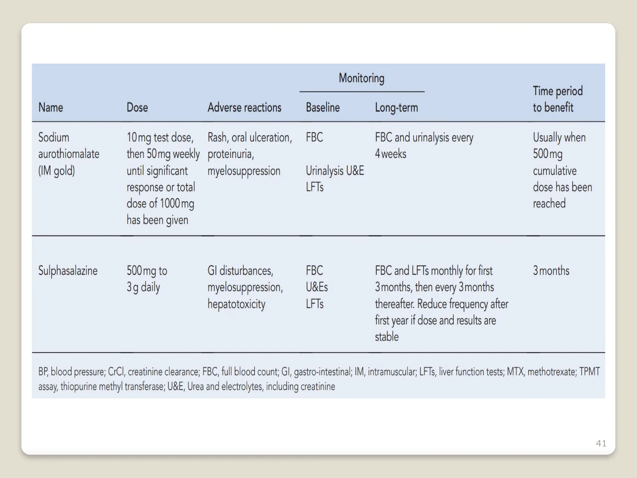
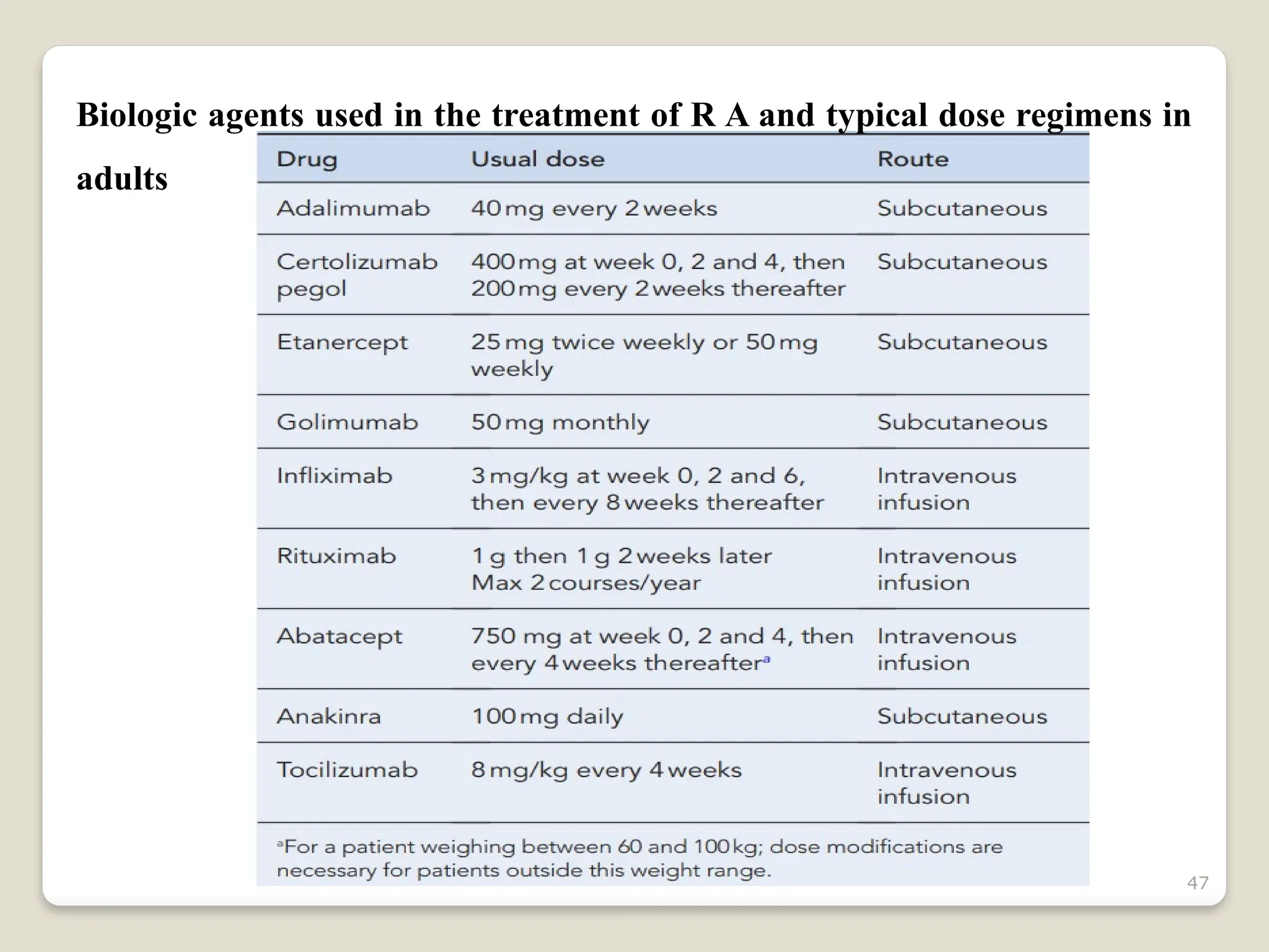
 Rheumatoid arthritis (RA) is a chronic and usually progressive
inflammatory disorder of unknown aetiology characterized by polyarticular
symmetrical joint involvement and systemic manifestations.
 Rheumatoid arthritis (RA) is a chronic, systemic autoimmune disease that
involves inflammation in the membrane lining of the joints and often
affects internal organs.
 It is the most common inflammatory arthritis affecting 1% of the
population.
 It affects 5% of women and 3% of men over 65years of age.
 The disease, if not treated early, will lead to progressive joint deformity
and increased morbidity and mortality.
3
4
Overview of joint changes in RA
5
6
1. SEROPOSITIVE RA
 If your blood tests positive for the protein called rheumatoid
factor (RF) or the antibody anti-cyclic citrullinated peptide (anti-
CCP), it means your body may be actively producing an immune
reaction to your normal tissues.
 Chance of developing RA is four times greater if parents or siblings test
positive for RF.
 According to Johns Hopkins Medicine, approximately 80 percent of
people who have RA are RF-positive.
7
2. SERONEGATIVE RA
 People who test negative for RF and anti-CCP in their blood can still
have RA.
 Diagnosis isn’t based on just these tests.
 Your doctor will also take into account clinical symptoms, X-rays, and
other laboratory tests.
 People who test negative for RF and anti-CCP tend to have a milder form
of RA than those who test positive.
8
3. JUVENILE RA (JUVENILE IDIOPATHIC
ARTHRITIS)
 The Mayo Clinic reports that juvenile RA is the most common type of
arthritis in children younger than age 17.
 Symptoms may be temporary or last for a lifetime.
 Like adult RA, symptoms of juvenile RA include joint inflammation,
stiffness, and pain.
 If the disease is severe, it can cause eye inflammation and interfere with
a child’s growth and development.
9
ETIOLOGY
 The cause of rheumatoid arthritis is unknown.
 It is believed that the tendency to develop rheumatoid arthritis may be
genetically inherited (hereditary).
 Smoking tobacco increases the risk of developing rheumatoid
arthritis.
 Environmental influences, such as infections or trauma, are thought to
trigger the development of RA.
 Genetic markers, such as human leukocyte antigen DR4 (HLA-DR4),
have been associated with triggering the inflammatory process in RA.
 Antigen-dependent activation of T lymphocytes leads to proliferation
of the synovial lining, activation of pro-inflammatory cells from the bone
marrow, cytokine and protease secretion, and autoantibody production.
10
 Anti- Citrullinated Proteins and peptides are high specific for RA.
 Tumour Necrosis Factor & (TNF-&), IL-1, IL-6, IL-8, and growth
factors propagate the inflammatory process, and agents found to alter
these cytokines show promise in reducing pain and deformity.
 Inflamed synovium is a hallmark of the pathophysiology of RA.
 Synovium proliferates abnormally, growing into the joint space and into
the bone, forming a Pannus.
 Pannus is a type of extra growth in your joints that can cause pain,
swelling, and damage to your bones, cartilage, and other tissue.
 The pannus migrates to the articular cartilage and into the sub-chondral
bone leading to destruction of cartilage, bone,
tendons, and blood vessels.
11
PREDISPOSING FACTORS
 Gender:
 Women before the menopause are affected three times more often than
men.
 After the menopause the frequency of onset is similar between the sexes,
suggesting an etiological role for sex hormones.
 The use of the oral contraceptive pill has shown no affect on RA overall, as
previously thought, but it may delay the onset of disease.
Familial:
 The disease is familial with an increased incidence in first degree relatives
and a high concordance amongst monozygotic twins (up to 15%) and
dizygotic twins (3.5%).
 In occasional families it affects several generations.
12
13
PATHOPHYSIOLOGY
 Antigen-presenting cells process and present antigens to T cells, which
may stimulate B cells to produce antibodies and osteoclasts to destroy
and remove bone.
 Macrophages stimulated by the immune response can stimulate T cells
and osteoclasts to promote inflammation.
 They also can stimulate fibroblasts, which produce matrix
metalloproteinase to degrade the bone matrix and produce
proinflammatory cytokines.
 Activated T cells and macrophages release factors that promote tissue
destruction, increase blood flow, and result in cellular invasion of
synovial tissue and joint fluid.
14
 (APC, antigen-presenting cell; IL, interleukin; MMP, matrix metalloproteinase;
TNF-α, tumor necrosis factor α.)
15
16
CLINICAL PRESENTATION
JOINT INVOLVEMENT
o Hands, wrists, ankles, and feet most commonly affected, often bilaterally
o Presence of warmth and swelling with or without pain
o Prolonged morning stiffness, often for longer than 30 minutes in duration
o Decreased functionality
o Symptoms present for 6 weeks or more
o Subluxations and deformities possible with advanced disease
EXTRA-ARTICULAR INVOLVEMENT
o Generalized fatigue, weakness, and decreased mood are nonspecific
implications of disease
o Rheumatoid nodules can be found on extensor or pleural lining surfaces.
17
o Interstitial lung disease or pleural disease
o Vasculitis
o Keratoconjunctivitis sicca, scleritis, or Sjögren’s syndrome
o Pericarditis, cardiac conduction abnormalities, or myocarditis
o Felty syndrome or anemia
GENERAL
•Fatigue and Pain.
•Stiffness, especially in early morning and after sitting a long period of time.
•Low Grade Fever, Weakness.
•Muscle pain and pain with prolonged sitting.
•Symmetrical, affects joints on both sides of the body.
•Rheumatoid nodules.
•Deformity of your joints over time.
•Raynaud's phenomenon.
18
Typical ulnar deviation, swan neck and boutonnière deformities
19
COMPLICATIONS
INFLAMMATORY
Eye involvement is common:
 Sjögren’s syndrome: The syndrome causes dry eyes and a dry mouth,
resulting from lymphocytic infiltration of the lachrymal and salivary
glands.
 Episcleritis: causing a localized or diffuse hyperaemia of the sclera
(white of the eye) is less common.
 Severe scleritis: which involves the deeper layers of the sclera, is
uncommon but more serious.
 Arteritis (vasculitis): may cause widespread obstructive vascular lesions
and is an indication of severe disease. Together with the formation of
myocardial nodules it is also the principal cardiovascular problem.
20
 Respiratory complications reflect diffuse inflammation and include
fibrosing alveolitis and, especially in men, pleurisy and pleural effusions
(‘rheumatoid lung’).
INFECTIVE
 Septic arthritis: due to joint infection by Staphylococcus aureus (in adults
and children) or Haemophilus influenzae (mostly in children), is a rare but
important complication.
SECONDARY TO ABNORMAL METABOLISM
 Mild anaemia occurs in about 80% of cases.
 Osteoporosis may lead to bone fractures.
 Amyloidosis, the widespread deposition in tissues of abnormal amyloid
protein, may occasionally cause clinical problems, notably nephrotic
syndrome
21
DIAGNOSIS
 A clinical diagnosis of rheumatoid arthritis is made based on the patient's
history, presenting symptoms and clinical findings.
 Family history is useful, as well as investigations including blood tests,
ultrasound for the presence of synovitis and X-rays.
 Rheumatoid factor (RF) is an autoantibody directed against the host
immunoglobulin and is most commonly found in rheumatoid arthritis.
 Routinely performed tests only detect immunoglobulin M rheumatoid
factor (IgM RF) which is present in 75–80% of patients with rheumatoid
arthritis (termed seropositive disease) and 5% of normal subjects.
 Those patients who do not have a detectable RF are said to be ‘sero-
negative’.
22
 RF is not specific to rheumatoid arthritis and is also present in patients with
chronic lung and liver disease, other connective tissue diseases, neoplasia,
infections (particularly bacterial endocarditis) and cryoglobulinaemia.
 Anti-cyclic citrullinated peptide antibodies (anti-CCP antibody) are a
more specific test for rheumatoid arthritis with a specificity of 90–96%
compared with the specificity of IgM RF of 85%.
 They are more useful for the early detection of RA in a patient with
inflammatory arthritis.
 The sensitivity of both anti-CCP antibody and IgM RF is approximately
70%.
 Antinuclear antibodies (ANA) and extractable nuclear antigens (ENA)
are useful for establishing the differential diagnosis, such as other
connective tissue diseases presenting or associated with an arthritis.
23
 ANA is almost universally positive in systemic lupus erythematosus and only
positive in 20% of patients with rheumatoid arthritis.
 Other abnormal laboratory tests include an elevated alkaline
phosphatase, an elevated platelet count, a decreased serum albumin
and a normochromic, normocytic anaemia.
 White cell count, particularly neutrophils, is elevated in patients with
infected joints and is also elevated whilst the patient is on steroids.
24
Typical results of investigations in rheumatoid arthritis
25
RADIOGRAPHIC EXAMINATION
 This can reveal the extent of bone erosion and cartilage loss. An MRI can
detect proliferative pannus.
 In early stages of RA, it may show soft-tissue swelling and joint space
narrowing.
 In late-stage disease, it may show joint subluxations, deviations.
26
American Rheumatism Association criteria for the diagnosis
of rheumatoid arthritis
 Morning stiffness in and around the joints for at least 6weeks, lasting at
least 1h before maximal improvement
 Swelling of three or more joints for at least 6weeks
 Swelling of the wrist, metacarpophalangeal or proximal interphalangeal
joints for at least 6weeks
 Symmetric joint swelling for at least 6weeks
 Hand X-ray changes typical of rheumatoid arthritis that must include
erosions or unequivocal bony decalcification around the joints
 Rheumatoid subcutaneous nodules
 Positive rheumatoid factor The presence of at least 4 of these indicates a
diagnosis of rheumatoid arthritis.
27
MANAGEMENT
There are four primary goals in the treatment of rheumatoid arthritis:
 Symptom relief including pain control
 Slowing or prevention of joint damage
 Preserving and improving functional ability
 Achieving and maintaining disease remission
28
NON- PHARMACOLOGICAL MANAGEMENT
 Diet
Being overweight puts an extra burden on your weight-bearing joints (back,
hips, knees, ankles and feet). Maintaining an appropriate weight
 Exercise regularly
Physical activity protects joints by strengthening the muscles around them.
Strengthening exercises maintain or increase muscle tone and protect your
joints.
 Acupuncture
 Herbal Medicines
 Massage
 Stress Reduction Techniques – prayer, meditation, yoga.
29
The most commonly observed vitamin and mineral deficiencies in
patients with RA are:
o Folic acid
o Vitamin C
o Vitamin D
o Vitamin B6
o Vitamin B12
o Vitamin E
o Calcium
o Magnesium
o Zinc
o selenium
30
31
PHARMACOLOGICAL MANAGEMENT
There are four types of medications used to treat RA:
 Non-steroidal anti-inflammatory drugs (NSAIDs)
 Disease-modifying anti-rheumatic drugs (DMARDS).
 Corticosteroids
 Biologic Response Modifiers (“Bioligicals”)
32
PHARMACOLOGICAL MANAGEMENT
33
The management of confirmed rheumatoid arthritis
34
ANTI-INFLAMMATORY DRUGS AND ANALGESICS
 NSAIDs have been the drugs of first choice for the treatment of mild RA
for many years because they possess both analgesic and anti-
inflammatory properties, and many are available
 They are also used as an adjunct to SAARDs (Slow Acting Anti-
Rheumatic Drugs) if symptomatic support is required until benefit is
obtained.
35
36
MOA:
37
38
39
DISEASE-MODIFYING ANTI-RHEUMATIC DRUGS
40
41
42
 Joint damage is known to occur early in rheumatoid arthritis and is
largely irreversible.
 The need for early intervention with DMARDs as part of an aggressive
approach to minimize disease progression has become standard practice
and is associated with better patient outcome.
 Early introduction of DMARDs also results in fewer adverse reactions
and withdrawals from therapy.
 Patients should be made aware that the DMARDs all have a slow onset of
action.
 DMARDs require regular blood monitoring.
43
CORTICOSTEROIDS
 These are the most potent anti-inflammatory agents available and are also
immunosuppressive.
 They produce a dramatic response.
 Mode of action: Corticosteroids down-regulate the production of LTs,
PGs, complement components, interferons, other cytokines and
histamine. However, they are not myelosuppressive because they act on
mature immune cells to prevent B cell and T cell clone proliferation. In
contrast, the anti-proliferative immuno-modulators act on immune cell
precursors in the bone marrow, and incidentally on all other haemopoietic
cells.
44
Side-effects
• Fluid retention and hypertension.
• Weight gain (additional to fluid retention).
• Loss of bone density (osteoporosis) and increased risk of fracture. •
Increased susceptibility to infections, e.g. shingles, chickenpox may be fatal.
• Reduced glucose tolerance.
• Cataract formation and glaucoma.
• Impaired wound healing.
• Loss of SC tissue.
• Cushing’s syndrome.
• Proximal myopathy.
• Steroid psychosis.
45
Interactions
• Corticosteroids reduce blood levels of salicylates.
• Thus, the introduction of steroids in a patient taking aspirin as an analgesic
is illogical.
Dose: 60–100 mg of prednisolone daily PO, or an equivalent IV or IM
injection of methylprednisolone, may be used in the short term for severe
uncontrolled rheumatoid disease or for serious systemic complications, e.g.
vasculitis.
• Pulsed high doses, e.g. up to 1 g of IV methylprednisolone on three
consecutive days, are sometimes used to avoid the corticosteroid
dependence that occurs with gradual progressive or prolonged regimens.
• Prednisolone, 5.0–7.5 mg/day
46
BIOLOGICAL THERAPIES
 The so-called biologics in rheumatoid arthritis are genetically engineered
monoclonal antibodies which selectively target different parts of the
inflammatory pathways.
 Activated T-cells release pro-inflammatory cytokines including TNF-α,
interleukin-1 and interleukin-6.
 Adalimumab, etanercept, golimumab, infliximab and certolizumab pegol
target TNF-α, anakinra and tocilizumab target the interleukins, whilst
abatacept and rituximab act on T-cells and B-cells, respectively.
 In current practice, biologics are used after a patient has failed DMARDs.
47
Biologic agents used in the treatment of R A and typical dose regimens in
adults
48
Biologic agents and their targets in the inflammatory pathway
49
Side Effects
• Increased appetite
• Weight gain
• Water/salt retention
• Increased blood pressure
• Thinning of skin
• Depression
• Mood swings
• Muscle weakness
• Osteoporosis
• Delayed wound healing
• Onset/worsening of diabetes
50
51
REFERENCES
1. Applied Therapeutics, the clinical use of drugs. 9th
Edition. Mary
Anne Koda-Kimble Lloyd Yee Young Brian K. Alldredge Robin L.
Corelli B. Joseph Guglielmo Wayne A. Kradjan Bradley R.
Williams. Page No. 1168-1240.
2. Pharmacotherapy: A Pathophysiologic approach, Joseph T
Dipiro, Thomas D Nolin.., 11th
edition. Page No: 4384-4405.
3. Clinical Pharmacy and Therapeutics. Roger Walker. 5TH
Edition.
Page No. 848-858.
4. Pathology and Therapeutics for Pharmacists: A basis for clinical
pharmacy practice, Third edition. Russell J Greene. Page No. 748-
787.
52

rheumatoidarthritis.pptxLLLLLLLLLLLLLLLLLLL

  • 1.
  • 2.
    2 RHEUMATOID ARTHRITIS  Rheumatoidarthritis (RA) is a chronic and usually progressive inflammatory disorder of unknown aetiology characterized by polyarticular symmetrical joint involvement and systemic manifestations.  Rheumatoid arthritis (RA) is a chronic, systemic autoimmune disease that involves inflammation in the membrane lining of the joints and often affects internal organs.  It is the most common inflammatory arthritis affecting 1% of the population.  It affects 5% of women and 3% of men over 65years of age.  The disease, if not treated early, will lead to progressive joint deformity and increased morbidity and mortality.
  • 3.
  • 4.
    4 Overview of jointchanges in RA
  • 5.
  • 6.
    6 1. SEROPOSITIVE RA If your blood tests positive for the protein called rheumatoid factor (RF) or the antibody anti-cyclic citrullinated peptide (anti- CCP), it means your body may be actively producing an immune reaction to your normal tissues.  Chance of developing RA is four times greater if parents or siblings test positive for RF.  According to Johns Hopkins Medicine, approximately 80 percent of people who have RA are RF-positive.
  • 7.
    7 2. SERONEGATIVE RA People who test negative for RF and anti-CCP in their blood can still have RA.  Diagnosis isn’t based on just these tests.  Your doctor will also take into account clinical symptoms, X-rays, and other laboratory tests.  People who test negative for RF and anti-CCP tend to have a milder form of RA than those who test positive.
  • 8.
    8 3. JUVENILE RA(JUVENILE IDIOPATHIC ARTHRITIS)  The Mayo Clinic reports that juvenile RA is the most common type of arthritis in children younger than age 17.  Symptoms may be temporary or last for a lifetime.  Like adult RA, symptoms of juvenile RA include joint inflammation, stiffness, and pain.  If the disease is severe, it can cause eye inflammation and interfere with a child’s growth and development.
  • 9.
    9 ETIOLOGY  The causeof rheumatoid arthritis is unknown.  It is believed that the tendency to develop rheumatoid arthritis may be genetically inherited (hereditary).  Smoking tobacco increases the risk of developing rheumatoid arthritis.  Environmental influences, such as infections or trauma, are thought to trigger the development of RA.  Genetic markers, such as human leukocyte antigen DR4 (HLA-DR4), have been associated with triggering the inflammatory process in RA.  Antigen-dependent activation of T lymphocytes leads to proliferation of the synovial lining, activation of pro-inflammatory cells from the bone marrow, cytokine and protease secretion, and autoantibody production.
  • 10.
    10  Anti- CitrullinatedProteins and peptides are high specific for RA.  Tumour Necrosis Factor & (TNF-&), IL-1, IL-6, IL-8, and growth factors propagate the inflammatory process, and agents found to alter these cytokines show promise in reducing pain and deformity.  Inflamed synovium is a hallmark of the pathophysiology of RA.  Synovium proliferates abnormally, growing into the joint space and into the bone, forming a Pannus.  Pannus is a type of extra growth in your joints that can cause pain, swelling, and damage to your bones, cartilage, and other tissue.  The pannus migrates to the articular cartilage and into the sub-chondral bone leading to destruction of cartilage, bone, tendons, and blood vessels.
  • 11.
    11 PREDISPOSING FACTORS  Gender: Women before the menopause are affected three times more often than men.  After the menopause the frequency of onset is similar between the sexes, suggesting an etiological role for sex hormones.  The use of the oral contraceptive pill has shown no affect on RA overall, as previously thought, but it may delay the onset of disease. Familial:  The disease is familial with an increased incidence in first degree relatives and a high concordance amongst monozygotic twins (up to 15%) and dizygotic twins (3.5%).  In occasional families it affects several generations.
  • 12.
  • 13.
    13 PATHOPHYSIOLOGY  Antigen-presenting cellsprocess and present antigens to T cells, which may stimulate B cells to produce antibodies and osteoclasts to destroy and remove bone.  Macrophages stimulated by the immune response can stimulate T cells and osteoclasts to promote inflammation.  They also can stimulate fibroblasts, which produce matrix metalloproteinase to degrade the bone matrix and produce proinflammatory cytokines.  Activated T cells and macrophages release factors that promote tissue destruction, increase blood flow, and result in cellular invasion of synovial tissue and joint fluid.
  • 14.
    14  (APC, antigen-presentingcell; IL, interleukin; MMP, matrix metalloproteinase; TNF-α, tumor necrosis factor α.)
  • 15.
  • 16.
    16 CLINICAL PRESENTATION JOINT INVOLVEMENT oHands, wrists, ankles, and feet most commonly affected, often bilaterally o Presence of warmth and swelling with or without pain o Prolonged morning stiffness, often for longer than 30 minutes in duration o Decreased functionality o Symptoms present for 6 weeks or more o Subluxations and deformities possible with advanced disease EXTRA-ARTICULAR INVOLVEMENT o Generalized fatigue, weakness, and decreased mood are nonspecific implications of disease o Rheumatoid nodules can be found on extensor or pleural lining surfaces.
  • 17.
    17 o Interstitial lungdisease or pleural disease o Vasculitis o Keratoconjunctivitis sicca, scleritis, or Sjögren’s syndrome o Pericarditis, cardiac conduction abnormalities, or myocarditis o Felty syndrome or anemia GENERAL •Fatigue and Pain. •Stiffness, especially in early morning and after sitting a long period of time. •Low Grade Fever, Weakness. •Muscle pain and pain with prolonged sitting. •Symmetrical, affects joints on both sides of the body. •Rheumatoid nodules. •Deformity of your joints over time. •Raynaud's phenomenon.
  • 18.
    18 Typical ulnar deviation,swan neck and boutonnière deformities
  • 19.
    19 COMPLICATIONS INFLAMMATORY Eye involvement iscommon:  Sjögren’s syndrome: The syndrome causes dry eyes and a dry mouth, resulting from lymphocytic infiltration of the lachrymal and salivary glands.  Episcleritis: causing a localized or diffuse hyperaemia of the sclera (white of the eye) is less common.  Severe scleritis: which involves the deeper layers of the sclera, is uncommon but more serious.  Arteritis (vasculitis): may cause widespread obstructive vascular lesions and is an indication of severe disease. Together with the formation of myocardial nodules it is also the principal cardiovascular problem.
  • 20.
    20  Respiratory complicationsreflect diffuse inflammation and include fibrosing alveolitis and, especially in men, pleurisy and pleural effusions (‘rheumatoid lung’). INFECTIVE  Septic arthritis: due to joint infection by Staphylococcus aureus (in adults and children) or Haemophilus influenzae (mostly in children), is a rare but important complication. SECONDARY TO ABNORMAL METABOLISM  Mild anaemia occurs in about 80% of cases.  Osteoporosis may lead to bone fractures.  Amyloidosis, the widespread deposition in tissues of abnormal amyloid protein, may occasionally cause clinical problems, notably nephrotic syndrome
  • 21.
    21 DIAGNOSIS  A clinicaldiagnosis of rheumatoid arthritis is made based on the patient's history, presenting symptoms and clinical findings.  Family history is useful, as well as investigations including blood tests, ultrasound for the presence of synovitis and X-rays.  Rheumatoid factor (RF) is an autoantibody directed against the host immunoglobulin and is most commonly found in rheumatoid arthritis.  Routinely performed tests only detect immunoglobulin M rheumatoid factor (IgM RF) which is present in 75–80% of patients with rheumatoid arthritis (termed seropositive disease) and 5% of normal subjects.  Those patients who do not have a detectable RF are said to be ‘sero- negative’.
  • 22.
    22  RF isnot specific to rheumatoid arthritis and is also present in patients with chronic lung and liver disease, other connective tissue diseases, neoplasia, infections (particularly bacterial endocarditis) and cryoglobulinaemia.  Anti-cyclic citrullinated peptide antibodies (anti-CCP antibody) are a more specific test for rheumatoid arthritis with a specificity of 90–96% compared with the specificity of IgM RF of 85%.  They are more useful for the early detection of RA in a patient with inflammatory arthritis.  The sensitivity of both anti-CCP antibody and IgM RF is approximately 70%.  Antinuclear antibodies (ANA) and extractable nuclear antigens (ENA) are useful for establishing the differential diagnosis, such as other connective tissue diseases presenting or associated with an arthritis.
  • 23.
    23  ANA isalmost universally positive in systemic lupus erythematosus and only positive in 20% of patients with rheumatoid arthritis.  Other abnormal laboratory tests include an elevated alkaline phosphatase, an elevated platelet count, a decreased serum albumin and a normochromic, normocytic anaemia.  White cell count, particularly neutrophils, is elevated in patients with infected joints and is also elevated whilst the patient is on steroids.
  • 24.
    24 Typical results ofinvestigations in rheumatoid arthritis
  • 25.
    25 RADIOGRAPHIC EXAMINATION  Thiscan reveal the extent of bone erosion and cartilage loss. An MRI can detect proliferative pannus.  In early stages of RA, it may show soft-tissue swelling and joint space narrowing.  In late-stage disease, it may show joint subluxations, deviations.
  • 26.
    26 American Rheumatism Associationcriteria for the diagnosis of rheumatoid arthritis  Morning stiffness in and around the joints for at least 6weeks, lasting at least 1h before maximal improvement  Swelling of three or more joints for at least 6weeks  Swelling of the wrist, metacarpophalangeal or proximal interphalangeal joints for at least 6weeks  Symmetric joint swelling for at least 6weeks  Hand X-ray changes typical of rheumatoid arthritis that must include erosions or unequivocal bony decalcification around the joints  Rheumatoid subcutaneous nodules  Positive rheumatoid factor The presence of at least 4 of these indicates a diagnosis of rheumatoid arthritis.
  • 27.
    27 MANAGEMENT There are fourprimary goals in the treatment of rheumatoid arthritis:  Symptom relief including pain control  Slowing or prevention of joint damage  Preserving and improving functional ability  Achieving and maintaining disease remission
  • 28.
    28 NON- PHARMACOLOGICAL MANAGEMENT Diet Being overweight puts an extra burden on your weight-bearing joints (back, hips, knees, ankles and feet). Maintaining an appropriate weight  Exercise regularly Physical activity protects joints by strengthening the muscles around them. Strengthening exercises maintain or increase muscle tone and protect your joints.  Acupuncture  Herbal Medicines  Massage  Stress Reduction Techniques – prayer, meditation, yoga.
  • 29.
    29 The most commonlyobserved vitamin and mineral deficiencies in patients with RA are: o Folic acid o Vitamin C o Vitamin D o Vitamin B6 o Vitamin B12 o Vitamin E o Calcium o Magnesium o Zinc o selenium
  • 30.
  • 31.
    31 PHARMACOLOGICAL MANAGEMENT There arefour types of medications used to treat RA:  Non-steroidal anti-inflammatory drugs (NSAIDs)  Disease-modifying anti-rheumatic drugs (DMARDS).  Corticosteroids  Biologic Response Modifiers (“Bioligicals”)
  • 32.
  • 33.
    33 The management ofconfirmed rheumatoid arthritis
  • 34.
    34 ANTI-INFLAMMATORY DRUGS ANDANALGESICS  NSAIDs have been the drugs of first choice for the treatment of mild RA for many years because they possess both analgesic and anti- inflammatory properties, and many are available  They are also used as an adjunct to SAARDs (Slow Acting Anti- Rheumatic Drugs) if symptomatic support is required until benefit is obtained.
  • 35.
  • 36.
  • 37.
  • 38.
  • 39.
  • 40.
  • 41.
  • 42.
    42  Joint damageis known to occur early in rheumatoid arthritis and is largely irreversible.  The need for early intervention with DMARDs as part of an aggressive approach to minimize disease progression has become standard practice and is associated with better patient outcome.  Early introduction of DMARDs also results in fewer adverse reactions and withdrawals from therapy.  Patients should be made aware that the DMARDs all have a slow onset of action.  DMARDs require regular blood monitoring.
  • 43.
    43 CORTICOSTEROIDS  These arethe most potent anti-inflammatory agents available and are also immunosuppressive.  They produce a dramatic response.  Mode of action: Corticosteroids down-regulate the production of LTs, PGs, complement components, interferons, other cytokines and histamine. However, they are not myelosuppressive because they act on mature immune cells to prevent B cell and T cell clone proliferation. In contrast, the anti-proliferative immuno-modulators act on immune cell precursors in the bone marrow, and incidentally on all other haemopoietic cells.
  • 44.
    44 Side-effects • Fluid retentionand hypertension. • Weight gain (additional to fluid retention). • Loss of bone density (osteoporosis) and increased risk of fracture. • Increased susceptibility to infections, e.g. shingles, chickenpox may be fatal. • Reduced glucose tolerance. • Cataract formation and glaucoma. • Impaired wound healing. • Loss of SC tissue. • Cushing’s syndrome. • Proximal myopathy. • Steroid psychosis.
  • 45.
    45 Interactions • Corticosteroids reduceblood levels of salicylates. • Thus, the introduction of steroids in a patient taking aspirin as an analgesic is illogical. Dose: 60–100 mg of prednisolone daily PO, or an equivalent IV or IM injection of methylprednisolone, may be used in the short term for severe uncontrolled rheumatoid disease or for serious systemic complications, e.g. vasculitis. • Pulsed high doses, e.g. up to 1 g of IV methylprednisolone on three consecutive days, are sometimes used to avoid the corticosteroid dependence that occurs with gradual progressive or prolonged regimens. • Prednisolone, 5.0–7.5 mg/day
  • 46.
    46 BIOLOGICAL THERAPIES  Theso-called biologics in rheumatoid arthritis are genetically engineered monoclonal antibodies which selectively target different parts of the inflammatory pathways.  Activated T-cells release pro-inflammatory cytokines including TNF-α, interleukin-1 and interleukin-6.  Adalimumab, etanercept, golimumab, infliximab and certolizumab pegol target TNF-α, anakinra and tocilizumab target the interleukins, whilst abatacept and rituximab act on T-cells and B-cells, respectively.  In current practice, biologics are used after a patient has failed DMARDs.
  • 47.
    47 Biologic agents usedin the treatment of R A and typical dose regimens in adults
  • 48.
    48 Biologic agents andtheir targets in the inflammatory pathway
  • 49.
    49 Side Effects • Increasedappetite • Weight gain • Water/salt retention • Increased blood pressure • Thinning of skin • Depression • Mood swings • Muscle weakness • Osteoporosis • Delayed wound healing • Onset/worsening of diabetes
  • 50.
  • 51.
    51 REFERENCES 1. Applied Therapeutics,the clinical use of drugs. 9th Edition. Mary Anne Koda-Kimble Lloyd Yee Young Brian K. Alldredge Robin L. Corelli B. Joseph Guglielmo Wayne A. Kradjan Bradley R. Williams. Page No. 1168-1240. 2. Pharmacotherapy: A Pathophysiologic approach, Joseph T Dipiro, Thomas D Nolin.., 11th edition. Page No: 4384-4405. 3. Clinical Pharmacy and Therapeutics. Roger Walker. 5TH Edition. Page No. 848-858. 4. Pathology and Therapeutics for Pharmacists: A basis for clinical pharmacy practice, Third edition. Russell J Greene. Page No. 748- 787.
  • 52.