Everything You Need to Know About Spondyloarthropathies
•Download as PPT, PDF•
1 like•271 views

spondyloarthropathies are group of disorders which impact spine.

Recommended
More Related Content
What's hot
What's hot (20)
Similar to Everything You Need to Know About Spondyloarthropathies
Similar to Everything You Need to Know About Spondyloarthropathies (20)
More from drshyamsundersharma
More from drshyamsundersharma (7)
Recently uploaded
Recently uploaded (20)
Everything You Need to Know About Spondyloarthropathies
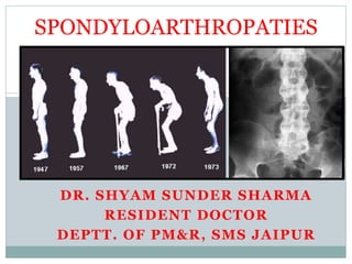
- 1. SPONDYLOARTHROPATIES DR. SHYAM SUNDER SHARMA RESIDENT DOCTOR DEPTT. OF PM&R, SMS JAIPUR
- 2. HISTORICAL BACKGROUND 1892 Bechterew- upper dorsal type case complicated by meningeal and spinal cord involvment “Bechterew’s disease” 1898 Marie- described the entity as separate from all other types of arthritis involving the spine
- 3. HISTORICAL BACKGROUND 1940-50s Discovery of the human leukocyte antigens (HLAs) and characterization of the MHCs 1960s- American Rheumatism Association (ARA) recognized the spondyloarthropathies as a distinct group of inflammatory arthritis 1973 Association found between HLA-B27 and the spondyloarthropathies
- 4. SPONDYLOARTHRITIDES These are interrelated group of rhematic diseases that are characterized by clinical features as inflammatory back pain, Asymmetric peripheral oligoarthritis, predominantly of lower limbs. Enthesitis. specific organ involvement such as anterior uveitis, psoriasis. chronic inflammatory bowel disease.
- 5. Juvenile Juvenile SpA SpA Reactive arthritis Reactive arthritis Reiter syndrome Reiter syndrome Arthritis Arthritis associated associated with with Crohn Crohn’ ’s s disease / UC disease / UC Psoriatic Psoriatic Arthritis Arthritis Ankylosing Ankylosing Spondylitis Spondylitis Sacroiliitis Sacroiliitis AAU Undifferentiated Undifferentiated SpA SpA Spondyloarthritides: The Spectrum IAVB
- 6. Group of diseases characterized by: SpAs comprise group of related inflammatory musculoskeletal diseases that show overlap in their clinical features and have a shared immunogenic association with HLA-27. Spondyloarthropathies: Definition Occurring in the absence of serum rheumatoid factor
- 7. I T I N C L U D E S ; •ANKYLOSING SPONDYLITIS (AS) •REACTIVE ARTHRITIS (RA) •PSORIATIC ARTHRITIS (PA) •ENTEROPATHIC SPONDYLOARTHRITIS •AXIAL SPONDYLOARTHRITIS Spondyloarthropathies
- 8. ESSG* Classification Criteria for SpA (*European Spondylarthropathy Study Group) Alternating buttock pain Sacroiliitis (x-rays) * Positive family history Psoriasis Inflammatory bowel disease Urethritis / acute diarrhea in the preceding 4 weeks Heel pain (enthesitis) Inflammatory Back Pain Synovitis Plus one of the following: • Asymmetrical • Lower extremities Dougados M, et al. Arthritis Rheum 1991;34:1218 *without sacroiliitis: sens =77%, spec 89% OR
- 9. ENTHESITIS The characteristic feature of AS and spondyloarthritides Inflammation at sites where tendons, ligaments or joint capsules attaches to the bone: primarily affects the sacroiliac joints & the axial skeleton in AS, but extra- articular or juxta-articular bony tenderness occurs from enthesitis at many other sites (e.g. costosternal junctions, spinous processes, greater femoral trochanters, iliac crests, ischial tuberosities, tibial tubercles, heels, etc.) Khan MA; Rheumatology. Eds. Hochberg M. London, Mosby. 3rd ed. 2003
- 10. Enthesitis Periosteal new bone formation Bone McGonagle D. Arthritis Rheum. 1999;42:1080-1086. Subchondral bone inflammation and resorption Tendon ©ACR Inflammatory Rheumatoid arthritis Ankylosing spondylitis Reactive arthritis Psoriatic arthritis Inflammatory bowel disease Lyme disease Late-onset Pauciarticular JRA Leprosy Mechanical/Degenerative Trauma Osteoarthritis Metabolic/Endocrine DISH Acromegaly Fluorosis Retinoid therapy Hypoparathyroidism Hyperparathyroidism POEMS syndrome X-linked hypophosphatemia DDx:
- 11. Seronegative spondyloarthropathy (or seronegative spondyloarthritis) is a group of diseases involving the axial skeleton and having a negative serostatus. "Seronegative" refers to the fact that these diseases are negative for rheumatoid factor , indicating a different patho- physiological mechanism of disease than what is commonly seen in rheumatoid arthritis. Seronegative spondyloarthropathy
- 12. The following conditions are typically included within the group of seronegative spondylarthropathies: Ankylosing Spondylitis Caucasians: 92% African-Americans: 50% Reactive arthritis (Reiter's syndrome) - 60 -80% Enteropathic spondylitis or spondylitis associated with inflammatory bowel disease (including Crohn's disease and ulcerative colitis)- 60%
- 13. Psoriatic arthritis -60% Isolated acute anterior uveitis -50% Undifferentiated spondyloarthropathy (USpA)- 20-25% Some sources also include Behcet's disease and Whipple's disease
- 14. These diseases have the following conditions in common: They are in relation to HLA-B27 Inflammatory arthritis,generally sacroiliitis and spondylitis Oligoarthritis, generally with asymmetrical presentation Enthesitis (inflammation of the entheses, the sites where tendons or ligaments insert into the bone)
- 15. These diseases have the following conditions in common: familial aggregation occurs rheumatoid factor is not present Extra-articular features, such as involvement of eyes, skin and genitourinary tract Overlap is likely between several of the causative conditions
- 20. Pathophysiology The primary pathology of the spondyloarthropathies is – Enthesitis with chronic inflammation, including CD4+ and CD8+ T lymphocytes and macrophages. Cytokines, particularly tumor necrosis factor-α (TNF-α) and transforming growth factor-β (TGF-β), are also important in the inflammatory process by leading to fibrosis and ossification at sites of enthesitis.
- 21. Pathogenesis Etiology/pathogenesis unclear Interplay of genetic, immunologic and environmental factors Interaction between MHC-I molecule HLA-B27 with T cell response believed to be key in pathogenesis. 94% of AS patients are HLA-B27+(OR 161 [CI 113-230]) Bacterial infections may trigger events M Tc HLAB27 Intense Inflammatory response Ag TCR McMichael A and Bowness P. Arthritis Res 2002;4 (suppl 3):S153-8.
- 22. Role of HLA-B27 Has a role in presenting protein antigens that have been synthesized in the cell (viral, tumor, self-derived) to cytotoxic T-cells HLA-B27 individuals are less efficient at elimination of intracellular organisms (e.g, Chlamydia) and certain enteric bacteria Unclear if disease susceptibility is due to presentation of an arthritogenic peptide vs. failure to eliminate intracellular organisms vs. itself acting as a source of Ag 25 different subtypes have been identified: B*2705, B*2704, B*2702 or B*2707 have accounted for most of the spondyloarthritides McMichael A and Bowness P. Arthritis Res 2002;4 (suppl 3):S153-8.
- 23. Ankylosing Spondylitis Rheumatoid Spondylitis, Spondylarthropathy, Spondylitis, Bechterew’s Disease, Bamboo Spine
- 24. Etiology Spondylitis – inflammation of the spine Ankylosing – fusion of the spine Chronic inflammation of the spine and SI joints Combination of genetic and environmental factors Genetic: Presence of HLA-B27 gene 5% of population has gene 90% of those with AS have gene
- 25. Incidence 3 males to every 1 female Worldwide annual incidence estimated to be 7.3 per 100,000 individuals US incidence in ages 16 and older: 8.9 per 100,000 individuals Prevalence in US is between 0.1% and 1.4%
- 26. Risk Factors Men > Women; 2-3 x more likely Family History; 6 x more likely Age 17-45 Presence of HLA-B27 gene detected in blood • Current research focuses on ARTS1 and IL23R, which play a role in immune function Frequent GI infections
- 27. The initial presentation of AS generally occurs in the SI joints; involvement of the SI joints is required to establish the diagnosis. SI joint involvement is followed by involvement of the diskovertebral, apophyseal, costovertebral, and costotransverse joints and the paravertebral ligaments. Early lesions include subchondral granulation tissue that erodes the joint and is replaced gradually by fibrocartilage and then ossification. This occurs in ligamentous and capsular attachment sites to bone and is called enthesitis. In the spine, this initial process occurs at the junction of the vertebrae and the anulus fibrosus of the intervertebral discs. The outer fibers of the discs eventually undergo ossification to form syndesmophytes. The condition progresses to the characteristic bamboo spine appearance.
- 28. Diagnosis of AS Modified New York criteria A definite diagnosis Radiological criterion and ≥ 1 clinical criterion Radiological criterion Sacroiliitis ≥ grade 2 bilaterally or grade 3 or 4 unilaterally. Clinical criteria Low back pain and stiffness for more than 3 months improves with exercise, not relieved by rest Limitation of motion of the lumbar spine in both the sagittal and frontal planes Limitation of chest expansion (correlated for age and sex)
- 29. Bath Ankylosing Spondylitis Functional Index (BASFI) Standing unsupported for 10 minutes without discomfort Climbing 12-15 steps without use of handrail or walking aid Looking over your shoulder without turning your body Doing physically demanding activities Doing a full days activities at home or at work
- 30. Ankylosing Spondylitis Differentiating Inflammatory vs Mechanical Back Pain Inflammatory Back Pain Features Mechanical Back Pain Prolonged > 60min. AM Stiffness Minor < 45 min. Early AM (wakes pt up) Max. Pain/Stiffness Late in day Improves Symptoms Exercise/activity Worsens Symptoms Chronic Duration Acute or Chronic 9-40 yrs. Age at Onset 20-65 yrs. sacroiliitis vertebral ankylosis syndesmophytes Radiographs osteophytes spondylolisthesis scoliosis
- 32. Imaging Radiograph (L-spine, AP pelvis, SI joints) Grade 2-4 sacroiliitis Equivocal Grade I sacroiliitis Normal MRI CT or MRI Bone scan Equivocal Multifocal disease suspected No further studies Koehler et al. Rheumatolgy 2000;39;360-8.
- 33. AS Clinical Features - axial Early AS Romanus lesion (This reflects subdiskal and marginal destruction of the vertebral ring) Advanced AS bony ankylosis
- 34. Severe Spinal Complications from AS Spinal stiffness/ankylosis in kyphotic position Spinal fractures (10- 20%) axial/T spine; increased 6-8 fold Cauda equina synd. (arachnoiditis)
- 45. Fused SI joints
- 46. Schober’s test
- 47. Lumbar Flexion (Schober) A mark is placed at L5, measure 10 cm cephalad and place another mark. Have the patient bends forward as far as possible with knees extended, the difference is recorded. Normal is >5 cm J Brandt, J Sieper
- 48. Wall to Tragus Distance Patient stands, heels and buttocks against the wall, the head is placed back as far as possible, keeping the chin horizontal J Brandt, J Sieper
- 51. Ocular (25-40%): uveitis, conjunctivitis Heart (29%): AI, heart block, aortitis Lung (10%): ILD, apical fibrosis, aspergilloma Kidney (14%): amyloidosis, nephritis, IgA nephropathy Bones: osteoporosis – bone resorption > bone formation Associated Extraarticular Features
- 52. Associated Extraarticular Features Gastrointestinal: oral ulcerations, asymptomatic gut inflammation, symptomatic colitis Genitourinary: urethritis, vaginitis, balanitis Cutaneous: keratoderma blennorrhagicum, psoriasis or nail lesions (onycholysis, dystrophy, pitting). Periarticular: tendonitis, dactylitis (sausage-digit)
- 53. Management Medication Exercise Posture Heat/Cold Surgery Other Symptom Management Tools • Acupuncture • Chiropractic Treatment • Massage • Yoga Therapy
- 55. Exercise Exercise is an integral part of any spondylitis program, along with good posture habits and medication to reduce pain and stiffness. Fitting exercise into your day can be tough, but it needs to be done. Exercise is such a high priority that it is important to make time for it each day (even 5-10 minutes during a work break is helpful). If you do, many benefits will result from your efforts. A spondylitis exercise program will help you maintain good posture, flexibility and eventually help to lessen pain. In many cases, good posture and mobility can even be regained with proper doses of medicine and exercise. Most people with spondylitis feel much better with exercise. The trick is to do enough but not too much. This can vary from day to day. Be good to yourself and never push to the point of pain or extreme fatigue.
- 56. Osteoporosis in AS Prevalence of vertebral osteoporosis in AS is between 20% to 60%1 Relative risk of fractures is 6 times in early AS compared with controls2 Risk factors: disease duration, severity, male sex Major etiologic factors: pro-inflammatory cytokines and spinal immobility No studies on treatment of osteoporosis in AS: ? role of pamidronate, anti-TNF 1. Bessant R, Keat A. J Rheumatol. 2002;29:1511-1519. 2. Mitra D, et al. Rheumatology. 2000;39:85-89.
- 58. PSORIATIC ARTHRITIS (PsA) Chronic inflammatory arthropathy in setting of psoriasis Sites of psoriasis: scalp, ears, gluteal fold, umbilicus, perineum, palms, soles, nails, extremities Nail changes: pitting, dystrophy, onycholysis 1-5% of US population has Psoriasis: 5-42% of these develop psoriatic arthritis
- 59. PSORIATIC ARTHRITIS (PsA) Frequency of PsA increases with disease severity and duration (estimated 350-400,000 patients in USA) Skin precedes nails, but 15% will have arthritis before skin disease Course: chronic, destructive arthritis in 30-50%
- 60. PsA Up to 40% of psoriasis patients develop IA Peak 35 to 50 years Male = female (spine involvement 3:1) 1/3 of cases arthritis precedes rash No association between severity of joint disease and psoriasis. Nail changes more commonly associated with arthritis
- 62. Classification of Psoriatic Arthritis 4/25/2021 1:30:23 PM 69 Type Key Clinical Features Incidence Asymmetric polyarthritis or oligoarthritis Morning stiffness, DIP and PIP involvement, nail disease, 4 joints involved 40% Symmetric polyarthritis Symmetric polyarthritis, RA-like distribution, but RF negative 25% Spondylitis Inflammatory low back pain, sacroilitis, axial involvement, 50% HLA-B27+ 20% Distal interphalangeal joint disease Nail changes, often bilateral joint involvement 15% Arthritis mutilans Destructive form of arthritis, telescoping digits, joint lysis, typically in phalanges and metacarpals <5%
- 64. Radiological changes 1. Soft tissue swelling (sausage digit) 2. Preserved bone density 3. Periarticular erosions 4. “Pencil in cup” deformity 5. Bone proliferation • Adjacent to erosions, tendon insertions 6. Periosteal new bone formation 7. Sacroiliitis
- 65. Pencil and Cup Deformity
- 67. (Modified from Rheumatology 3rd Edition, 2003)
- 70. Classification of Psoriatic Arthritis (CASPAR) Taylor et al, A&R, 2006 Inflammatory articular disease (joint, spine or entheseal) With ≥ 3 points 1. Current psoriasis*, a personal history of psoriasis, or a family history of psoriasis. 2. Typical psoriatic nail dystrophy including onycholysis, pitting and hyperkeratosis. 3. A negative rheumatoid factor. 4. Current dactylitis or a history of dactylitis. 5. Radiographic evidence of juxtaarticular new bone formation, ill- defined ossification near joint margins (exclude osteophyte formation) on plain radiographs of the hand or foot. * Current psoriasis scores 2, all other items score 1.
- 71. TREATMENT OF PSORIATIC ARTHRITIS The approach to the treatment of PsA includes therapy for both skin and joint disease. Methotrexate- only limited observational datas showing efficacy. Non steroidal inflamatory drugs.
- 72. TREATMENT OF PSORIATIC ARTHRITIS Leflunomide; Promising results with leflunomide were noted in uncontrolled trials. Leflunomide is also effective in controlling skin disease.
- 73. TREATMENT OF PSORIATIC ARTHRITIS TNF Inhibitors; Clinical trials have proven the efficacy of TNF Inhibitors in PsA.
- 75. REACTIVE ARTHRITIS Acute inflammatory arthritis occuring 1-4 weeks after infectious event (GU, GI, idiopathic) TRIAD: aseptic arthritis + urethritis (vaginitis) + conjunctivitis (classic triad found in < one-third of pts) Usually asymmetric oligoarticular + extraarticular Sxs Arthritis recurrent in 15-30%, more in chlamydia associated arthritis HLA-B27+ in 75-80% Caucasians
- 76. REACTIVE ARTHRITIS Post-venereal onset: more common Sex 5:1 M:F Post-dysenteric: less, equal M=F Course: self limiting (< 6 mos), chronic, intermittent Complications: Acute anterior uveitis 5%, carditis, fasciitis
- 77. REACTIVE ARTHRITIS Conjunctivitis may occur in the same time as flares of arthritis Average duration of arthritis 4-5 months; 2/3 will have mild musculosketal symptoms for more than a year Recurrence more common with chlamydia-induced reactive arthritis (15-30%)
- 78. REACTIVE ARTHRITIS Decreasing incidence in the HIV era. HIV itself can produce a reactive type of arthritis rate of spondyloarthritis is 180/100,000 in HIV infected individuals– 12 times higher than non- HIV persons)
- 79. COMMON PATHOGENS Enteric Infections Shigella flexneri (0.2-2%) Salmonella typhimurium, S. enteritidis (1-3%) Yersinia enterocololitica, Y. pseudotuberculosis Campylobacter jejuni Clostridium difficile Urogenital Infections Chlamydia trachomatis, C. pneumoniae Ureaplasma Urealyticum BCG when instilled into the bladder (for treatment of bladder carcinoma) Infectious Triggers for Reactive Arthritis
- 80. GU involvement • Urethritis • Prostatitis • Orchitis • Balanitis • Vaginitis • Cervicitis Sausage Digits = periostitis + enthesitis + synovitis. Seen in SpA, JRA, MCTD
- 82. Treatment Non-medical OT/Physio/Education Medical Analgesia NSAIDs Glucocorticoids – IV, IM, intra-articular, oral.
- 83. DMARD Vaccinations – before DMARD Flu vaccine – once a year Pneumococcal vaccination - Pneumovax – every 3 to 5 years Live vaccines
- 84. Methotrexate Folate antagonist Monitoring: FBC, U&E and LFT every 2 weeks until dose is stable for 6 weeks. Then monthly. Side effects Minor: mouth ulcers, nausea, hair loss Major: bone marrow suppression, liver damage, pneumonitis Teratogenic
- 85. Sulfasalazine Unknown mode of action Monitoring: FBC + LFT every 2 weeks for 8 weeks, monthly for 3 months, then 3 monthly. Year 2 - every 6 months then stop. Side effects Minor: nausea, rash Major: leukopaenia, liver damage, fibrosing alveolitis
- 86. Other DMARDS and Immunosuppressants Azathioprine Ciclosporin
- 87. Biologic Therapy Immunotherapy Anti-TNF Etanercept Infliximab Adalimumab Cost £10,000/yr
- 88. Biologic Therapy Very effective in clinical trials Suppress disease activity Slow onset of erosions Improve quality of life TB and infection Long term safety - unknown BUT very expensive and NICE controlled!
- 89. Inflammatory Bowel Disease (UC and CD) and Inflammatory Arthritis (IA)
- 90. IBD and IA Arthropathies – 4% to 23% Type I arthropathy (oligoarthritis <5 joints). Occurs with active IBD Weight bearing joints Self limiting (settles as IBD activity decreases) No joint damage
- 91. IBD and IA Type II arthropathy (Polyarthritis ≥ 5 joints) Small joints of both hands Symmetrical Independent of IBD activity. Orchard R et al Gut 1998; 42:387-91
- 92. Inflammatory Bowel Disease and Inflammatory Arthritis Axial involvement Similar to other spondyloarthropathy Up to 75% HLA-B27 + Steer S et al, J Rheumatol 2003; 30:518-22 Asymptomatic sacroiliitis 11 - 52% Orchard R et al Gut 1998; 42:387-91. De Vos, J Gastroenterol Hepatol. 2008;23(1):132-7 Enthesopathy
- 93. Treatment Summary Seronegative spondyloarthropathies Spinal involvement - Anti-TNF (DMARD not useful) Peripheral joints (DMARD and anti-TNF) Entheses (Anti-TNF) Gut – CD (Etanercept not work) Eye – uveitis (?Etanercept not as good) Skin – psorasis (DMARD and anti-TNF)
- 94. SUMMARY Seronegative spondyloarthropathies Spine - inflammatory back pain Peripheral joints Entheses Gut - IBD Eye - uveitis Skin - psorasis
- 95. Summary: Spondyloarthritides Beware of the pitfalls: these disease are more common than previously thought and are often diagnosed late, causing significant functional disability Consider the differential diagnosis Recognize the role of labs/imaging Traditional therapies provide symptomatic control, DMARDs have no role in spinal disease Osteoporosis is common in AS Anti-TNF- therapy improves symptoms and productivity with the potential to modify structural outcomes in AS
- 96. REFERENCES KELLY RHEMATOLOGY. DAVIDSON MEDICINE 23RD EDITION. RADIOGRAPHS BY DIFFERENT INTERNET BROWSERS.
- 97. Thank You