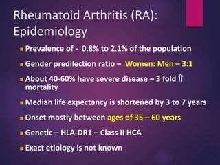
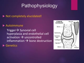
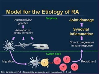
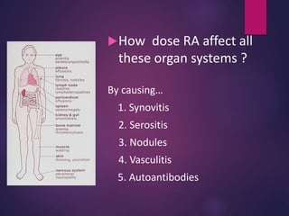
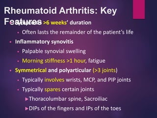
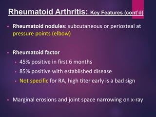
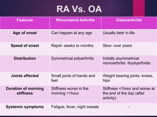
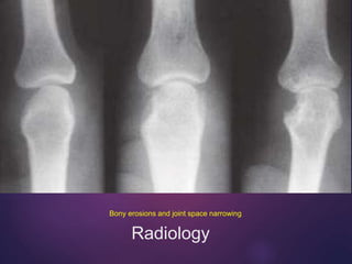
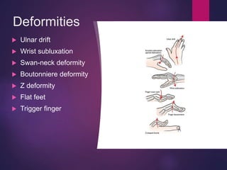
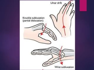
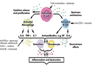
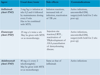
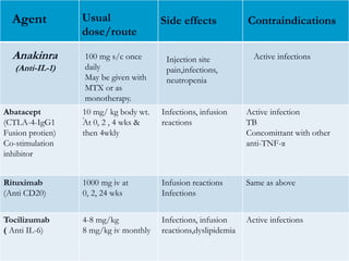
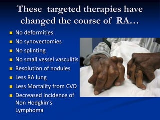
Rheumatoid
arthritis
DR. D. ANANDADURAI M.D (GEN MED)
Rheumatoid Arthritis (RA):
Epidemiology
 Prevalence of - 0.8% to 2.1% of the population
 Gender predilection ratio – Women: Men – 3:1
 About 40-60% have severe disease – 3 fold 
mortality
 Median life expectancy is shortened by 3 to 7 years
 Onset mostly between ages of 35 – 60 years
 Genetic – HLA-DR1 – Class II HCA
 Exact etiology is not known
Features
 Chronic inflammatory disease of unknown etiology
marked by a symmetric, peripheral polyarthritis
 Systemic disease
 Cigarette smoking is associated with increased risk
Pathophysiology
 Not completely elucidated!
 Autoimmune
Trigger Synovial cell
hyperplasia and endothelial cell
activation  uncontrolled
inflammation  bone destruction
 Genetics
Synovial PANNUS
How dose RA affect all
these organ systems ?
By causing…
1. Synovitis
2. Serositis
3. Nodules
4. Vasculitis
5. Autoantibodies
Rheumatoid Arthritis: Key
Features
• Symptoms >6 weeks’ duration
• Often lasts the remainder of the patient’s life
• Inflammatory synovitis
• Palpable synovial swelling
• Morning stiffness >1 hour, fatigue
• Symmetrical and polyarticular (>3 joints)
• Typically involves wrists, MCP, and PIP joints
• Typically spares certain joints
Thoracolumbar spine, Sacroiliac
DIPs of the fingers and IPs of the toes
Rheumatoid Arthritis: Key Features (cont’d)
• Rheumatoid nodules: subcutaneous or periosteal at
pressure points (elbow)
• Rheumatoid factor
• 45% positive in first 6 months
• 85% positive with established disease
• Not specific for RA, high titer early is a bad sign
• Marginal erosions and joint space narrowing on x-ray
Rheumatoid
arthritis
Osteoarthritis
RA Vs. OA
Features Rheumatoid Arthritis Osteoarthritis
Age of onset Can happen at any age Usually later in life
Speed of onset Rapid- weeks to months Slow- over years
Distribution Symmetrical polyarthritis Initially asymmetrical
monoarthritis polyarthritis
Joints affected Small joints of hands and
feet
Weight bearing joints- knees,
hips
Duration of morning
stiffness
Stiffness worse in the
morning >1hour
Stiffness <1hour and worse at
the end of the day (after
activity)
Systemic symptoms Fatigue, fever, night sweats -
Pathology
Radiology
Bony erosions and joint space narrowing
Deformities
 Ulnar drift
 Wrist subluxation
 Swan-neck deformity
 Boutonniere deformity
 Z deformity
 Flat feet
 Trigger finger
Rheumatoid Arthritis: PIP Swelling
 Swelling is confined to the area of the joint capsule
Rheumatoid Arthritis:
Ulnar Deviation and MCP Swelling
 Prominent ulnar deviation in the right hand
 MCP and PIP swelling in both hands
 Synovitis of left wrist
Cockup toes
 Feet are involved 90% / MTP’s
 ( Although patients get Hallux valgus – the 1st MTP is spared in RA)
Rheumatoid nodule
•These are small subcutaneous nodules
present at the extensor surfaces of hand,
wrist, elbow and back in rheumatoid
arthritis patients.
•Characteristics feature of rheumatoid
arthritis
•A marker of disease activity
Nodules
Systemic
Disease
 Constitutional symptoms ( most common)
 Rheumatoid nodules(30%)
 Hematological-
 normocytic normochromic anemia
 leucocytosis /leucopenia
 thrombocytosis
 Felty’s syndrome-
 Chronic nodular Rheumatoid Arthritis
 Splenomegaly
 Neutropenia
Extraarticular Involvement
 Respiratory- pleural effusion, pneumonitis , pleuro-
pulmonary nodules, ILD
 CVS-asymptomatic pericarditis , pericardial effusion,
cardiomyopathy
 Rheumatoid vasculitis- mononeuritis multiplex,
cutaneous ulceration, digital gangrene, visceral
infarction
 CNS- peripheral neuropathy, cord-compression from
atlantoaxial/midcervical spine subluxation,
entrapment neuropathies
 EYE- kerato-conjunctivitis sicca, episcleritis, scleritis
RA Systemic disease—Sicca
syndrome – dry eye and mouth
 Major cause of
Secondary
Sjogren’s
disease
Scleromalacia- thinning of
sclera
Blue choroid coming through thinned sclera
Rheumatoid lung - most serious visceral
organ affected by Rheumatoid arthritis
**LUNG
 Most common small
bilateral pleural effusions
 Cause of lung fibrosis
 Pulmonary vasculitis
 Caplan’s syndrome
Rheumatoid nodules
Caplan’s syndrome
 First reported in coal
miners (Pneumoconiosis)
with RA
 RA nodules
Neurologic
Entrapment Carpal tunnel
Wrist Synovitis in RA
C1 C2 subluxation synovial
involvement of the ligament
over the odontoid
Rheumatoid vasculitis
 Digital infarcts
warning sign of small
vessel vasculitis in RA or
impending digital
gangrene
 Small vessel Vasculitis
Result in Lung
hemorrhage or GI
bleeding
ACR-
EULAR
criteria
Clinical Course of RA
Type 1 = Self-limited—5% to 20%
Type 2 = Minimally progressive—5% to 20%
Type 3 = Progressive—60% to 90%
0
1
2
3
4
0 0.5 1 2 3 4 6 8 16
Type 1
Type 2
Type 3
Years
Severity
of
Arthritis
Rheumatoid Arthritis: Typical Course
• Damage occurs early in most patients
• 50% show joint space narrowing or erosions in
the first 2 years
• By 10 years, 50% of young working patients are
disabled
• Death comes early
• Multiple causes
• Compared to general population
Women lose 10 years, men lose 4 years
Critical Elements of a Treatment
Plan: Assessment
• Assess current activity
• Morning stiffness, synovitis, ESR
• Document the degree of damage
• Restriction of movt and deformities
• Joint space narrowing and erosions on x-ray
• Document extra-articular manifestations
• Nodules, pulmonary fibrosis, vasculitis
Diagnosis
 Blood investigation
 X-Ray of involved joints
 CT/MRI scans
 Direct arthroscopy
 Synovial/Fluid aspirate
 Synovial membrane biopsy
 Arthrocentesis
No single test is specific to Rheumatoid Arthritis
Inflammatory Markers: ESR and
CRPTest
ESR rates for men: 0-15mm/hr
ESR rates for women: 0-20mm/hr
The level of CRP in the blood is normally low
Increasing amount
suggests inflammation
Antibody Tests:
Rheumatoid Factor and Anti CCP Ab
Other blood tests check for the presence of antibodies
that are not normally present in the human body
Rheumatoid Factor (RF)
 IgM antibodies Fc portion of IgG
 Positive in 5% of normal persons and in only 70-80% of RA /
negative in 30% cases of RA – Sero negative RA
 85% of patients with RA over the first 2 years become RF+
• A negative RF may be repeated 4-6 monthly for the first two year of
disease, since some patients may take 18-24 months to become
seropositive.
• PROGNOSTIC VALUE- Patients with high titres of RF, in general,
tend to have POOR PROGNOSIS, MORE EXTRA ARTICULAR
MANIFESTATION.
Causes of positive test for RF
 Rheumatoid arthritis
 Sjogrens syndrome
 Vasculitis such as polyarteritis nodosa
 Sarcoidosis
 Systemic lupus erythematosus
 Cryoglobulinemia
 Chronic liver disease
 Infections- tuberculosis , bacterial endocarditis,
infectious mononucleosis, leprosy, syphilis, leishmaniasis.
 Malignancies
 Old age(5% women aged above 60)
Anti-CCP
Anti Cyclic Citrulinated Peptide Ab
IgG against synovial membrane
peptides damaged via inflammation
Sensitivity (65%) & Specificity (95%)
Both diagnostic & prognostic value
Predictive of Erosive Disease
Disease severity
Radiologic progression
Poor functional outcomes
Acute Phase Reactants
Positive acute phase reactants () Negative acute phase reactants ()
Mild elevations
– Ceruloplasmin
– Complement C3 & C4
Moderate elevations
– Haptoglobulin
– Fibrinogen (ESR)
– 1 – acid glycoprotein
– 1 – proteinase inhibitor
Marked elevations
– C-reactive protein (CRP)
– Serum amyloid A protein
– Albumin
– Transferrin
Other Lab Abnormalities
Elevated APRs( ESR, CRP )
Thrombocytosis
Leukocytosis
ANA
30-40%
Inflammatory synovial fluid
Hypoalbuminemia
Direct arthroscopy
Benefits
•Minimally invasive
•Less tissue damage
•Fewer complications
•Reduced pain
•Quicker recovery time
•Outpatient basis
Synovial/Fluid aspirate
Synovial membrane biopsy
Arthrocentesis
Athrocentesis: synovial fluid is aspirated and analysed for inflammatory components
Abnormal synovial fluid: cloudy, milky, or dark yellow containing leukocytes
X-Ray
X-rays are an important diagnostic test for monitoring the disease progression
Patients may reveal NO changes on an X-ray in the early stages
Arthography
A radiopaque substance or air is injected
into the joint, which outlines soft tissue
structures surrounding the joint
CT/MRI scans
MRI is particularly sensitive for the early and subtle features of RA
(Radiopaedia, 2010; Dat et al., 2010)
Used for better visualization of soft tissue
Can detect changes of Rheumatoid Arthritis prior to an X-Ray
Management
Medical
management
 NSAIDs
 Glucocorticoids
 Conventional DMARDs
 Biologics (Biologic
Response Modifiers)
Surgical
management
 Arthroplasty
RA: Drug Treatment Options
• NSAIDs
• Symptomatic relief, improved function
• No change in disease progression
• Low-dose prednisone (10 mg OD)
• Used as bridge therapy, For extra-articular RA like
rheumatoid vasculitis and interstitial lung disease
• If used long term, consider prophylactic treatment
for osteoporosis
• Intra-articular steroids
• Useful for flares
NSAIDS
Non-Steroidal anti-inflammatories (NSAIDS) / Coxibs for symptom control
1) Reduce pain and swelling by inhibiting COX
2) Do not alter course of the disease.
3) Chronic use should be minimised.
4) Most common side effect related to GI tract.
DMARDs
Conventional
 Methotrexate
 Sulfasalazine
 Leflunomide
 Hydroxychloroquine
Biological
 TNF- inhibitors
infliximab,
etanercept,
adalimumab,
golimumab and
certolizumab
 Anakinra
 Abatacept
 Rituximab
 Tocilizumab
DMARDs
Commonly used Less commonly used
Methotrexate Chloroquine
Hydroxychloroquine Gold(parenteral & oral)
Sulphasalazine Cyclosporine A
Leflunomide D-penicillamine/bucillamine
Minocycline/Doxycycline
Levamisole
Azathioprine,cyclophosphami
Disease Modifying Anti-rheumatic Agents
 Drugs that actually alter the disease course .
 Should be used as soon as diagnosis is made.
 Appearance of benefit delayed for weeks to
months.
 NSAIDS/Steroids must be continued with them
until true remission is achieved .
 Induction of true remission is unusual .
Clinical information about DMARDs
NAME DOSE SIDE EFFECTS MONITORING ONSET OF
ACTION
1) Hydroxycloro
quine
200mg twice
daily x 3 months,
then once daily
Skin
pigmentation ,
retinopathy
,nausea,
psychosis,
myopathy
Fundoscopy &
perimetry yearly
2-4 months
2) Methotrexate 7.5-25 mg once a
week orally
GI upset,
hepatotoxicity,
Bone marrow
suppression,
Pulmonary
fibrosis
Blood counts,LFT
6-8 weekly,Chest
x-ray annually,
urea/creatinine 3
monthly;
Liver biopsy
1-2 months
Clinical information about DMARDs contd..
NAME DOSE SIDE EFFECTS MONITORING ONSET OF
ACTION
3)Sulphasalazine 2gm daily p.o Rash,
myelosuppressio
n, may reduce
sperm count
Blood counts ,LFT
6-8 weekly
1-2 months
4)Leflunomide Loading 100 mg
daily x 3 days,
then 10-20 mg
daily p.o
Nausea,diarrhoe
a,alopecia,
hepatotoxicity
LFT 6-8 weekly 1-2 months
Methotrexate
 First used in 1947 for childhood leukaemia
 Probably the most effective DMARD
 Convenient ONCE weekly dosing
 Faster onset of action (6 weeks to 3 months)
(compared to other DMARDs)
 Mode of action - unclear!!
 Dose – 7.5 mg per week
 Remember – Folic acid
Side Effects of MTX
 Nausea, stomatitis
 Haematological toxicity
 Hepatic toxicity
LFTs, cirrhosis, hepatic fibrosis
 Pulmonary toxicity
Pneumonitis, Fibrosis
 Teratogenic (ova and sperm)
Limitations of conventional DMARDs
1) The onset of action takes several months.
2) The remission induced in many cases is partial.
3) There may be substantial toxicity which
requires careful monitoring.
4) DMARDs have a tendency to lose effectiveness
with time-(slip out).
 These drawbacks have made researchers look
for alternative treatment strategies for RA- The
Biologic Response Modifiers.
Immunosuppresive therapy
Agent Usual dose/route Side effects
Azathioprine 50-150 mg orally GI side effects ,
myelosuppression, infection,
Cyclosporin A 3-5 mg/kg/day Nephrotoxic , hypertension ,
hyperkalemia
Cyclophosphamide
(used in Vasculitis)
50 -150 mg orally Myelosuppression , gonadal
toxicity ,hemorrhagic cystitis ,
bladder cancer
.
.
Agent Usual dose/route Side effects Contraindications
Infliximab
(Anti-TNF)
3 mg/kg i.v infusion at
wks 0,2 and 6 followed
by maintainence dosing
every 8 wks
Has to be combined
with MTX.
Infusion reactions,
increased risk of
infection, reactivation
of TB ,etc
Active infections,
uncontrolled DM,
surgery(with hold for 2 wks
post op)
Etanercept
(Anti-TNF)
Active infections,
uncontrolled DM,
surgery(with hold for 2 wks
post op)
Adalimumab
(Anti-TNF)
40 mg s/c every 2
wks(fornightly)
May be given with MTX
or as monotherapy
Same as that of
infliximab
Active infections
.
25 mg s/c twice a wk
May be given with MTX
or as monotherapy.
Injection site
reaction,URTI ,
reactivation of
TB,development of
ANA,exacerbation
of demyelenating
disease.
Abatacept
(CTLA-4-IgG1
Fusion protien)
Co-stimulation
inhibitor
10 mg/ kg body wt.
At 0, 2 , 4 wks &
then 4wkly
Infections, infusion
reactions
Active infection
TB
Concomittant with other
anti-TNF-α
Rituximab
(Anti CD20)
1000 mg iv at
0, 2, 24 wks
Infusion reactions
Infections
Same as above
Tocilizumab
( Anti IL-6)
4-8 mg/kg
8 mg/kg iv monthly
Infections, infusion
reactions,dyslipidemia
Active infections
Agent Usual
dose/route
Side effects
.
Anakinra 100 mg s/c once
daily
May be given with
MTX or as
monotherapy.
Injection site
pain,infections,
neutropenia
Active infections
Contraindications
(Anti-IL-1)
Biologics: Relative
Contraindications
 Active Hepatitis B Infection
 Multiple sclerosis, optic neuritis
 Active serious infections
 Chronic or recurrent infections
 Current neoplasia
 History of TB or evidence of Koch’s
 Congestive heart failure (Class III or IV)
These targeted therapies have
changed the course of RA…
 No deformities
 No synovectomies
 No splinting
 No small vessel vasculitis
 Resolution of nodules
 Less RA lung
 Less Mortality from CVD
 Decreased incidence of
Non Hodgkin’s
Lymphoma
2012 ACR Update
Recent developments
Emergence of
methotrexate as the
DMARD of choice for
early disease
1
Development of
novel highly
efficacious
biologicals
2
Proven superiority of
combination DMARD
regimens over
methotrexate alone
3
How to measure Rheumatoid Arthritis?
28 Joint Count
DAS (28) Score
Swollen and Tender Joint Count
ESR
Global health assessment by patient
DAS28 =
0.56 x sqrt(tender28) + 0.28 x
sqrt(swollen28) + 0.70 x ln(ESR) +
0.014 x GH
DAS in Practice
“An objective method for measuring
disease activity”
>5.1 = Active disease
<3.2 = low disease activity
<2.6 = Remission
Surgical Approaches
 Synovectomy is ordinarily not recommended for patients
with rheumatoid arthritis, primarily because relief is only
transient.
However, an exception is synovectomy of the wrist, which is
recommended if intense synovitis is persistent despite medical
treatment over 6 to 12 months. Persistent synovitis involving the
dorsal compartments of the wrist can lead to extensor tendon
sheath rupture resulting in severe disability of hand function.
 Total joint arthroplasties , particularly of the knee, hip,
wrist, and elbow, are highly successful.
 Other operations include release of nerve entrapments
(e.g., carpal tunnel syndrome), arthroscopic procedures,
and, occasionally, removal of a symptomatic rheumatoid
nodule.
Thank you!

Rheumatoid Arthritis.pptx

  • 1.
  • 2.
    Rheumatoid Arthritis (RA): Epidemiology Prevalence of - 0.8% to 2.1% of the population  Gender predilection ratio – Women: Men – 3:1  About 40-60% have severe disease – 3 fold  mortality  Median life expectancy is shortened by 3 to 7 years  Onset mostly between ages of 35 – 60 years  Genetic – HLA-DR1 – Class II HCA  Exact etiology is not known
  • 3.
    Features  Chronic inflammatorydisease of unknown etiology marked by a symmetric, peripheral polyarthritis  Systemic disease  Cigarette smoking is associated with increased risk
  • 4.
    Pathophysiology  Not completelyelucidated!  Autoimmune Trigger Synovial cell hyperplasia and endothelial cell activation  uncontrolled inflammation  bone destruction  Genetics
  • 8.
  • 9.
    How dose RAaffect all these organ systems ? By causing… 1. Synovitis 2. Serositis 3. Nodules 4. Vasculitis 5. Autoantibodies
  • 10.
    Rheumatoid Arthritis: Key Features •Symptoms >6 weeks’ duration • Often lasts the remainder of the patient’s life • Inflammatory synovitis • Palpable synovial swelling • Morning stiffness >1 hour, fatigue • Symmetrical and polyarticular (>3 joints) • Typically involves wrists, MCP, and PIP joints • Typically spares certain joints Thoracolumbar spine, Sacroiliac DIPs of the fingers and IPs of the toes
  • 11.
    Rheumatoid Arthritis: KeyFeatures (cont’d) • Rheumatoid nodules: subcutaneous or periosteal at pressure points (elbow) • Rheumatoid factor • 45% positive in first 6 months • 85% positive with established disease • Not specific for RA, high titer early is a bad sign • Marginal erosions and joint space narrowing on x-ray
  • 12.
  • 13.
  • 14.
    RA Vs. OA FeaturesRheumatoid Arthritis Osteoarthritis Age of onset Can happen at any age Usually later in life Speed of onset Rapid- weeks to months Slow- over years Distribution Symmetrical polyarthritis Initially asymmetrical monoarthritis polyarthritis Joints affected Small joints of hands and feet Weight bearing joints- knees, hips Duration of morning stiffness Stiffness worse in the morning >1hour Stiffness <1hour and worse at the end of the day (after activity) Systemic symptoms Fatigue, fever, night sweats -
  • 15.
  • 16.
    Radiology Bony erosions andjoint space narrowing
  • 17.
    Deformities  Ulnar drift Wrist subluxation  Swan-neck deformity  Boutonniere deformity  Z deformity  Flat feet  Trigger finger
  • 20.
    Rheumatoid Arthritis: PIPSwelling  Swelling is confined to the area of the joint capsule
  • 21.
    Rheumatoid Arthritis: Ulnar Deviationand MCP Swelling  Prominent ulnar deviation in the right hand  MCP and PIP swelling in both hands  Synovitis of left wrist
  • 23.
    Cockup toes  Feetare involved 90% / MTP’s  ( Although patients get Hallux valgus – the 1st MTP is spared in RA)
  • 24.
    Rheumatoid nodule •These aresmall subcutaneous nodules present at the extensor surfaces of hand, wrist, elbow and back in rheumatoid arthritis patients. •Characteristics feature of rheumatoid arthritis •A marker of disease activity
  • 25.
  • 26.
  • 27.
     Constitutional symptoms( most common)  Rheumatoid nodules(30%)  Hematological-  normocytic normochromic anemia  leucocytosis /leucopenia  thrombocytosis  Felty’s syndrome-  Chronic nodular Rheumatoid Arthritis  Splenomegaly  Neutropenia Extraarticular Involvement
  • 28.
     Respiratory- pleuraleffusion, pneumonitis , pleuro- pulmonary nodules, ILD  CVS-asymptomatic pericarditis , pericardial effusion, cardiomyopathy  Rheumatoid vasculitis- mononeuritis multiplex, cutaneous ulceration, digital gangrene, visceral infarction  CNS- peripheral neuropathy, cord-compression from atlantoaxial/midcervical spine subluxation, entrapment neuropathies  EYE- kerato-conjunctivitis sicca, episcleritis, scleritis
  • 29.
    RA Systemic disease—Sicca syndrome– dry eye and mouth  Major cause of Secondary Sjogren’s disease
  • 30.
    Scleromalacia- thinning of sclera Bluechoroid coming through thinned sclera
  • 31.
    Rheumatoid lung -most serious visceral organ affected by Rheumatoid arthritis **LUNG  Most common small bilateral pleural effusions  Cause of lung fibrosis  Pulmonary vasculitis  Caplan’s syndrome Rheumatoid nodules
  • 32.
    Caplan’s syndrome  Firstreported in coal miners (Pneumoconiosis) with RA  RA nodules
  • 33.
    Neurologic Entrapment Carpal tunnel WristSynovitis in RA C1 C2 subluxation synovial involvement of the ligament over the odontoid
  • 34.
    Rheumatoid vasculitis  Digitalinfarcts warning sign of small vessel vasculitis in RA or impending digital gangrene  Small vessel Vasculitis Result in Lung hemorrhage or GI bleeding
  • 35.
  • 36.
    Clinical Course ofRA Type 1 = Self-limited—5% to 20% Type 2 = Minimally progressive—5% to 20% Type 3 = Progressive—60% to 90% 0 1 2 3 4 0 0.5 1 2 3 4 6 8 16 Type 1 Type 2 Type 3 Years Severity of Arthritis
  • 37.
    Rheumatoid Arthritis: TypicalCourse • Damage occurs early in most patients • 50% show joint space narrowing or erosions in the first 2 years • By 10 years, 50% of young working patients are disabled • Death comes early • Multiple causes • Compared to general population Women lose 10 years, men lose 4 years
  • 38.
    Critical Elements ofa Treatment Plan: Assessment • Assess current activity • Morning stiffness, synovitis, ESR • Document the degree of damage • Restriction of movt and deformities • Joint space narrowing and erosions on x-ray • Document extra-articular manifestations • Nodules, pulmonary fibrosis, vasculitis
  • 39.
    Diagnosis  Blood investigation X-Ray of involved joints  CT/MRI scans  Direct arthroscopy  Synovial/Fluid aspirate  Synovial membrane biopsy  Arthrocentesis No single test is specific to Rheumatoid Arthritis
  • 40.
    Inflammatory Markers: ESRand CRPTest ESR rates for men: 0-15mm/hr ESR rates for women: 0-20mm/hr The level of CRP in the blood is normally low Increasing amount suggests inflammation
  • 41.
    Antibody Tests: Rheumatoid Factorand Anti CCP Ab Other blood tests check for the presence of antibodies that are not normally present in the human body
  • 42.
    Rheumatoid Factor (RF) IgM antibodies Fc portion of IgG  Positive in 5% of normal persons and in only 70-80% of RA / negative in 30% cases of RA – Sero negative RA  85% of patients with RA over the first 2 years become RF+ • A negative RF may be repeated 4-6 monthly for the first two year of disease, since some patients may take 18-24 months to become seropositive. • PROGNOSTIC VALUE- Patients with high titres of RF, in general, tend to have POOR PROGNOSIS, MORE EXTRA ARTICULAR MANIFESTATION.
  • 43.
    Causes of positivetest for RF  Rheumatoid arthritis  Sjogrens syndrome  Vasculitis such as polyarteritis nodosa  Sarcoidosis  Systemic lupus erythematosus  Cryoglobulinemia  Chronic liver disease  Infections- tuberculosis , bacterial endocarditis, infectious mononucleosis, leprosy, syphilis, leishmaniasis.  Malignancies  Old age(5% women aged above 60)
  • 44.
    Anti-CCP Anti Cyclic CitrulinatedPeptide Ab IgG against synovial membrane peptides damaged via inflammation Sensitivity (65%) & Specificity (95%) Both diagnostic & prognostic value Predictive of Erosive Disease Disease severity Radiologic progression Poor functional outcomes
  • 45.
    Acute Phase Reactants Positiveacute phase reactants () Negative acute phase reactants () Mild elevations – Ceruloplasmin – Complement C3 & C4 Moderate elevations – Haptoglobulin – Fibrinogen (ESR) – 1 – acid glycoprotein – 1 – proteinase inhibitor Marked elevations – C-reactive protein (CRP) – Serum amyloid A protein – Albumin – Transferrin
  • 46.
    Other Lab Abnormalities ElevatedAPRs( ESR, CRP ) Thrombocytosis Leukocytosis ANA 30-40% Inflammatory synovial fluid Hypoalbuminemia
  • 47.
    Direct arthroscopy Benefits •Minimally invasive •Lesstissue damage •Fewer complications •Reduced pain •Quicker recovery time •Outpatient basis
  • 48.
    Synovial/Fluid aspirate Synovial membranebiopsy Arthrocentesis Athrocentesis: synovial fluid is aspirated and analysed for inflammatory components Abnormal synovial fluid: cloudy, milky, or dark yellow containing leukocytes
  • 49.
    X-Ray X-rays are animportant diagnostic test for monitoring the disease progression Patients may reveal NO changes on an X-ray in the early stages
  • 50.
    Arthography A radiopaque substanceor air is injected into the joint, which outlines soft tissue structures surrounding the joint
  • 51.
    CT/MRI scans MRI isparticularly sensitive for the early and subtle features of RA (Radiopaedia, 2010; Dat et al., 2010) Used for better visualization of soft tissue Can detect changes of Rheumatoid Arthritis prior to an X-Ray
  • 52.
    Management Medical management  NSAIDs  Glucocorticoids Conventional DMARDs  Biologics (Biologic Response Modifiers) Surgical management  Arthroplasty
  • 53.
    RA: Drug TreatmentOptions • NSAIDs • Symptomatic relief, improved function • No change in disease progression • Low-dose prednisone (10 mg OD) • Used as bridge therapy, For extra-articular RA like rheumatoid vasculitis and interstitial lung disease • If used long term, consider prophylactic treatment for osteoporosis • Intra-articular steroids • Useful for flares
  • 54.
    NSAIDS Non-Steroidal anti-inflammatories (NSAIDS)/ Coxibs for symptom control 1) Reduce pain and swelling by inhibiting COX 2) Do not alter course of the disease. 3) Chronic use should be minimised. 4) Most common side effect related to GI tract.
  • 55.
    DMARDs Conventional  Methotrexate  Sulfasalazine Leflunomide  Hydroxychloroquine Biological  TNF- inhibitors infliximab, etanercept, adalimumab, golimumab and certolizumab  Anakinra  Abatacept  Rituximab  Tocilizumab
  • 56.
    DMARDs Commonly used Lesscommonly used Methotrexate Chloroquine Hydroxychloroquine Gold(parenteral & oral) Sulphasalazine Cyclosporine A Leflunomide D-penicillamine/bucillamine Minocycline/Doxycycline Levamisole Azathioprine,cyclophosphami
  • 57.
    Disease Modifying Anti-rheumaticAgents  Drugs that actually alter the disease course .  Should be used as soon as diagnosis is made.  Appearance of benefit delayed for weeks to months.  NSAIDS/Steroids must be continued with them until true remission is achieved .  Induction of true remission is unusual .
  • 58.
    Clinical information aboutDMARDs NAME DOSE SIDE EFFECTS MONITORING ONSET OF ACTION 1) Hydroxycloro quine 200mg twice daily x 3 months, then once daily Skin pigmentation , retinopathy ,nausea, psychosis, myopathy Fundoscopy & perimetry yearly 2-4 months 2) Methotrexate 7.5-25 mg once a week orally GI upset, hepatotoxicity, Bone marrow suppression, Pulmonary fibrosis Blood counts,LFT 6-8 weekly,Chest x-ray annually, urea/creatinine 3 monthly; Liver biopsy 1-2 months
  • 59.
    Clinical information aboutDMARDs contd.. NAME DOSE SIDE EFFECTS MONITORING ONSET OF ACTION 3)Sulphasalazine 2gm daily p.o Rash, myelosuppressio n, may reduce sperm count Blood counts ,LFT 6-8 weekly 1-2 months 4)Leflunomide Loading 100 mg daily x 3 days, then 10-20 mg daily p.o Nausea,diarrhoe a,alopecia, hepatotoxicity LFT 6-8 weekly 1-2 months
  • 60.
    Methotrexate  First usedin 1947 for childhood leukaemia  Probably the most effective DMARD  Convenient ONCE weekly dosing  Faster onset of action (6 weeks to 3 months) (compared to other DMARDs)  Mode of action - unclear!!  Dose – 7.5 mg per week  Remember – Folic acid
  • 62.
    Side Effects ofMTX  Nausea, stomatitis  Haematological toxicity  Hepatic toxicity LFTs, cirrhosis, hepatic fibrosis  Pulmonary toxicity Pneumonitis, Fibrosis  Teratogenic (ova and sperm)
  • 63.
    Limitations of conventionalDMARDs 1) The onset of action takes several months. 2) The remission induced in many cases is partial. 3) There may be substantial toxicity which requires careful monitoring. 4) DMARDs have a tendency to lose effectiveness with time-(slip out).  These drawbacks have made researchers look for alternative treatment strategies for RA- The Biologic Response Modifiers.
  • 64.
    Immunosuppresive therapy Agent Usualdose/route Side effects Azathioprine 50-150 mg orally GI side effects , myelosuppression, infection, Cyclosporin A 3-5 mg/kg/day Nephrotoxic , hypertension , hyperkalemia Cyclophosphamide (used in Vasculitis) 50 -150 mg orally Myelosuppression , gonadal toxicity ,hemorrhagic cystitis , bladder cancer . .
  • 66.
    Agent Usual dose/routeSide effects Contraindications Infliximab (Anti-TNF) 3 mg/kg i.v infusion at wks 0,2 and 6 followed by maintainence dosing every 8 wks Has to be combined with MTX. Infusion reactions, increased risk of infection, reactivation of TB ,etc Active infections, uncontrolled DM, surgery(with hold for 2 wks post op) Etanercept (Anti-TNF) Active infections, uncontrolled DM, surgery(with hold for 2 wks post op) Adalimumab (Anti-TNF) 40 mg s/c every 2 wks(fornightly) May be given with MTX or as monotherapy Same as that of infliximab Active infections . 25 mg s/c twice a wk May be given with MTX or as monotherapy. Injection site reaction,URTI , reactivation of TB,development of ANA,exacerbation of demyelenating disease.
  • 67.
    Abatacept (CTLA-4-IgG1 Fusion protien) Co-stimulation inhibitor 10 mg/kg body wt. At 0, 2 , 4 wks & then 4wkly Infections, infusion reactions Active infection TB Concomittant with other anti-TNF-α Rituximab (Anti CD20) 1000 mg iv at 0, 2, 24 wks Infusion reactions Infections Same as above Tocilizumab ( Anti IL-6) 4-8 mg/kg 8 mg/kg iv monthly Infections, infusion reactions,dyslipidemia Active infections Agent Usual dose/route Side effects . Anakinra 100 mg s/c once daily May be given with MTX or as monotherapy. Injection site pain,infections, neutropenia Active infections Contraindications (Anti-IL-1)
  • 68.
    Biologics: Relative Contraindications  ActiveHepatitis B Infection  Multiple sclerosis, optic neuritis  Active serious infections  Chronic or recurrent infections  Current neoplasia  History of TB or evidence of Koch’s  Congestive heart failure (Class III or IV)
  • 69.
    These targeted therapieshave changed the course of RA…  No deformities  No synovectomies  No splinting  No small vessel vasculitis  Resolution of nodules  Less RA lung  Less Mortality from CVD  Decreased incidence of Non Hodgkin’s Lymphoma
  • 70.
  • 71.
    Recent developments Emergence of methotrexateas the DMARD of choice for early disease 1 Development of novel highly efficacious biologicals 2 Proven superiority of combination DMARD regimens over methotrexate alone 3
  • 72.
    How to measureRheumatoid Arthritis? 28 Joint Count
  • 73.
    DAS (28) Score Swollenand Tender Joint Count ESR Global health assessment by patient DAS28 = 0.56 x sqrt(tender28) + 0.28 x sqrt(swollen28) + 0.70 x ln(ESR) + 0.014 x GH
  • 74.
    DAS in Practice “Anobjective method for measuring disease activity” >5.1 = Active disease <3.2 = low disease activity <2.6 = Remission
  • 75.
    Surgical Approaches  Synovectomyis ordinarily not recommended for patients with rheumatoid arthritis, primarily because relief is only transient. However, an exception is synovectomy of the wrist, which is recommended if intense synovitis is persistent despite medical treatment over 6 to 12 months. Persistent synovitis involving the dorsal compartments of the wrist can lead to extensor tendon sheath rupture resulting in severe disability of hand function.  Total joint arthroplasties , particularly of the knee, hip, wrist, and elbow, are highly successful.  Other operations include release of nerve entrapments (e.g., carpal tunnel syndrome), arthroscopic procedures, and, occasionally, removal of a symptomatic rheumatoid nodule.
  • 76.