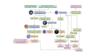
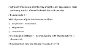
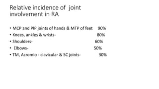
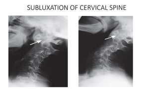
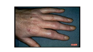
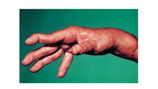
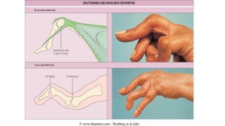
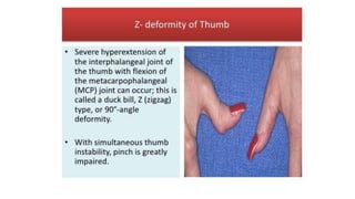
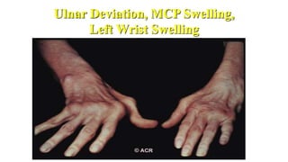
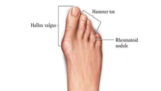
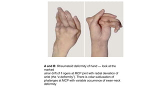
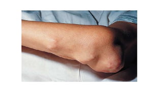
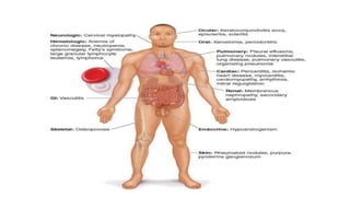
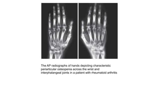
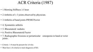
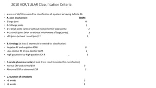
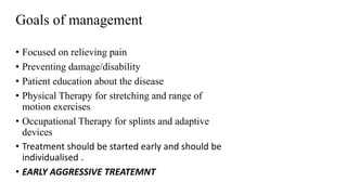
RHEUMATOID ARTHRITIS
BY- DR. HIMALAYA SINGH
INTRODUCTION
Guillaume de Baillou (1538–1616) was the first to use the
word “rheumatism” in the modern sense. RA is a chronic,
inflammatory, autoimmune disease that may involve
multiple organ systems but mainly targets the synovial
joints.
It is more common in women than men (ratio of
2–3:1) and has peak incidence somewhere between 35–50
years.
RA is diagnosed in individuals with symmetrical
inflammatory polyarthritis affecting the small joints of the
hands, feet and wrists persisting for more than 6 weeks
with characteristic laboratory abnormalities.
Patients have swollen joints and early morning stiffness
typically lasting longer than 1 hour.
The most specific laboratory finding for RA is the
presence of anticyclic citrullinated peptide (Anti-CCP)
antibodies seen in 60–70% patients.
Anti-CCP antibody may be present before rheumatoid
factor is detected. The majority of patients afflicted with
RA have
ongoing, progressive disease with periods of reduced
activity.
ETIOLOGY
Infections, genetic susceptibility and environmental
factors have been implicated in etiology. The concordance
rate in monozygotic twins is high (12–15%) compared
to a prevalence of 1% in the general population.
Major histocompatibility complex class II loci is the prime
culprit
that hosts the DR4 chains (DRB*0401 and DRB*0404)
having greatest association with RA. Other loci that may
have a role include the major histocompatibility complex
class III genes for TNF-, heat shock protein 70 (HSP70),
and
PATHOGENESIS
Antigen-mediated activation of T-cells initiates a cascade
activation of endothelial cells, proliferation of Types A and B
synoviocytes, and recruitment of additional inflammatory cells
from the bone marrow.
TNF- and IL-1 stimulate inflammatory cells to release
oncostatin M
into synovial fluid.
TNF- and oncostatin M work together to induce production of
collagenases called MMPs by the diseased synoviocytes and
chondrocytes resulting in excessive collagen degradation and
When the immune response is triggered by the
antigen, additional antigens may be created and recognized
by the T-cell to create an ongoing immune response.
The major population of T-cells in the rheumatoid synovium
is
composed of CD4+ memory T-cells. The antibody, a gamma
globulin (rheumatoid factor), is produced in lymph nodes
and coats the cell membranes of lymphocytes and plasma
cells, which travel to the synovium where they react with
the antigen causing acute inflammatory reaction.
The cytokines IL-1 and TNF play key role.
The synovitis and vascular proliferation invade adjacent
cartilage and bone.
These further degrade cartilage and stimulate
osteoclasts to promote periarticular bony erosion and
osteopenia
Although Rheumatoid arthritis may present at any age, patients most
commonly are first affected in the third to sixth decades.
Female: male 3:1
Initial pattern of joint involvement could be:-
1) Polyarticular : most common
2) Oligoarticular
3) Monoarticular
Morning joint stiffness > 1 hour and easing with physical activity is
characteristic.
Small joints of hand and feet are typically involved.
• MCP and PIP joints of hands & MTP of feet 90%
• Knees, ankles & wrists- 80%
• Shoulders- 60%
• Elbows- 50%
• TM, Acromio - clavicular & SC joints- 30%
Relative incidence of joint
involvement in RA
SUBLUXATION OF CERVICAL SPINE
Ulnar Deviation, MCP Swelling,
Left Wrist Swelling
A and B: Rheumatoid deformity of hand — look at the
marked
ulnar drift of fi ngers at MCP joint with radial deviation of
wrist (the “z-deformity”). There is volar subluxation of
phalanges at MCP with variable occurrence of swan-neck
deformity
• Constitutional symptoms ( most common)
• Rheumatoid nodules(30%)
• Hematological-
• normocytic normochromic anemia
• leucocytosis /leucopenia
• thrombocytosis
• Felty’s syndrome-
• Chronic nodular Rheumatoid Arthritis
• Splenomegaly
 Neutropenia
oCaplan Syndrome-
• Pneumoconiosis following silica exposure
• Rheumatoid Arthritis
Extraarticular Involvement
Laboratory Evaluation
• CBC ( TLC, DLC, Hb )
• Acute phase reactants ( ESR, CRP )
• Rheumatoid Factor (RF).
• Anti- CCP antibodies (most specific ).
Rheumatoid Factor
These are nearly 59% sensitive and 93% specific for diagnosis
of rheumatoid arthritis. These are autoantibodies to the
epitopes in the Fc portion of IgG.
These autoantibodies may be of the IgM, IgA, IgG or IgE
isotype but commonly the IgM portion of rheumatoid factor is
tested which is present in 60–70% of patient population.
Both IgM RF and IgG RF can enhance complement activation
(both alternate and classic pathways) by IgG complexes. RF
may directly activate natural killer (NK) cells stimulating the
secretion of TNF and IL-1.
• Antibodies that recognize Fc portion of IgG.
• Can be IgM , IgG , IgA
• 85% of patients with RA over the first 2 years become RF positive.
• A negative RF may be repeated 4-6 monthly for the first two year
of disease, since some patients may take 18-24 months to become
seropositive.
• PROGNISTIC VALUE- Patients with high titres of RF, in
general, tend to have POOR PROGNOSIS, MORE EXTRA
ARTICULAR MANIFESTATION.
• The presence of serum anti-CCP antibodies has about the
same sensitivity as serum RF for the diagnosis of RA.
However, its specificity approaches 95%.
• A positive test for anti-CCP antibodies in the setting of an
early inflammatory arthritis is useful for distinguishing RA
from other forms of arthritis.
• There is some incremental value in testing for the
presence of both RF and anti-CCP, as some patients with
RA are positive for RF but negative for anti-CCP and visa
versa.
• The presence of RF or anti-CCP antibodies also has
prognostic significance, with anti-CCP antibodies showing
the most value for predicting worse outcomes.
serum anti-CCP antibodies
•Elevated APRs( ESR, CRP )
•Thrombocytosis
•Leukocytosis
•ANA
• 30-40%
•Inflammatory synovial fluid
•Hypoalbuminemia
Other Lab Abnormalities :
oRadiographic Features
 Peri-articular osteopenia
Uniform symmetric joint space narrowing
Marginal subchondral erosions
 Joint Subluxations
Joint destruction
Collapse
• Ultrasound detects early soft tissue lesions.
• MRI has greatest sensitivity to detect synovitis and
marrow changes.
The AP radiographs of hands depicting characteristic
periarticular osteopenia across the wrist and
interphalangeal joints in a patient with rheumatoid arthritis
DIAGNOSIS
• 1.Morning Stiffness ≥1 hour
• 2.Arthritis of ≥ 3 joints observed by physician.
• 3.Arthritis of hand joints-PIP,MCP,wrist
• 4. Symmetric arthritis
• 5. Rheumatoid nodules
• 6. Positive Rheumatoid Factor
• 7. Radiographic Erosions or periarticular osteopenia in hand or wrist
joints .
• Criteria 1-4 must be present for ≥6 wks
• Must have ≥4 criteria to meet diagnosis of RA
ACR Criteria (1987)
• a score of ≥6/10 is needed for classification of a patient as having definite RA
• A. Joint involvement SCORE
• 1 large joint 0
• 2−10 large joints 1
• 1−3 small joints (with or without involvement of large joints) 2
• 4−10 small joints (with or without involvement of large joints) 3
• >10 joints (at least 1 small joint)†† 5
• B. Serology (at least 1 test result is needed for classification)
• Negative RF and negative ACPA 0
• Low-positive RF or low-positive ACPA 2
• High-positive RF or high-positive ACP A , 3
• C. Acute-phase reactants (at least 1 test result is needed for classification)
• Normal CRP and normal ESR 0
• Abnormal CRP or abnormal ESR 1
• D. Duration of symptoms
• <6 weeks 0
• ≥6 weeks 1
2010 ACR/EULAR Classification Criteria
Management
• Focused on relieving pain
• Preventing damage/disability
• Patient education about the disease
• Physical Therapy for stretching and range of
motion exercises
• Occupational Therapy for splints and adaptive
devices
• Treatment should be started early and should be
individualised .
• EARLY AGGRESSIVE TREATEMNT
Goals of management
Treatment modalities for RA
 NSAIDS
 Steroids
 DMARDs
 Biological therapies
 Surgery
Non-Steroidal anti-inflammatories (NSAIDS) for
symptom control :
1) Reduce pain and swelling by inhibiting COX
2) Do not alter course of the disease.
3) Chronic use should be minimised.
4) Most common side effect related to GI tract.
NSAIDS
Corticosteroids , both systemic and intra-articular are
important adjuncts in management of RA.
Indications for systemic steroids are:-
 For treatment of rheumatoid flares.
 For extra-articular RA like rheumatoid vasculitis and interstitial
lung disease.
 As bridge therapy for 6-8 weeks before the action of DMARDs
begin.
 Maintainence dose of 10mg or less of predinisolone daily in
patients with active RA.
 Sometimes in pregnancy when other DMARDs cannot be used.
Corticosteroids in RA
Disease Modifying Anti-rheumatic Agents
• Drugs that actually alter the disease course .
• Should be used as soon as diagnosis is made.
• Appearance of benefit delayed for weeks to months.
• NSAIDS must be continued with them until true
remission is achieved .
• Induction of true remission is unusual .
[Previously Called Second Line Agents (They Are Now the
First Line Agents), Slow-acting Antirheumatic Drugs]—Now
Th ey Are Called “Synthetic” DMARDs (in View of
Availability
of Biological Agents)
Commonly used Less commonly used
Methotrexate Chloroquine
Hydroxychloroquine Gold(parenteral &oral)
Sulphasalazine CyclosporineA
Leflunomide D-penicillamine/bucillamine
Minocycline/Doxycycline
Levamisole
Azathioprine,cyclophosphamide,
chlorambucil
NAME DOSE SIDE EFFECTS MONITORING ONSET OF
ACTION
1) Hydroxycloro
quine
200mg twice
daily x 3 months,
then once daily
Skin
pigmentation ,
retinopahy
,nausea,
psychosis,
myopathy
Fundoscopy&
perimetry yearly
2-4 months
2) Methotrexate 7.5-25 mg once a
week orally,s/c or
i/m
GI upset,
hepatotoxicity,
Bone marrow
suppression,
pulmonary
fibrosis
Blood counts,LFT
6-8 weekly,Chest
x-ray annually,
urea/creatinine 3
monthly;
Liver biopsy
1-2 months
NAME DOSE SIDE EFFECTS MONITORING ONSET OF
ACTION
3)Sulphasalazine 2gm daily p.o Rash,
myelosuppressio
n, may reduce
sperm count
Blood counts ,LFT
6-8 weekly
1-2 months
4)Leflunomide Loading 100 mg
daily x 3 days,
then 10-20 mg
daily p.o
Nausea,diarrhoe
a,alopecia,
hepatotoxicity
LFT 6-8 weekly 1-2 months
When to start DMARDs?
• DMARDs are indicated in all patients with RA who
continue to have active disease even after 3 months of
NSAIDS use.
• The period of 3 months is arbitary & has been chosen
since a small percentage of patients may go in
spontaneous remission.
• The vast majority , however , need DMARDs and many
rheumatologists start DMARDs from Day 1.
How to select DMARDs?
• There are no strict guidelines about which DMARDs to start
first in an individual.
• Methotrexate has rapid onset of action than other DMARD.
• Taking in account patient tolerance, cost considerations and
ease of once weekly oral administration METHOTREXATE is
the DMARD of choice, most widely prescribed in the world.
Should DMARDs be used singly or in
combination?
• Since single DMARD therapy (in conjunction with NSAIDS) is
often only modestly effective , combination therapy has an
inherent approach .
• DMARD combination is specially effective if they include
methotrexate as an anchor drug.
• Combination of methotrexate with leflunamide are
synergestic since there mode of action is different.
Limitations of conventional DMARDs
1) The onset of action takes several months.
2) The remission induced in many cases is partial.
3) There may be substantial toxicity which
requires careful monitoring.
4) DMARDs have a tendency to lose effectiveness
with time-(slip out).
 These drawbacks have made researchers look
for alternative treatment strategies for RA-
The Biologic Response Modifiers.
BIOLOGICS IN RA
• Cytokines such as TNF-α ,IL-1,IL-10 etc. are key
mediators of immune function in RA and have been
major targets of therapeutic manipulations in RA.
• Of the various cytokines,TNF-α has attaracted maximum
attention.
• Various biologicals approved in RA are:-
1) Anti TNF agents : Infliximab Etanercept Adalimumab
2) IL-1 receptor antagonist : Anakinra
3) IL-6 receptor antagonist : Tocilizumab
4) Anti CD20 antibody : Rituximab
5) T cell costimulatory inhibitor : Abatacept
Agent Usual dose/route Side effects Contraindications
Infliximab
(Anti-TNF)
3 mg/kg i.v infusion at
wks 0,2 and 6 followed
by maintainence
dosing every 8 wks
Has to be combined
with MTX.
Infusion reactions,
increased risk of
infection, reactivation
of TB ,etc
Active
infections,uncontrolled
DM,surgery(with hold for 2
wks post op)
Etanercept
(Anti-TNF)
25 mg s/c twice a wk
May be given with
MTX or as
monotherapy.
Injection site
reaction,URTI ,
reactivation of
TB,development of
ANA,exacerbation of
demyelenating
disease.
Active
infections,uncontrolled
DM,surgery(with hold for 2
wks post op)
Adalimumab
(Anti-TNF)
40 mg s/c every 2
wks(fornightly)
May be given with
MTX or as
monotherapy
Same as that of
infliximab
Active infections
100 mg s/c once
daily
May be given with
MTX or as
monotherapy.
Injection site
pain,infections,
neutropenia
Active infections
Abatacept
(CTLA-4-IgG1 Fusion
protien)
Co-stimulation
inhibitor
10 mg/ kg body wt.
At 0, 2 , 4 wks &
then 4wkly
Infections, infusion
reactions
Active infection
TB
Concomittant with other
anti-TNF-α
Rituximab
(Anti CD20)
1000 mg iv at
0, 2, 24 wks
Infusion reactions,
Infections
Same as above
Tocilizumab
( Anti IL-6)
4-8 mg/kg
8 mg/kg iv monthly
Infections, infusion
reactions,dyslipidemi
a
Active infections
Anakinra
IL – 1
receptor
antagonist
How to monitor Tt in RA?
• Disease activity is assesed by several parameters…
duration of morning stiffness,tender joint count,swollen
joint count, visual analogue scale for pain, health
assessment questionnaire,ESR,NSAID pill count,DAS score
etc..
• Patient on MTX,SSZ or leflunamide show clinical
improvement in 6-8 wks.
• Patient should be observed for 6 months before declaring
a DMARD ineffective.
How long should Tt. be continued?
• Once remission is achieved , maintenance dose for long
period is recommended.
• Relapse occurs in 3-5 months (1-2 months in case of MTX) if
drug is discontinued in most instances.
• DMARDs are discontinued by patients because of toxicity or
secondary failure(common after 1-2 yrs) and such patients
might have to shift over different DMARDs over 5-10 yrs.
• Disease flare may require escalation of DMARD dose with
short course of steroids.
Surgical Approaches
• Synovectomy is ordinarily not recommended for patients
with rheumatoid arthritis, primarily because relief is only
transient.
• However, an exception is synovectomy of the wrist, which is
recommended if intense synovitis is persistent despite
medical treatment over 6 to 12 months.
• Total joint arthroplasties , particularly of the knee, hip, wrist,
and elbow, are highly successful.
• Other operations include release of nerve entrapments
(e.g., carpal tunnel syndrome), arthroscopic procedures,
and, occasionally, removal of a symptomatic rheumatoid
nodule.
Various joint reconstruction surgeries performed in Rheumatoid arthritis. (A)
MCP joint arthroplasty;
(B) Arthrodesis of 1st MTP joint; (C) Total knee replacement for advanced
RA with secondary degeneration of knee
Rheumatoid arthritis in pregnancy
Most patients with RA go into remission during pregnancy.
Methotrexate and leflunomide should be discontinued for at least
3 months before trying to conceive.
Paracetamol is the oral analgesic of choice during pregnancy.
Corticosteroids may be used to control disease flares
DMARDs that may be used: sulfasalazine, hydroxychloroquine,
azathioprine or ciclosporin if required to control inflammation.
DMARDs that must be avoided: methotrexate,
leflunomide,cyclophosphamide, gold and penicillamine.
Biological therapies: safety during pregnancy is currently
unclear.
Rheumatoid arthritis in elderly
Older individuals may receive less aggressive treatment due
to concerns about increased risks of drug toxicity.
Conventional DMARDs and biologic agents are equally
effective and safe in younger and older patients.
Due to comorbidities,many elderly patients have an
increased risk of infection.
Aging also leads to a gradual decline in renal function that
may raise the risk for side effects from NSAIDs and some
DMARDS, such as methotrexate.
Renal function must be taken into consideration before
prescribing methotrexate, which is mostly cleared by the
kidneys.

RHEUMATOID ARTHRITIS with dmards classification.pptx

  • 1.
  • 2.
    INTRODUCTION Guillaume de Baillou(1538–1616) was the first to use the word “rheumatism” in the modern sense. RA is a chronic, inflammatory, autoimmune disease that may involve multiple organ systems but mainly targets the synovial joints. It is more common in women than men (ratio of 2–3:1) and has peak incidence somewhere between 35–50 years.
  • 3.
    RA is diagnosedin individuals with symmetrical inflammatory polyarthritis affecting the small joints of the hands, feet and wrists persisting for more than 6 weeks with characteristic laboratory abnormalities. Patients have swollen joints and early morning stiffness typically lasting longer than 1 hour.
  • 4.
    The most specificlaboratory finding for RA is the presence of anticyclic citrullinated peptide (Anti-CCP) antibodies seen in 60–70% patients. Anti-CCP antibody may be present before rheumatoid factor is detected. The majority of patients afflicted with RA have ongoing, progressive disease with periods of reduced activity.
  • 5.
    ETIOLOGY Infections, genetic susceptibilityand environmental factors have been implicated in etiology. The concordance rate in monozygotic twins is high (12–15%) compared to a prevalence of 1% in the general population. Major histocompatibility complex class II loci is the prime culprit that hosts the DR4 chains (DRB*0401 and DRB*0404) having greatest association with RA. Other loci that may have a role include the major histocompatibility complex class III genes for TNF-, heat shock protein 70 (HSP70), and
  • 6.
    PATHOGENESIS Antigen-mediated activation ofT-cells initiates a cascade activation of endothelial cells, proliferation of Types A and B synoviocytes, and recruitment of additional inflammatory cells from the bone marrow. TNF- and IL-1 stimulate inflammatory cells to release oncostatin M into synovial fluid. TNF- and oncostatin M work together to induce production of collagenases called MMPs by the diseased synoviocytes and chondrocytes resulting in excessive collagen degradation and
  • 7.
    When the immuneresponse is triggered by the antigen, additional antigens may be created and recognized by the T-cell to create an ongoing immune response. The major population of T-cells in the rheumatoid synovium is composed of CD4+ memory T-cells. The antibody, a gamma globulin (rheumatoid factor), is produced in lymph nodes and coats the cell membranes of lymphocytes and plasma cells, which travel to the synovium where they react with the antigen causing acute inflammatory reaction.
  • 8.
    The cytokines IL-1and TNF play key role. The synovitis and vascular proliferation invade adjacent cartilage and bone. These further degrade cartilage and stimulate osteoclasts to promote periarticular bony erosion and osteopenia
  • 10.
    Although Rheumatoid arthritismay present at any age, patients most commonly are first affected in the third to sixth decades. Female: male 3:1 Initial pattern of joint involvement could be:- 1) Polyarticular : most common 2) Oligoarticular 3) Monoarticular Morning joint stiffness > 1 hour and easing with physical activity is characteristic. Small joints of hand and feet are typically involved.
  • 11.
    • MCP andPIP joints of hands & MTP of feet 90% • Knees, ankles & wrists- 80% • Shoulders- 60% • Elbows- 50% • TM, Acromio - clavicular & SC joints- 30% Relative incidence of joint involvement in RA
  • 12.
  • 17.
    Ulnar Deviation, MCPSwelling, Left Wrist Swelling
  • 19.
    A and B:Rheumatoid deformity of hand — look at the marked ulnar drift of fi ngers at MCP joint with radial deviation of wrist (the “z-deformity”). There is volar subluxation of phalanges at MCP with variable occurrence of swan-neck deformity
  • 20.
    • Constitutional symptoms( most common) • Rheumatoid nodules(30%) • Hematological- • normocytic normochromic anemia • leucocytosis /leucopenia • thrombocytosis • Felty’s syndrome- • Chronic nodular Rheumatoid Arthritis • Splenomegaly  Neutropenia oCaplan Syndrome- • Pneumoconiosis following silica exposure • Rheumatoid Arthritis Extraarticular Involvement
  • 23.
    Laboratory Evaluation • CBC( TLC, DLC, Hb ) • Acute phase reactants ( ESR, CRP ) • Rheumatoid Factor (RF). • Anti- CCP antibodies (most specific ).
  • 24.
    Rheumatoid Factor These arenearly 59% sensitive and 93% specific for diagnosis of rheumatoid arthritis. These are autoantibodies to the epitopes in the Fc portion of IgG. These autoantibodies may be of the IgM, IgA, IgG or IgE isotype but commonly the IgM portion of rheumatoid factor is tested which is present in 60–70% of patient population. Both IgM RF and IgG RF can enhance complement activation (both alternate and classic pathways) by IgG complexes. RF may directly activate natural killer (NK) cells stimulating the secretion of TNF and IL-1.
  • 25.
    • Antibodies thatrecognize Fc portion of IgG. • Can be IgM , IgG , IgA • 85% of patients with RA over the first 2 years become RF positive. • A negative RF may be repeated 4-6 monthly for the first two year of disease, since some patients may take 18-24 months to become seropositive. • PROGNISTIC VALUE- Patients with high titres of RF, in general, tend to have POOR PROGNOSIS, MORE EXTRA ARTICULAR MANIFESTATION.
  • 26.
    • The presenceof serum anti-CCP antibodies has about the same sensitivity as serum RF for the diagnosis of RA. However, its specificity approaches 95%. • A positive test for anti-CCP antibodies in the setting of an early inflammatory arthritis is useful for distinguishing RA from other forms of arthritis. • There is some incremental value in testing for the presence of both RF and anti-CCP, as some patients with RA are positive for RF but negative for anti-CCP and visa versa. • The presence of RF or anti-CCP antibodies also has prognostic significance, with anti-CCP antibodies showing the most value for predicting worse outcomes. serum anti-CCP antibodies
  • 27.
    •Elevated APRs( ESR,CRP ) •Thrombocytosis •Leukocytosis •ANA • 30-40% •Inflammatory synovial fluid •Hypoalbuminemia Other Lab Abnormalities :
  • 28.
    oRadiographic Features  Peri-articularosteopenia Uniform symmetric joint space narrowing Marginal subchondral erosions  Joint Subluxations Joint destruction Collapse • Ultrasound detects early soft tissue lesions. • MRI has greatest sensitivity to detect synovitis and marrow changes.
  • 29.
    The AP radiographsof hands depicting characteristic periarticular osteopenia across the wrist and interphalangeal joints in a patient with rheumatoid arthritis
  • 30.
  • 31.
    • 1.Morning Stiffness≥1 hour • 2.Arthritis of ≥ 3 joints observed by physician. • 3.Arthritis of hand joints-PIP,MCP,wrist • 4. Symmetric arthritis • 5. Rheumatoid nodules • 6. Positive Rheumatoid Factor • 7. Radiographic Erosions or periarticular osteopenia in hand or wrist joints . • Criteria 1-4 must be present for ≥6 wks • Must have ≥4 criteria to meet diagnosis of RA ACR Criteria (1987)
  • 32.
    • a scoreof ≥6/10 is needed for classification of a patient as having definite RA • A. Joint involvement SCORE • 1 large joint 0 • 2−10 large joints 1 • 1−3 small joints (with or without involvement of large joints) 2 • 4−10 small joints (with or without involvement of large joints) 3 • >10 joints (at least 1 small joint)†† 5 • B. Serology (at least 1 test result is needed for classification) • Negative RF and negative ACPA 0 • Low-positive RF or low-positive ACPA 2 • High-positive RF or high-positive ACP A , 3 • C. Acute-phase reactants (at least 1 test result is needed for classification) • Normal CRP and normal ESR 0 • Abnormal CRP or abnormal ESR 1 • D. Duration of symptoms • <6 weeks 0 • ≥6 weeks 1 2010 ACR/EULAR Classification Criteria
  • 33.
  • 34.
    • Focused onrelieving pain • Preventing damage/disability • Patient education about the disease • Physical Therapy for stretching and range of motion exercises • Occupational Therapy for splints and adaptive devices • Treatment should be started early and should be individualised . • EARLY AGGRESSIVE TREATEMNT Goals of management
  • 35.
    Treatment modalities forRA  NSAIDS  Steroids  DMARDs  Biological therapies  Surgery
  • 36.
    Non-Steroidal anti-inflammatories (NSAIDS)for symptom control : 1) Reduce pain and swelling by inhibiting COX 2) Do not alter course of the disease. 3) Chronic use should be minimised. 4) Most common side effect related to GI tract. NSAIDS
  • 37.
    Corticosteroids , bothsystemic and intra-articular are important adjuncts in management of RA. Indications for systemic steroids are:-  For treatment of rheumatoid flares.  For extra-articular RA like rheumatoid vasculitis and interstitial lung disease.  As bridge therapy for 6-8 weeks before the action of DMARDs begin.  Maintainence dose of 10mg or less of predinisolone daily in patients with active RA.  Sometimes in pregnancy when other DMARDs cannot be used. Corticosteroids in RA
  • 38.
    Disease Modifying Anti-rheumaticAgents • Drugs that actually alter the disease course . • Should be used as soon as diagnosis is made. • Appearance of benefit delayed for weeks to months. • NSAIDS must be continued with them until true remission is achieved . • Induction of true remission is unusual .
  • 39.
    [Previously Called SecondLine Agents (They Are Now the First Line Agents), Slow-acting Antirheumatic Drugs]—Now Th ey Are Called “Synthetic” DMARDs (in View of Availability of Biological Agents)
  • 40.
    Commonly used Lesscommonly used Methotrexate Chloroquine Hydroxychloroquine Gold(parenteral &oral) Sulphasalazine CyclosporineA Leflunomide D-penicillamine/bucillamine Minocycline/Doxycycline Levamisole Azathioprine,cyclophosphamide, chlorambucil
  • 41.
    NAME DOSE SIDEEFFECTS MONITORING ONSET OF ACTION 1) Hydroxycloro quine 200mg twice daily x 3 months, then once daily Skin pigmentation , retinopahy ,nausea, psychosis, myopathy Fundoscopy& perimetry yearly 2-4 months 2) Methotrexate 7.5-25 mg once a week orally,s/c or i/m GI upset, hepatotoxicity, Bone marrow suppression, pulmonary fibrosis Blood counts,LFT 6-8 weekly,Chest x-ray annually, urea/creatinine 3 monthly; Liver biopsy 1-2 months
  • 42.
    NAME DOSE SIDEEFFECTS MONITORING ONSET OF ACTION 3)Sulphasalazine 2gm daily p.o Rash, myelosuppressio n, may reduce sperm count Blood counts ,LFT 6-8 weekly 1-2 months 4)Leflunomide Loading 100 mg daily x 3 days, then 10-20 mg daily p.o Nausea,diarrhoe a,alopecia, hepatotoxicity LFT 6-8 weekly 1-2 months
  • 43.
    When to startDMARDs? • DMARDs are indicated in all patients with RA who continue to have active disease even after 3 months of NSAIDS use. • The period of 3 months is arbitary & has been chosen since a small percentage of patients may go in spontaneous remission. • The vast majority , however , need DMARDs and many rheumatologists start DMARDs from Day 1.
  • 44.
    How to selectDMARDs? • There are no strict guidelines about which DMARDs to start first in an individual. • Methotrexate has rapid onset of action than other DMARD. • Taking in account patient tolerance, cost considerations and ease of once weekly oral administration METHOTREXATE is the DMARD of choice, most widely prescribed in the world.
  • 45.
    Should DMARDs beused singly or in combination? • Since single DMARD therapy (in conjunction with NSAIDS) is often only modestly effective , combination therapy has an inherent approach . • DMARD combination is specially effective if they include methotrexate as an anchor drug. • Combination of methotrexate with leflunamide are synergestic since there mode of action is different.
  • 46.
    Limitations of conventionalDMARDs 1) The onset of action takes several months. 2) The remission induced in many cases is partial. 3) There may be substantial toxicity which requires careful monitoring. 4) DMARDs have a tendency to lose effectiveness with time-(slip out).  These drawbacks have made researchers look for alternative treatment strategies for RA- The Biologic Response Modifiers.
  • 47.
    BIOLOGICS IN RA •Cytokines such as TNF-α ,IL-1,IL-10 etc. are key mediators of immune function in RA and have been major targets of therapeutic manipulations in RA. • Of the various cytokines,TNF-α has attaracted maximum attention. • Various biologicals approved in RA are:- 1) Anti TNF agents : Infliximab Etanercept Adalimumab 2) IL-1 receptor antagonist : Anakinra 3) IL-6 receptor antagonist : Tocilizumab 4) Anti CD20 antibody : Rituximab 5) T cell costimulatory inhibitor : Abatacept
  • 48.
    Agent Usual dose/routeSide effects Contraindications Infliximab (Anti-TNF) 3 mg/kg i.v infusion at wks 0,2 and 6 followed by maintainence dosing every 8 wks Has to be combined with MTX. Infusion reactions, increased risk of infection, reactivation of TB ,etc Active infections,uncontrolled DM,surgery(with hold for 2 wks post op) Etanercept (Anti-TNF) 25 mg s/c twice a wk May be given with MTX or as monotherapy. Injection site reaction,URTI , reactivation of TB,development of ANA,exacerbation of demyelenating disease. Active infections,uncontrolled DM,surgery(with hold for 2 wks post op) Adalimumab (Anti-TNF) 40 mg s/c every 2 wks(fornightly) May be given with MTX or as monotherapy Same as that of infliximab Active infections
  • 49.
    100 mg s/conce daily May be given with MTX or as monotherapy. Injection site pain,infections, neutropenia Active infections Abatacept (CTLA-4-IgG1 Fusion protien) Co-stimulation inhibitor 10 mg/ kg body wt. At 0, 2 , 4 wks & then 4wkly Infections, infusion reactions Active infection TB Concomittant with other anti-TNF-α Rituximab (Anti CD20) 1000 mg iv at 0, 2, 24 wks Infusion reactions, Infections Same as above Tocilizumab ( Anti IL-6) 4-8 mg/kg 8 mg/kg iv monthly Infections, infusion reactions,dyslipidemi a Active infections Anakinra IL – 1 receptor antagonist
  • 51.
    How to monitorTt in RA? • Disease activity is assesed by several parameters… duration of morning stiffness,tender joint count,swollen joint count, visual analogue scale for pain, health assessment questionnaire,ESR,NSAID pill count,DAS score etc.. • Patient on MTX,SSZ or leflunamide show clinical improvement in 6-8 wks. • Patient should be observed for 6 months before declaring a DMARD ineffective.
  • 52.
    How long shouldTt. be continued? • Once remission is achieved , maintenance dose for long period is recommended. • Relapse occurs in 3-5 months (1-2 months in case of MTX) if drug is discontinued in most instances. • DMARDs are discontinued by patients because of toxicity or secondary failure(common after 1-2 yrs) and such patients might have to shift over different DMARDs over 5-10 yrs. • Disease flare may require escalation of DMARD dose with short course of steroids.
  • 53.
    Surgical Approaches • Synovectomyis ordinarily not recommended for patients with rheumatoid arthritis, primarily because relief is only transient. • However, an exception is synovectomy of the wrist, which is recommended if intense synovitis is persistent despite medical treatment over 6 to 12 months. • Total joint arthroplasties , particularly of the knee, hip, wrist, and elbow, are highly successful. • Other operations include release of nerve entrapments (e.g., carpal tunnel syndrome), arthroscopic procedures, and, occasionally, removal of a symptomatic rheumatoid nodule.
  • 54.
    Various joint reconstructionsurgeries performed in Rheumatoid arthritis. (A) MCP joint arthroplasty; (B) Arthrodesis of 1st MTP joint; (C) Total knee replacement for advanced RA with secondary degeneration of knee
  • 55.
    Rheumatoid arthritis inpregnancy Most patients with RA go into remission during pregnancy. Methotrexate and leflunomide should be discontinued for at least 3 months before trying to conceive. Paracetamol is the oral analgesic of choice during pregnancy. Corticosteroids may be used to control disease flares DMARDs that may be used: sulfasalazine, hydroxychloroquine, azathioprine or ciclosporin if required to control inflammation. DMARDs that must be avoided: methotrexate, leflunomide,cyclophosphamide, gold and penicillamine. Biological therapies: safety during pregnancy is currently unclear.
  • 56.
    Rheumatoid arthritis inelderly Older individuals may receive less aggressive treatment due to concerns about increased risks of drug toxicity. Conventional DMARDs and biologic agents are equally effective and safe in younger and older patients. Due to comorbidities,many elderly patients have an increased risk of infection. Aging also leads to a gradual decline in renal function that may raise the risk for side effects from NSAIDs and some DMARDS, such as methotrexate. Renal function must be taken into consideration before prescribing methotrexate, which is mostly cleared by the kidneys.