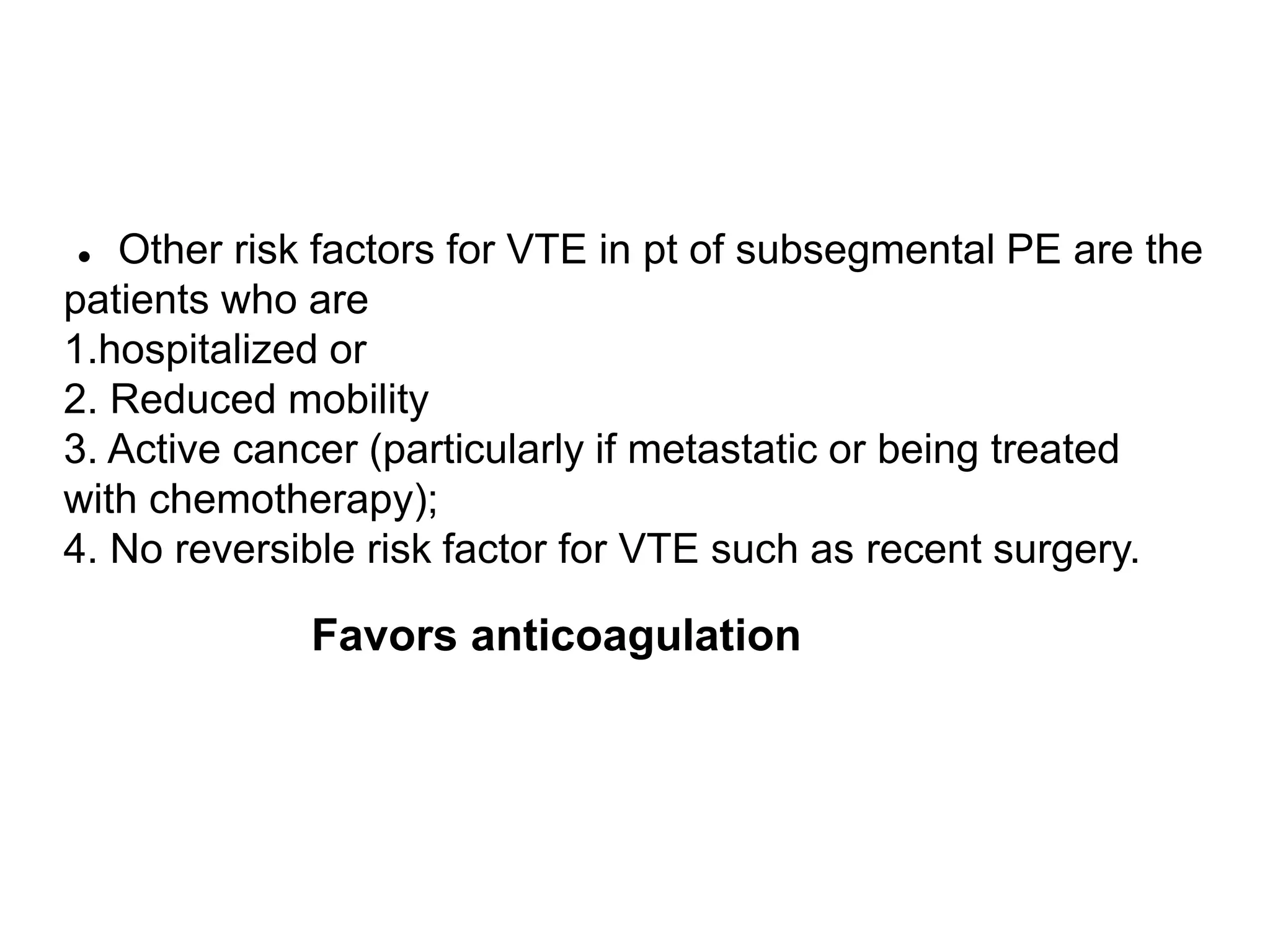
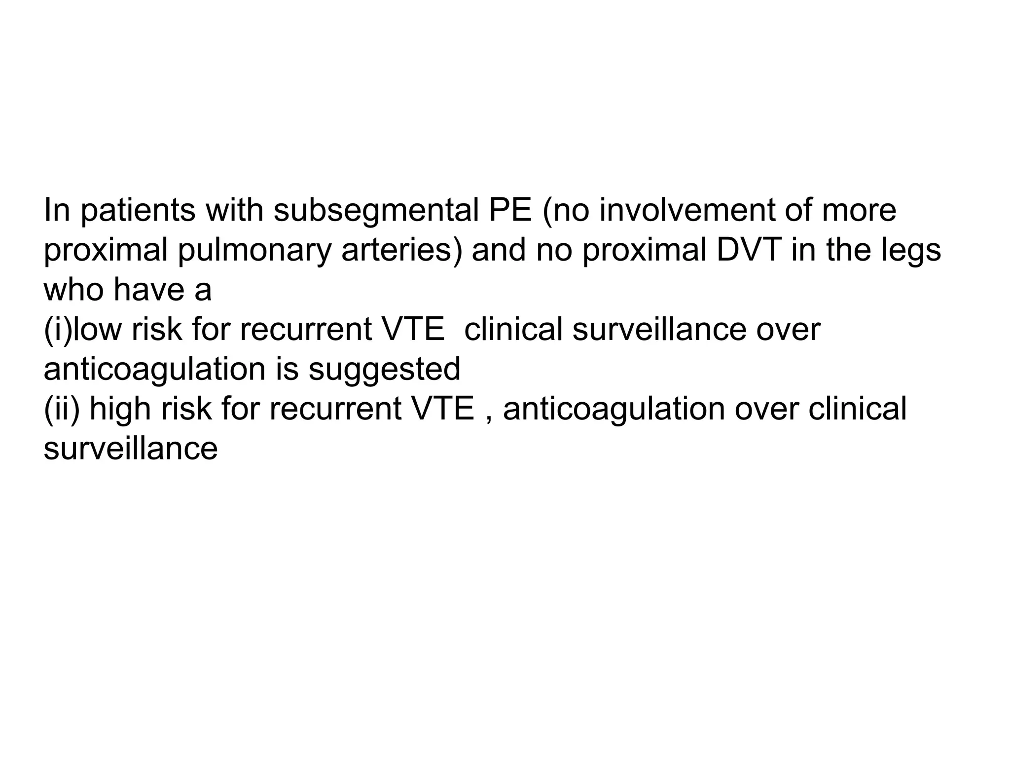
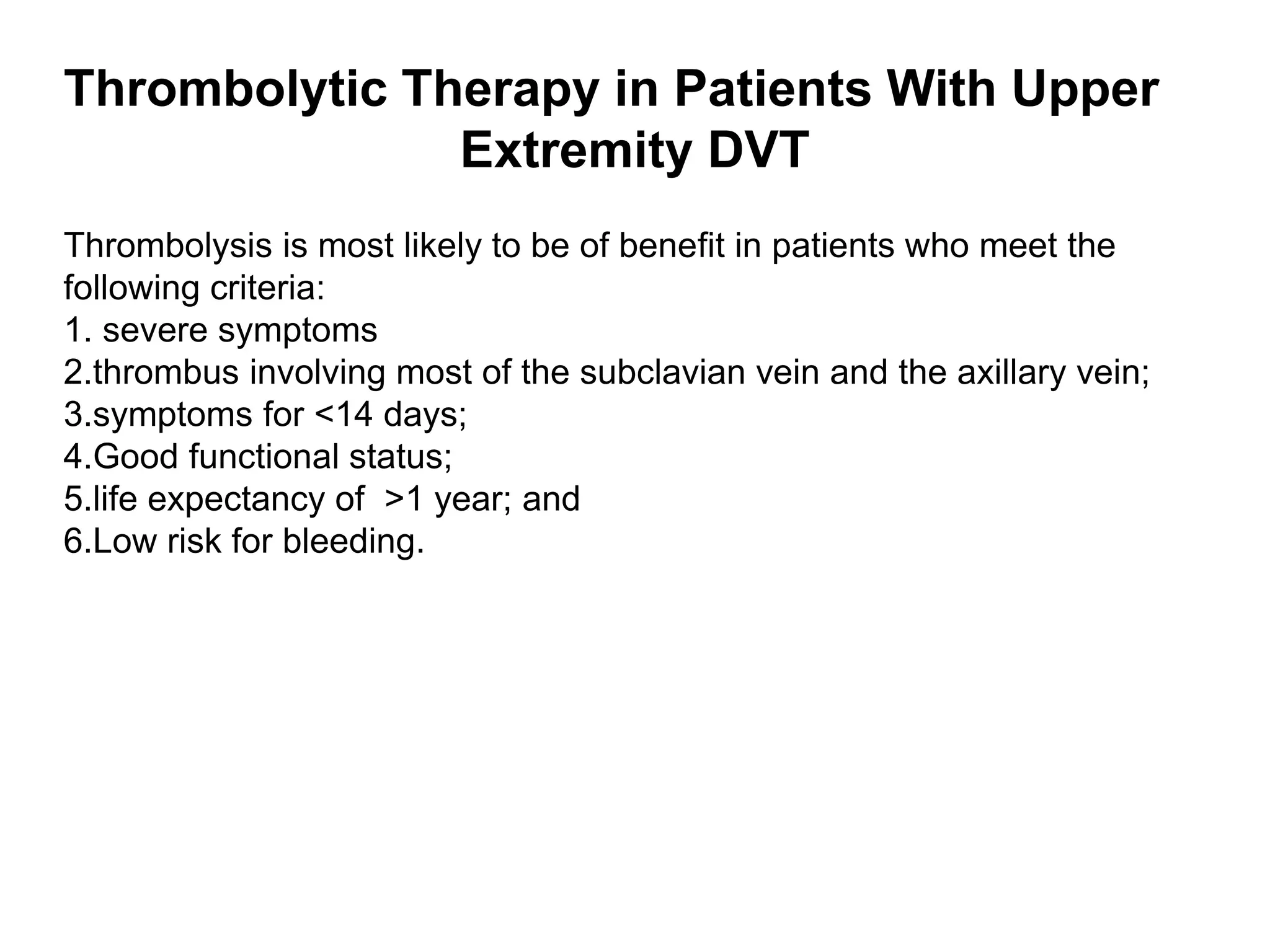
1) The document discusses guidelines for antithrombotic therapy for venous thromboembolism (VTE) from the CHEST guidelines in 2016.
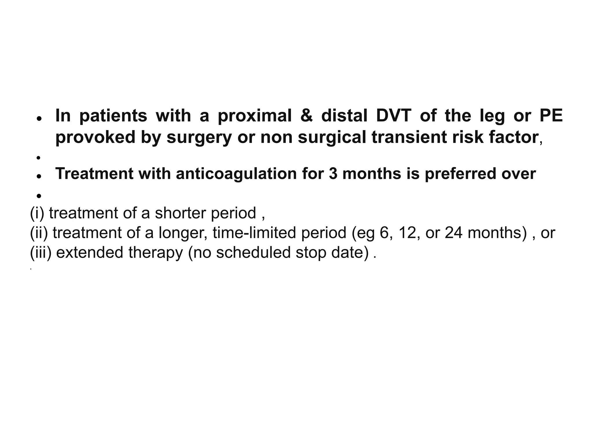
2) It reviews subgroups of VTE, classifications of anticoagulants including novel oral anticoagulants, and recommendations for duration of anticoagulant therapy based on provoking factors and bleeding risk.
3) For patients with unprovoked proximal DVT or PE, extended anticoagulant therapy indefinitely is recommended over shorter, time-limited courses for those with low or moderate bleeding risk.