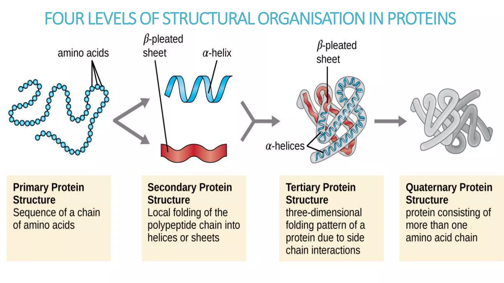
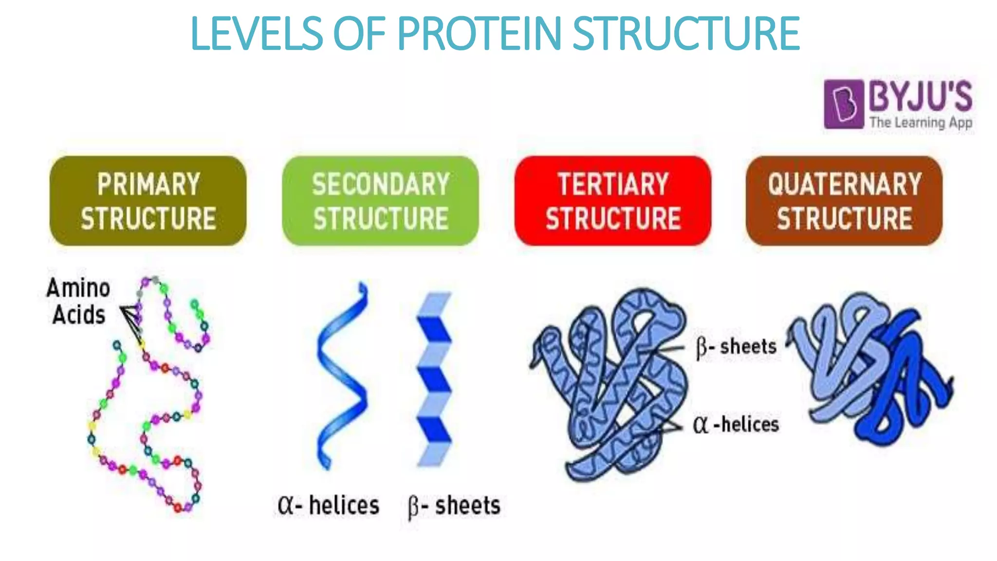
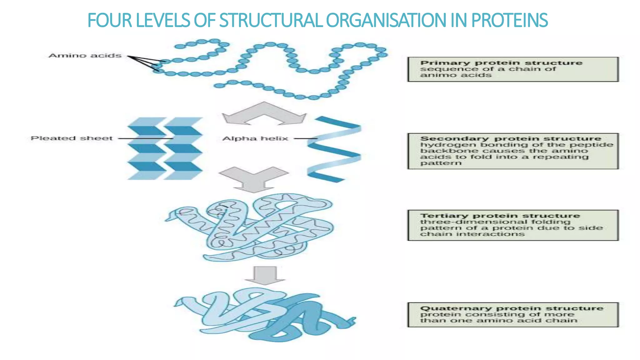
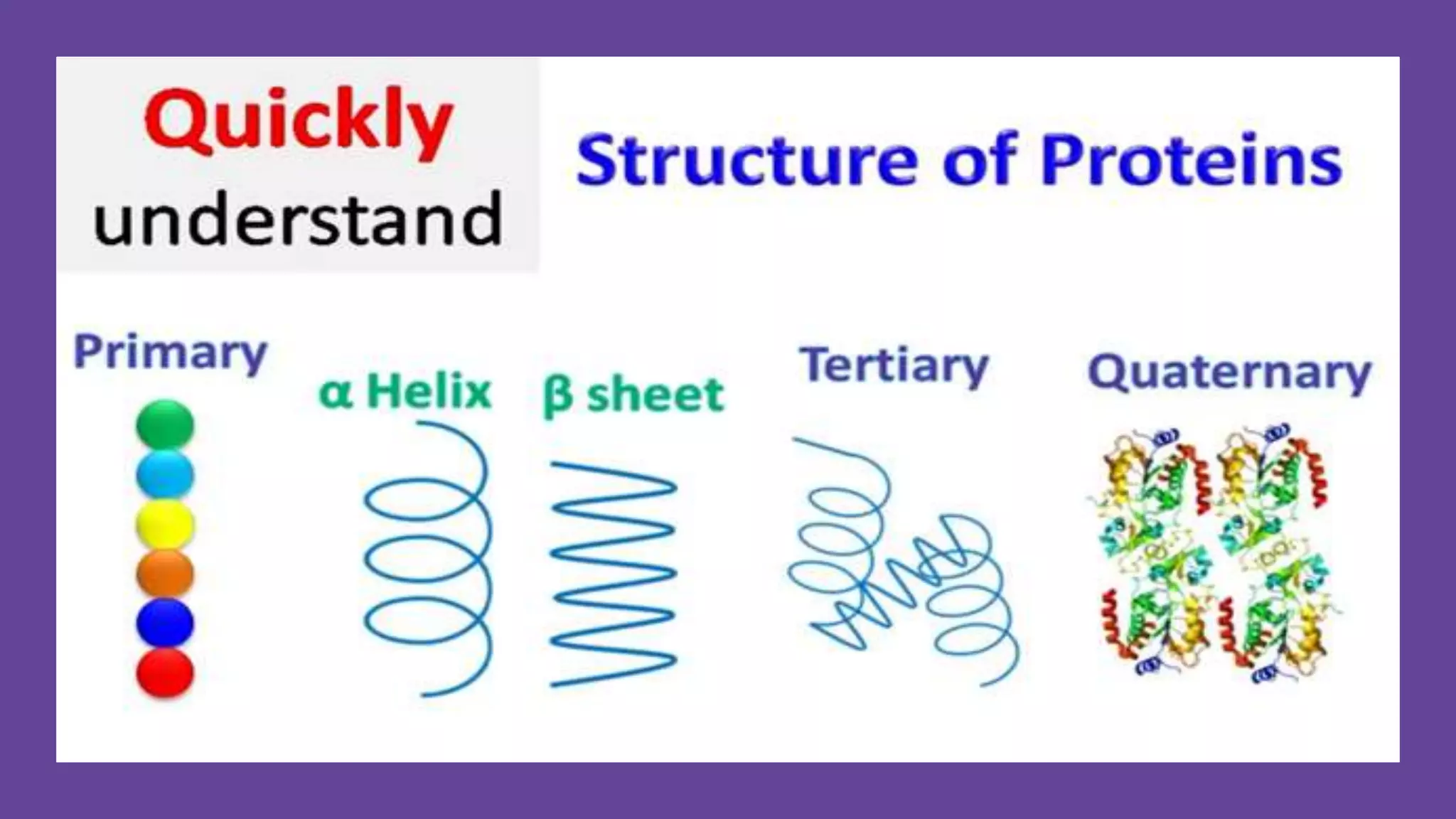
UNIVERSITYOFAGRICULTUREANDENVIRONMENTALSCIENCES,UMUAGWO
FACULTYOFSCIENCE&COMPUTING
BCH 201:GENERALBIOCHEMISTRY
PROTEINSAND
LEVELSOFPROTEINSTRUCTURALCONFORMATION
ADAKU VIVIEN IWUEKE
DEPARTMENT OF BIOLOGICAL SCIENCES
PROTEINS
Spider silk is a fibrous protein that exhibits unmatched
strength and toughness.
WHAT ARE PROTEINS ?
Protein is a macronutrient that is essential to building muscle
mass.
It is commonly found in animal products, though it is also present in
other sources, such as nuts and legumes.
Proteins makes up about 15% of a person's body weight.
PROTEINS-INTRODUCTION
In 1839 a Dutch chemist GJ Mulder while investigating
certain substances found in milk and egg discovered that they
could be coagulated on heating and were nitrogenous
compounds.
Swedish scientist JJ Berzelius suggested to Mulder that
these substances should be called proteins.
The term ‘Protein’ is derived from the Greek word Proteios
meaning “primary”, or “holding first place” or “pre-eminent”
because Berzelius thought them to be the most important of
biological substances.
And now we know that proteins are fundamental structural
components of the body.
PROTEINS-INTRODUCTION
Proteins are more or less the most important of all biological
compounds.
Chemically, proteins are unbranched polymers of amino acids
They are nitrogenous “macromolecules” composed of many
amino acids. linked head to tail, from carboxyl group to amino
group, through formation of covalent amide linkages called
peptide bonds.
Proteins are a very important class of biological macro
molecules which are the polymers of amino acids; containing
Carbon(C), hydrogen(H), Oxygen(O), and a high nitrogen(N)
content. They may also contain other elements like S, Zn, P,
Cu, Fe.
PROTEINS-INTRODUCTION
For a particular protein function, the presence of specific amino acids at a
specific positions is very important.
A polypeptide chain is formed by the linking of a number of amino acids
through peptide bonds. In the polypeptide chain each amino acid unit is
termed as residue.
There are four levels of structural organization in proteins: –
 Primary structure –
Secondary structure –
Tertiary structure –
 Quaternary structure--
BEHAVIOUR OF AMINO ACIDS AT DIFFERENT pH levels
AMIDE(PEPTIDE)BONDFORMATION
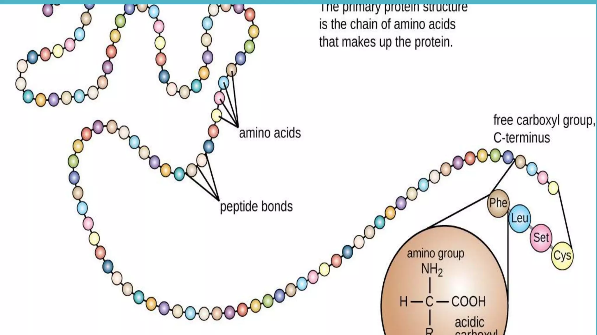
 The primary sequence of a protein is formed by the linkage of
the carboxylic acid group of Amino acid 1 with the amine
functional group of the Amino acid 2 to form an amide
(Peptide)linkage [through a dehydration synthesis (loss of
water)] .
 Similarly, the reverse reaction is hydrolysis and requires the
incorporation of a water molecule to separate two amino acids
and break the amide bond.
 Notably, peptidyl transferase (in the ribosome) serves as the
enzyme that mediates the dehydration synthesis reactions
required to build protein molecules, whereas a class of
enzymes called proteases are required for protein hydrolysis.
AMIDEBONDFORMATION
NUMBERING/NAMINGOFPEPTIDES
 Over 200 amino acids occur in nature, but only about 1/10th of these
are proteinogenic.
 Most of the naturally occurring polypeptide chains contain amino
acid residues ranging between 50 and 2000.
 They are commonly known as proteins.
 In contrast when the number of amino acids in a peptide chain is
less than 50, they are known as oligopeptides or simply peptides.
 Dipeptide: (2 amino acids & 1 peptide bond).
 Tripeptide: (3 amino acids & 2 peptide bonds)
 Oligopeptides: several amino acids (up to 20)
 Polypeptides: (more than 20 amino acids).
 All proteins are polypeptides.
Namingofaminoacidsinapolypeptidechain
The amino acid residue’s mean molecular weight is about 110. One
Dalton is equal to one atomic mass unit.
The mass of a protein is expressed in Daltons units.
 By changing the suffix “ine” to “yl” E.g. A Tripeptide like Glutathione
is named
(glutamyl-cysteinyl-glycine); A Pseudopeptide - NH2-Gly-Ala-Val-COOH
is named glycyl-alanyl-valine)
 The dipeptide L-aspartyl-L-phenylalanine is of considerable
commercial importance. Its methyl ester- Aspartame is a
sweetener.
POLYPEPTIDE CHAIN SEQUENCE
 A polypeptide chain is polar (+ -)because its ends are different, with
an α-amino group at one end and an α-carboxyl group at the other.
While writing the amino acid sequence of a polypeptide chain, the
amino terminal residue is written first.
Eg: pentapeptide Tyr-Gly-Gly-Phe-Leu (YGGFL)
Tyrosine is the amino-terminal (N-terminal) residue and leucine is the
carboxyl-terminal (C- terminal) residue.
Leu-Phe-Gly-Gly-Tyr (LFGGY) is a different pentapeptide, with different
chemical properties.
A polypeptide chain comprises of a regularly repeated part known as
the main chain or backbone, and a variable part consisting of the
characteristic side chains. The polypeptide backbone has rich
hydrogen bonding ability.
Primary structureof protein
FUNCTIONS OF PROTEINS
 Many types of proteins exist, and they perform a
variety of crucial functions which include the following:
1. Structure In animals, structural proteins contribute to
the high tensile strength of skin, bones, hair, and nails.
Two important structural proteins are collagen and
keratin.
2. Catalysis Virtually all the reactions that take place in
living organisms are catalyzed by proteins called
enzymes. Without enzymes, the reactions would take
place so slowly as to be useless.
FUNCTIONS OF PROTEINS
3. Storage Some proteins store materials in the way that
starch and glycogen store energy. For example, casein in
milk and ovalbumin in eggs store nutrients for newborn
mammals and birds. Ferritin, a protein in the liver, stores
iron.
4. Movement Every time we crook a finger, climb stairs, or
blink an eye, we use our muscles. Muscle expansion and
contraction are involved in every movement we make.
Muscles are made up of protein molecules called myosin
and actin.
FUNCTIONS OF PROTEINS
5. Hormones Many hormones are proteins, including
insulin, erythropoietin, and human growth hormone.
Chemical messengers, sending signals into the
bloodstream and tissues.
6. Protection When a protein from an outside source or
some other foreign substance (called an antigen) enters
the body, the body makes its own proteins (called
antibodies) to counteract the foreign protein. This
antibody production is one of the major mechanisms that
the body uses to fight disease. Blood clotting is another
protective function carried out by a protein, called
fibrinogen. Without blood clotting, we would bleed to
death from any small wound.
FUNCTIONS OF PROTEINS
7. Transport A large number of proteins perform
transportation functions. For example, hemoglobin, a protein
in the blood, carries oxygen from the lungs to the cells in
which it is used and carbon dioxide from the cells to the
lungs. Other proteins transport molecules across cell
membranes.
8. Regulation Some proteins not only control the
expression of genes, thereby regulating the kind of proteins
synthesized in a particular cell, but also dictate when such
manufacture takes place.
These are not the only functions of proteins, but they
are among the most important.
Clearly, any individual needs a great many proteins to
carry out these varied functions. A typical cell contains
about 9000 different proteins; the entire human body has
CLASSIFICATION OF
PROTEINS
 Proteins can be classified
into two major types:
fibrous proteins, which
are insoluble in water and
are used mainly for
structural purposes, and
globular proteins, which
are soluble in water and
are used mainly for
nonstructural purposes.
 Spider silk is a fibrous
protein that exhibits
unmatched strength
and toughness
CLASSIFICATION OF PROTEINS
Conjugated Proteins
1.Conjugated proteins are complex proteins that, when hydrolyzed,
release amino acids, inorganic, and organic components.
2.The prosthetic group is the non-amino acid portion of conjugated
proteins.
3.The chemical composition of the prosthetic group can be used to
further divide conjugated proteins.
They are –
1.Lipoproteins – proteins and lipid
2.Phosphoproteins – proteins with a phosphoric acid group
3.Nucleoproteins – protein and nucleic acid
4.Metalloproteins – metal-binding proteins with zinc, iron, or copper
5.Mucoproteins and glycoproteins – protein and carbohydrates
6.Chromoproteins – proteins and a colored pigment
FOURLEVELSOFSTRUCTURALORGANISATIONINPROTEINS
LEVELS OF PROTEIN STRUCTURE
FOURLEVELSOFSTRUCTURALORGANISATIONINPROTEINS
Primary
structure of
protein
Definition: The unique liner sequence of amino acids joined
by peptide bonds (amide bond) that make up a protein or a
polypeptide chain is known as its primary structure.
(Backbone)
Importance of Primary structure:
 Higher levels of organization of Proteins are dependent on the
primary structure.
Even a single amino acid change (mutation) in the linear
sequence can cause a serious disease eg: Sickle Cell Disease.
In HbA (normal Hemoglobin) glutamic acid is the 6th amino
acid in the beta chain. In HbS (sickle cell anemia) glutamic acid
is changed to valine.
The Primary structure shows the number and sequence of
amino acids in the peptide chain and location of disulfide
bonds if present.
The primary structure is maintained by the covalent peptide
bond
Peptide bonds are not broken down by conditions that
denature proteins such as high temperatures.
Example of primary structure of protein:
Structure of insulin
Secondary structure of protein
This the spatial arrangement of amino acid residues that are adjacent in
the primary structure by twisting and folding of the polypeptide chain.
It is maintained by:
Hydrogen bonds,
Disulphide bonds,
Hydrophobic bonds,
Van der waals forces.
Types of secondary structure: There are three main types of Secondary
structure of proteins based on the Number of Polypeptide chains present
in the polypeptide molecule.
 {α helix, β pleated sheet, loops and turns}
1) The α helix – Having One Polypeptide Chain
2) The β-pleated sheet – Having Two Polypeptide chains
3) The Triple helical structure – Having Three Polypeptide chains
Secondary structure of protein
Not all part of the polypeptide chain take an alpha-helix or a beta
conformation (secondary structure). There are also bends, loops or
turns.
Example: Carboxypeptidase shows 38 % of the amino acids
forming alpha-helix and 27 % forming beta structures and around
35 % of the residues are not included in these secondary structures.
Some regions of the long chain polypeptide will form alpha
helices and other regions will form beta pleated sheet structure.
These two types of shapes combine together and give the protein
its final shape.
Finally, the function of the protein is determined by this shape.
CARBOXYPEPTIDASE
1.ALPHAHELICALSTRUCTURE
•Thealphahelixconsistsofonepolypeptidechain.
•Itispresentbothinthefibrousproteinsandinglobularproteins.
•Itcharacterizeskeratinaswellasfibrinogenandmyosin.
2. B-PleatedSheetStructure
The β-pleated sheet structure (beta-sheet structure)
was proposed by 2 scientists, Pauling and Corey.
The β-pleated sheet structure has two Polypeptide
chains.
It consists of the juxtaposition of β strands, with a
highly stretched chain conformation.
Chains are presented in “Pleated sheets “(to take the
first topographical sense- a succession of “roofs”).
Fewer hydrogen bonds are found between the strands
2. B-PleatedSheetStructure
The beta-pleated sheet structure can be divided into
two types based on the orientation of peptide chains in a
sheet, it may be parallel or antiparallel.
In Parallel sheet structure, the orientation of the two
polypeptide chains is in the same direction. The Amino
groups (-NH2) in the two polypeptide chains are in the
same direction. Eg: β-Keratin
In Anti-Parallel sheet structure, the orientation of the
two polypeptide chains is in the opposite direction. The
Amino groups (-NH2) in the two polypeptide chains are
in the opposite direction. Eg: Silk Fibroin
Betapleatedsheet(secondarystructureofproteins)
3. The Triple Helical Structure of Proteins
In the triple helical structure, three polypeptide chains are
twisted around each other itself.
The Secondary structure of collagen is a Rod-shaped
molecule and the most abundant protein of mammals.
The structure of Collagen is a Triple helix.
It is the principal structural element of the human body and
makes up 25% to 33% of all the body proteins.
It is found in the connective tissues such as tendons,
cartilages, the organic matrix of bones and the cornea of the
eye.
Every third amino acid is Glycine in the Collagen helix.
3. The Triple Helical Structure of Proteins
OtherSecondarystructureof protein – Loops
A Loop is an example of secondary structure of protein apart
from alpha helix and beta pleated sheets.
It connects the secondary structure elements. Most proteins contain
combinations of α-helix and β- pleated sheets which are connected by
loops.
Often found at the surface of proteins. They generally have
hydrophilic residues.
Have Irregular length and shape.
Backbone groups in the loops do not form hydrogen bond to each
other. But do form hydrogen bond to water. The lack of hydrogen
bonds make loops in a protein flexible than helix and sheet structure.
Loops with only 4 or 5 residues of amino acids are called turns.
Long loops are also called random coils.
Secondarystructureofprotein–Loops
Loops participate in the formation of binding site of an enzyme active site.
Apart from connecting function, Loops can be the part of the binding site of
a ligand or a receptor.
The flexibility of loops allows them to take different local conformation. By
changing conformation (open or closed conformation) they can either block
or allow the protein function.
Classes of Loops
Hairpin loops: loop regions connecting two adjacent sheets (anti-parallel
ones).
The shortest Hairpin Loops (2-5 amino acids long) are called reverse
turns or u turns.
Omega loop: Name came from shape resemblance with Greek letter omega
The beginning and end of the loop sequence will be close together and the
middle part is open.
SUPERSECONDARYSTRUCTURE(MOTIFS)
ExamplesofSupersecondaryStructures
(A)β-hairpin-βstructuresarecharacterizedbyasharphairpinturnthatdoesnotdisruptthehydrogen
bondingofthetwoβ-pleatedsheetstructures.
(B)Proposedhelix-turn-helixstructureoftheTaspase1protein
(C)α-αcornerstructurepresentintheMyoglobinprotein.
 Produced by packing side chains from adjacent secondary structural elements close to each
other.
β – Turns
Definition: Turns and bends refer to short segments of amino acids that join two
units of secondary structure such as two adjacent strands of an antiparallel β
sheet.
Other names: β bend, reverse turns or hairpin turns
Location: Beta-turns have several unsaturated backbone hydrogen bond donors
and acceptors. The peptide groups of the central two amino acid residues in the
turn can hydrogen-bond with water. So beta turns are often found near the surface
of a protein (it is polar).
Structure: A β turn involves four amino acid residues, in which the carbonyl oxygen
of first residue is hydrogen-bonded to the hydrogen of N-H group of fourth
residue, resulting in a tight 180-degree turn.
The peptides groups of the central two residues do not participate in any inter
residue hydrogen bonding.
Amino acids commonly found: Gly (small and flexible) and Pro (Peptide bonds
involving the imino nitrogen of proline readily assume the cis
configuration) residues often occur in turns.
FUNCTIONSOFΒ–Turns
Functions: Permits the change of direction of the peptide chain to get
a folded structure. It connects the ends of two adjacent segments of
an antiparallel sheet.
Turns are classified into Types I and Type II according to the (phi, psi)
angles of the two central residues (residue 2 and 3).
Why are they are called β turns?: Usually they connect adjacent beta
strands in a beta sheet.
Why are they called reverse turns?: Because the polypeptide chain
makes a 1800 change in direction when they are connected by turns.
Why they are included under secondary structure?: Because they are
highly ordered structures stabilized by internal hydrogen bonds.
3. TERTIARY STRUCTURE
Tertiary structure denotes three-
dimensional arrangement (structure)
of whole protein.
 It defines steric relationship of
amino acids which are far apart from
each other in linear sequence, but are
close in three-dimensional aspect.
It is thermodynamically very stable. It
refers to folding of domains and to the
final arrangement of domains in the
polypeptide.
 It is maintained by:
 Hydrogen bonds,
Disulphide bonds,
 Hydrophobic bonds,
 Van der waals forces.
TERTIARY STRUCTURE CONT’
DOMAINS are fundamental functional and three
dimensional structural units of polypeptides.
 The core of domain is built from combinations of
super secondary structural elements (motifs).
 Domain is a compact globular unit of protein.
These are connected with relatively flexible areas of
protein.
STRUCTURE-FUNCTIONRELATIONSHIPINPROTEINS(FIBROUS&GLOBULARPROTEINS)
TERTIARY STRUCTURE cont’d
There are two general classes of proteins:
 Fibrous and Globular.
Fibrous proteins: Serve mainly structural roles. They have simple repeating
elements of secondary structure.
Globular proteins: They have more complicated tertiary structures. They often
contain several types of secondary structure in the same polypeptide chain.
The first globular protein structure to be determined is Myoglobin (using x-ray
diffraction methods).
Tertiary structures may contain common patterns or motifs of secondary
structures.
The complex structures of globular proteins can be analyzed by examining stable
substructures called supersecondary structure motifs or folds.
STRUCTURE - FUNCTION RELATIONSHIP IN PROTEINS
EXAMPLES OF FIBROUS PROTEINS
MORE
EXAMPLESOF
FIBROUS
PROTEINS
COMPARISON
AMINO ACIDSEQUENCE/STRUCTUREIN COLLAGEN&ELASTIN
Collagen is rich in Proline and Glycine, both of which are important
in the formation of the triple-helix (its ring structure, the smallest
amino acid, respectively).
The Glycine residues are part of the repeating sequence, (-Gly-X-Y-
), where X is frequently Proline and Y is often Hydroxyproline (Hyp)
or Hydroxylysine (Hyl). Thus, the α-chain can be regarded as a
polypeptide whose sequence can be represented as (- Gly-X-Y-)333.
Triple-helical structure: Collagen has an elongated, triple-helical
structure that places many of its amino acid side chains on the
surface of the molecule.
 - Hydroxyproline (Hyp) & Hydroxylysine (Hyl): Collagen contains
Hyp and Hyl, which result from the hydroxylation of some of the
Proline and Lysine residues.
SEQUENCE OF AMINO ACIDS IN PROTEINS
 In contrast to collagen, elastin is a connective
tissue protein with rubber-like properties.
 Elastin fibers composed of elastin and
glycoprotein micro fibrils are found in the lungs,
and walls of large arteries.
They can be stretched to several times their
normal length, but recoil to their original shape
when the stretching force is relaxed.
COLLAGENDEFICIENCYDISEASES
4. QUATERNARY STRUCTURE OF PROTEINS
 It describes polypeptide subunits aggregating to form one functional unit. .
 It is maintained by Hydrogen bonds, Electrostatic bonds, Hydrophobic
bonds, Van der waals forces.
 Proteins having more than one polypeptide chains: Oligomeric proteins e.g
A multi subunit protein is also referred to as a multimer. Multimeric proteins
can have from two to hundreds of subunits.
Each polypeptide chain in the oligomeric protein is called subunits or
monomers.
 Depending on the number of polypeptide chains, a protein is categorised
as:
A. Monomer,
B. Dimer E.g.; Creatine kinase
C. Tetramer E.g. i) Hemoglobin, (ii) Immunoglobulin.
DENATURATIONOFPROTEINS
Proteins are subject to environmental damages like oxidation
proteolysis, denaturation and other irreversible modifications.
Denaturation involves the destruction of the higher level structural
organization (20, 30 and 40) of protein with the retention of the primary
structure by denaturing agents.
A denatured protein loses its native physico-chemical and biological
properties since the bonds that stabilize the protein are broken down.
Thus the polypeptide chain unfolds itself and remain in solution in the
unfolded state. The denatured protein may retain its biological activity
by refolding (renaturing) when the denaturing agent is removed
PROTEIN DENATURATION
PROTEIN(EGG)DENATURATION
 Denaturation of a protein simply implies converting it
from the native state to a denatured state.
 When you heat an egg to about 60° C, the albumin
proteins denature and aggregate.
You are not breaking bonds when you boil an egg - you
are changing and rearranging the molecular interactions.
The aggregated protein forms large assemblies that
scatter white light, giving the egg a white colour
Denaturing agents cause the protein to lose its biological activity.
1.Physical factors
Temperature,
Pressure,
Mechanical shear force,
 Ultrasonic vibration and
 Ionizing radiation
2. Chemical factors
Acids and alkalis,
Organic solvents (acetone, ethanol),
Detergents (cleaning agents),
certain amides like urea,
Guanidine hydrochloride, alkaloids, and
Heavy metal salts (Hg, Cu, Ba, Zn, Cd...)
Factors that Affect Denaturation
STRUCTURE - FUNCTION RELATIONSHIPIN PROTEINS
MYOGLOBIN: STRUCTURE AND FUNCTION
Myoglobin is a monomeric protein and binds molecular oxygen and
carry to muscle tissues.
Muscle cells use myoglobin to exchange oxygen during active
respiration.
 Myoglobin consists of 8 right handed α-helices and each protein
molecule contains one heme prosthetic group
Each heme residue contains one central coordinately bound iron
atom.
 Oxygen is bound directly to the iron atom of the heme prosthetic
group.
It transports and stores oxygen. Binds oxygen more tightly and easily.
HAEMOGLOBIN
HAEMOGLOBIN: STRUCTURE AND FUNCTION
Hemoglobin is a tetrameric protein and
binds molecular oxygen on RBCs.
 Being a tetramer it binds four
oxygen molecules and distribute them
throughout the whole body.
 It serves to deliver the oxygen needed
for cellular metabolism and removes
the resulting waste product, carbon
dioxide from the body tissues.
 Human hemoglobin is composed of
two α (alpha) and two β (beta) subunits.
Each α-subunit has 144 residues, and
each β-subunit has 146 residues.
 Structural characteristics of both α (alpha)
and β (beta) subunits are similar to
myoglobin. It transport oxygen.
Concentration of hemoglobin is high in RBCs.
Binds oxygen loosely and with
difficulty. Hemoglobin in its deoxygenated
state has a low affinity for oxygen compared
to myoglobin.
When oxygen is bound to the first subunit of
hemoglobin it leads to subtle changes in the
quaternary structure of the protein.
This in turn makes it easier for a subsequent
molecule of oxygen to bind to the next
subunit.
Thus, with the initial oxygen binding to a
subunit, the remaining unbound subunits
become more receptive to oxygen.
PROTEINS STUDYQUESTIONS
1. Discuss the Structural Organization of Proteins.
2A) Proteins can be classified based on various factors. Discuss.
2B)What are i) Metaloproteins. (ii) Nucleoproteins give examples
3.Write short notes on the following using two (2) specific examples each:
Fibrous Proteins
Globular Proteins
4. Mention and briefly describe the bonds responsible for the formation of protein
molecules.
5.What role do proteins play in humans.
RECOMMENDED TEXT BOOKS
Biochemistry – U. Satyanarayana
Advanced organic chemistry – Bahl and Bahl
Principles of biochemistry by by Albert L. Lehninger, David L.
Nelson and Michael M. Cox
A textbook on biochemistry – MN Chatterjee
Biochemistry by Reginald H. Garrett and Charles M. Grisham
Biochemistry by by Donald Voet and Judith G. Voet
Lippincott's Illustrated Reviews: Biochemistry by Pamela C.
Champe,
Richard A. Harvey, Denise R. Ferrier
END
GOOD LUCK

PROTEINS & LEVELS OF STRUCTURAL CONFORMATION pptx.pptx

  • 1.
  • 2.
  • 3.
    Spider silk isa fibrous protein that exhibits unmatched strength and toughness.
  • 4.
    WHAT ARE PROTEINS? Protein is a macronutrient that is essential to building muscle mass. It is commonly found in animal products, though it is also present in other sources, such as nuts and legumes. Proteins makes up about 15% of a person's body weight.
  • 5.
    PROTEINS-INTRODUCTION In 1839 aDutch chemist GJ Mulder while investigating certain substances found in milk and egg discovered that they could be coagulated on heating and were nitrogenous compounds. Swedish scientist JJ Berzelius suggested to Mulder that these substances should be called proteins. The term ‘Protein’ is derived from the Greek word Proteios meaning “primary”, or “holding first place” or “pre-eminent” because Berzelius thought them to be the most important of biological substances. And now we know that proteins are fundamental structural components of the body.
  • 6.
    PROTEINS-INTRODUCTION Proteins are moreor less the most important of all biological compounds. Chemically, proteins are unbranched polymers of amino acids They are nitrogenous “macromolecules” composed of many amino acids. linked head to tail, from carboxyl group to amino group, through formation of covalent amide linkages called peptide bonds. Proteins are a very important class of biological macro molecules which are the polymers of amino acids; containing Carbon(C), hydrogen(H), Oxygen(O), and a high nitrogen(N) content. They may also contain other elements like S, Zn, P, Cu, Fe.
  • 7.
    PROTEINS-INTRODUCTION For a particularprotein function, the presence of specific amino acids at a specific positions is very important. A polypeptide chain is formed by the linking of a number of amino acids through peptide bonds. In the polypeptide chain each amino acid unit is termed as residue. There are four levels of structural organization in proteins: –  Primary structure – Secondary structure – Tertiary structure –  Quaternary structure--
  • 8.
    BEHAVIOUR OF AMINOACIDS AT DIFFERENT pH levels
  • 9.
    AMIDE(PEPTIDE)BONDFORMATION  The primarysequence of a protein is formed by the linkage of the carboxylic acid group of Amino acid 1 with the amine functional group of the Amino acid 2 to form an amide (Peptide)linkage [through a dehydration synthesis (loss of water)] .  Similarly, the reverse reaction is hydrolysis and requires the incorporation of a water molecule to separate two amino acids and break the amide bond.  Notably, peptidyl transferase (in the ribosome) serves as the enzyme that mediates the dehydration synthesis reactions required to build protein molecules, whereas a class of enzymes called proteases are required for protein hydrolysis.
  • 10.
  • 11.
    NUMBERING/NAMINGOFPEPTIDES  Over 200amino acids occur in nature, but only about 1/10th of these are proteinogenic.  Most of the naturally occurring polypeptide chains contain amino acid residues ranging between 50 and 2000.  They are commonly known as proteins.  In contrast when the number of amino acids in a peptide chain is less than 50, they are known as oligopeptides or simply peptides.  Dipeptide: (2 amino acids & 1 peptide bond).  Tripeptide: (3 amino acids & 2 peptide bonds)  Oligopeptides: several amino acids (up to 20)  Polypeptides: (more than 20 amino acids).  All proteins are polypeptides.
  • 12.
    Namingofaminoacidsinapolypeptidechain The amino acidresidue’s mean molecular weight is about 110. One Dalton is equal to one atomic mass unit. The mass of a protein is expressed in Daltons units.  By changing the suffix “ine” to “yl” E.g. A Tripeptide like Glutathione is named (glutamyl-cysteinyl-glycine); A Pseudopeptide - NH2-Gly-Ala-Val-COOH is named glycyl-alanyl-valine)  The dipeptide L-aspartyl-L-phenylalanine is of considerable commercial importance. Its methyl ester- Aspartame is a sweetener.
  • 13.
    POLYPEPTIDE CHAIN SEQUENCE A polypeptide chain is polar (+ -)because its ends are different, with an α-amino group at one end and an α-carboxyl group at the other. While writing the amino acid sequence of a polypeptide chain, the amino terminal residue is written first. Eg: pentapeptide Tyr-Gly-Gly-Phe-Leu (YGGFL) Tyrosine is the amino-terminal (N-terminal) residue and leucine is the carboxyl-terminal (C- terminal) residue. Leu-Phe-Gly-Gly-Tyr (LFGGY) is a different pentapeptide, with different chemical properties. A polypeptide chain comprises of a regularly repeated part known as the main chain or backbone, and a variable part consisting of the characteristic side chains. The polypeptide backbone has rich hydrogen bonding ability.
  • 14.
  • 15.
    FUNCTIONS OF PROTEINS Many types of proteins exist, and they perform a variety of crucial functions which include the following: 1. Structure In animals, structural proteins contribute to the high tensile strength of skin, bones, hair, and nails. Two important structural proteins are collagen and keratin. 2. Catalysis Virtually all the reactions that take place in living organisms are catalyzed by proteins called enzymes. Without enzymes, the reactions would take place so slowly as to be useless.
  • 16.
    FUNCTIONS OF PROTEINS 3.Storage Some proteins store materials in the way that starch and glycogen store energy. For example, casein in milk and ovalbumin in eggs store nutrients for newborn mammals and birds. Ferritin, a protein in the liver, stores iron. 4. Movement Every time we crook a finger, climb stairs, or blink an eye, we use our muscles. Muscle expansion and contraction are involved in every movement we make. Muscles are made up of protein molecules called myosin and actin.
  • 17.
    FUNCTIONS OF PROTEINS 5.Hormones Many hormones are proteins, including insulin, erythropoietin, and human growth hormone. Chemical messengers, sending signals into the bloodstream and tissues. 6. Protection When a protein from an outside source or some other foreign substance (called an antigen) enters the body, the body makes its own proteins (called antibodies) to counteract the foreign protein. This antibody production is one of the major mechanisms that the body uses to fight disease. Blood clotting is another protective function carried out by a protein, called fibrinogen. Without blood clotting, we would bleed to death from any small wound.
  • 18.
    FUNCTIONS OF PROTEINS 7.Transport A large number of proteins perform transportation functions. For example, hemoglobin, a protein in the blood, carries oxygen from the lungs to the cells in which it is used and carbon dioxide from the cells to the lungs. Other proteins transport molecules across cell membranes. 8. Regulation Some proteins not only control the expression of genes, thereby regulating the kind of proteins synthesized in a particular cell, but also dictate when such manufacture takes place. These are not the only functions of proteins, but they are among the most important. Clearly, any individual needs a great many proteins to carry out these varied functions. A typical cell contains about 9000 different proteins; the entire human body has
  • 21.
    CLASSIFICATION OF PROTEINS  Proteinscan be classified into two major types: fibrous proteins, which are insoluble in water and are used mainly for structural purposes, and globular proteins, which are soluble in water and are used mainly for nonstructural purposes.  Spider silk is a fibrous protein that exhibits unmatched strength and toughness
  • 22.
  • 23.
    Conjugated Proteins 1.Conjugated proteinsare complex proteins that, when hydrolyzed, release amino acids, inorganic, and organic components. 2.The prosthetic group is the non-amino acid portion of conjugated proteins. 3.The chemical composition of the prosthetic group can be used to further divide conjugated proteins. They are – 1.Lipoproteins – proteins and lipid 2.Phosphoproteins – proteins with a phosphoric acid group 3.Nucleoproteins – protein and nucleic acid 4.Metalloproteins – metal-binding proteins with zinc, iron, or copper 5.Mucoproteins and glycoproteins – protein and carbohydrates 6.Chromoproteins – proteins and a colored pigment
  • 24.
  • 25.
  • 26.
  • 28.
    Primary structure of protein Definition: Theunique liner sequence of amino acids joined by peptide bonds (amide bond) that make up a protein or a polypeptide chain is known as its primary structure. (Backbone) Importance of Primary structure:  Higher levels of organization of Proteins are dependent on the primary structure. Even a single amino acid change (mutation) in the linear sequence can cause a serious disease eg: Sickle Cell Disease. In HbA (normal Hemoglobin) glutamic acid is the 6th amino acid in the beta chain. In HbS (sickle cell anemia) glutamic acid is changed to valine. The Primary structure shows the number and sequence of amino acids in the peptide chain and location of disulfide bonds if present. The primary structure is maintained by the covalent peptide bond Peptide bonds are not broken down by conditions that denature proteins such as high temperatures.
  • 29.
    Example of primarystructure of protein: Structure of insulin
  • 30.
    Secondary structure ofprotein This the spatial arrangement of amino acid residues that are adjacent in the primary structure by twisting and folding of the polypeptide chain. It is maintained by: Hydrogen bonds, Disulphide bonds, Hydrophobic bonds, Van der waals forces. Types of secondary structure: There are three main types of Secondary structure of proteins based on the Number of Polypeptide chains present in the polypeptide molecule.  {α helix, β pleated sheet, loops and turns} 1) The α helix – Having One Polypeptide Chain 2) The β-pleated sheet – Having Two Polypeptide chains 3) The Triple helical structure – Having Three Polypeptide chains
  • 31.
    Secondary structure ofprotein Not all part of the polypeptide chain take an alpha-helix or a beta conformation (secondary structure). There are also bends, loops or turns. Example: Carboxypeptidase shows 38 % of the amino acids forming alpha-helix and 27 % forming beta structures and around 35 % of the residues are not included in these secondary structures. Some regions of the long chain polypeptide will form alpha helices and other regions will form beta pleated sheet structure. These two types of shapes combine together and give the protein its final shape. Finally, the function of the protein is determined by this shape.
  • 32.
  • 33.
  • 34.
    2. B-PleatedSheetStructure The β-pleatedsheet structure (beta-sheet structure) was proposed by 2 scientists, Pauling and Corey. The β-pleated sheet structure has two Polypeptide chains. It consists of the juxtaposition of β strands, with a highly stretched chain conformation. Chains are presented in “Pleated sheets “(to take the first topographical sense- a succession of “roofs”). Fewer hydrogen bonds are found between the strands
  • 35.
    2. B-PleatedSheetStructure The beta-pleatedsheet structure can be divided into two types based on the orientation of peptide chains in a sheet, it may be parallel or antiparallel. In Parallel sheet structure, the orientation of the two polypeptide chains is in the same direction. The Amino groups (-NH2) in the two polypeptide chains are in the same direction. Eg: β-Keratin In Anti-Parallel sheet structure, the orientation of the two polypeptide chains is in the opposite direction. The Amino groups (-NH2) in the two polypeptide chains are in the opposite direction. Eg: Silk Fibroin
  • 36.
  • 38.
    3. The TripleHelical Structure of Proteins In the triple helical structure, three polypeptide chains are twisted around each other itself. The Secondary structure of collagen is a Rod-shaped molecule and the most abundant protein of mammals. The structure of Collagen is a Triple helix. It is the principal structural element of the human body and makes up 25% to 33% of all the body proteins. It is found in the connective tissues such as tendons, cartilages, the organic matrix of bones and the cornea of the eye. Every third amino acid is Glycine in the Collagen helix.
  • 39.
    3. The TripleHelical Structure of Proteins
  • 40.
    OtherSecondarystructureof protein –Loops A Loop is an example of secondary structure of protein apart from alpha helix and beta pleated sheets. It connects the secondary structure elements. Most proteins contain combinations of α-helix and β- pleated sheets which are connected by loops. Often found at the surface of proteins. They generally have hydrophilic residues. Have Irregular length and shape. Backbone groups in the loops do not form hydrogen bond to each other. But do form hydrogen bond to water. The lack of hydrogen bonds make loops in a protein flexible than helix and sheet structure. Loops with only 4 or 5 residues of amino acids are called turns. Long loops are also called random coils.
  • 41.
    Secondarystructureofprotein–Loops Loops participate inthe formation of binding site of an enzyme active site. Apart from connecting function, Loops can be the part of the binding site of a ligand or a receptor. The flexibility of loops allows them to take different local conformation. By changing conformation (open or closed conformation) they can either block or allow the protein function. Classes of Loops Hairpin loops: loop regions connecting two adjacent sheets (anti-parallel ones). The shortest Hairpin Loops (2-5 amino acids long) are called reverse turns or u turns. Omega loop: Name came from shape resemblance with Greek letter omega The beginning and end of the loop sequence will be close together and the middle part is open.
  • 42.
  • 43.
    β – Turns Definition:Turns and bends refer to short segments of amino acids that join two units of secondary structure such as two adjacent strands of an antiparallel β sheet. Other names: β bend, reverse turns or hairpin turns Location: Beta-turns have several unsaturated backbone hydrogen bond donors and acceptors. The peptide groups of the central two amino acid residues in the turn can hydrogen-bond with water. So beta turns are often found near the surface of a protein (it is polar). Structure: A β turn involves four amino acid residues, in which the carbonyl oxygen of first residue is hydrogen-bonded to the hydrogen of N-H group of fourth residue, resulting in a tight 180-degree turn. The peptides groups of the central two residues do not participate in any inter residue hydrogen bonding. Amino acids commonly found: Gly (small and flexible) and Pro (Peptide bonds involving the imino nitrogen of proline readily assume the cis configuration) residues often occur in turns.
  • 44.
    FUNCTIONSOFΒ–Turns Functions: Permits thechange of direction of the peptide chain to get a folded structure. It connects the ends of two adjacent segments of an antiparallel sheet. Turns are classified into Types I and Type II according to the (phi, psi) angles of the two central residues (residue 2 and 3). Why are they are called β turns?: Usually they connect adjacent beta strands in a beta sheet. Why are they called reverse turns?: Because the polypeptide chain makes a 1800 change in direction when they are connected by turns. Why they are included under secondary structure?: Because they are highly ordered structures stabilized by internal hydrogen bonds.
  • 45.
    3. TERTIARY STRUCTURE Tertiarystructure denotes three- dimensional arrangement (structure) of whole protein.  It defines steric relationship of amino acids which are far apart from each other in linear sequence, but are close in three-dimensional aspect. It is thermodynamically very stable. It refers to folding of domains and to the final arrangement of domains in the polypeptide.  It is maintained by:  Hydrogen bonds, Disulphide bonds,  Hydrophobic bonds,  Van der waals forces.
  • 47.
    TERTIARY STRUCTURE CONT’ DOMAINSare fundamental functional and three dimensional structural units of polypeptides.  The core of domain is built from combinations of super secondary structural elements (motifs).  Domain is a compact globular unit of protein. These are connected with relatively flexible areas of protein.
  • 48.
  • 49.
    TERTIARY STRUCTURE cont’d Thereare two general classes of proteins:  Fibrous and Globular. Fibrous proteins: Serve mainly structural roles. They have simple repeating elements of secondary structure. Globular proteins: They have more complicated tertiary structures. They often contain several types of secondary structure in the same polypeptide chain. The first globular protein structure to be determined is Myoglobin (using x-ray diffraction methods). Tertiary structures may contain common patterns or motifs of secondary structures. The complex structures of globular proteins can be analyzed by examining stable substructures called supersecondary structure motifs or folds.
  • 50.
    STRUCTURE - FUNCTIONRELATIONSHIP IN PROTEINS
  • 51.
  • 52.
  • 53.
    AMINO ACIDSEQUENCE/STRUCTUREIN COLLAGEN&ELASTIN Collagenis rich in Proline and Glycine, both of which are important in the formation of the triple-helix (its ring structure, the smallest amino acid, respectively). The Glycine residues are part of the repeating sequence, (-Gly-X-Y- ), where X is frequently Proline and Y is often Hydroxyproline (Hyp) or Hydroxylysine (Hyl). Thus, the α-chain can be regarded as a polypeptide whose sequence can be represented as (- Gly-X-Y-)333. Triple-helical structure: Collagen has an elongated, triple-helical structure that places many of its amino acid side chains on the surface of the molecule.  - Hydroxyproline (Hyp) & Hydroxylysine (Hyl): Collagen contains Hyp and Hyl, which result from the hydroxylation of some of the Proline and Lysine residues.
  • 54.
    SEQUENCE OF AMINOACIDS IN PROTEINS  In contrast to collagen, elastin is a connective tissue protein with rubber-like properties.  Elastin fibers composed of elastin and glycoprotein micro fibrils are found in the lungs, and walls of large arteries. They can be stretched to several times their normal length, but recoil to their original shape when the stretching force is relaxed.
  • 55.
  • 56.
    4. QUATERNARY STRUCTUREOF PROTEINS  It describes polypeptide subunits aggregating to form one functional unit. .  It is maintained by Hydrogen bonds, Electrostatic bonds, Hydrophobic bonds, Van der waals forces.  Proteins having more than one polypeptide chains: Oligomeric proteins e.g A multi subunit protein is also referred to as a multimer. Multimeric proteins can have from two to hundreds of subunits. Each polypeptide chain in the oligomeric protein is called subunits or monomers.  Depending on the number of polypeptide chains, a protein is categorised as: A. Monomer, B. Dimer E.g.; Creatine kinase C. Tetramer E.g. i) Hemoglobin, (ii) Immunoglobulin.
  • 57.
    DENATURATIONOFPROTEINS Proteins are subjectto environmental damages like oxidation proteolysis, denaturation and other irreversible modifications. Denaturation involves the destruction of the higher level structural organization (20, 30 and 40) of protein with the retention of the primary structure by denaturing agents. A denatured protein loses its native physico-chemical and biological properties since the bonds that stabilize the protein are broken down. Thus the polypeptide chain unfolds itself and remain in solution in the unfolded state. The denatured protein may retain its biological activity by refolding (renaturing) when the denaturing agent is removed
  • 58.
  • 59.
    PROTEIN(EGG)DENATURATION  Denaturation ofa protein simply implies converting it from the native state to a denatured state.  When you heat an egg to about 60° C, the albumin proteins denature and aggregate. You are not breaking bonds when you boil an egg - you are changing and rearranging the molecular interactions. The aggregated protein forms large assemblies that scatter white light, giving the egg a white colour
  • 60.
    Denaturing agents causethe protein to lose its biological activity. 1.Physical factors Temperature, Pressure, Mechanical shear force,  Ultrasonic vibration and  Ionizing radiation 2. Chemical factors Acids and alkalis, Organic solvents (acetone, ethanol), Detergents (cleaning agents), certain amides like urea, Guanidine hydrochloride, alkaloids, and Heavy metal salts (Hg, Cu, Ba, Zn, Cd...) Factors that Affect Denaturation
  • 61.
    STRUCTURE - FUNCTIONRELATIONSHIPIN PROTEINS
  • 62.
    MYOGLOBIN: STRUCTURE ANDFUNCTION Myoglobin is a monomeric protein and binds molecular oxygen and carry to muscle tissues. Muscle cells use myoglobin to exchange oxygen during active respiration.  Myoglobin consists of 8 right handed α-helices and each protein molecule contains one heme prosthetic group Each heme residue contains one central coordinately bound iron atom.  Oxygen is bound directly to the iron atom of the heme prosthetic group. It transports and stores oxygen. Binds oxygen more tightly and easily.
  • 63.
  • 64.
    HAEMOGLOBIN: STRUCTURE ANDFUNCTION Hemoglobin is a tetrameric protein and binds molecular oxygen on RBCs.  Being a tetramer it binds four oxygen molecules and distribute them throughout the whole body.  It serves to deliver the oxygen needed for cellular metabolism and removes the resulting waste product, carbon dioxide from the body tissues.  Human hemoglobin is composed of two α (alpha) and two β (beta) subunits. Each α-subunit has 144 residues, and each β-subunit has 146 residues.  Structural characteristics of both α (alpha) and β (beta) subunits are similar to myoglobin. It transport oxygen. Concentration of hemoglobin is high in RBCs. Binds oxygen loosely and with difficulty. Hemoglobin in its deoxygenated state has a low affinity for oxygen compared to myoglobin. When oxygen is bound to the first subunit of hemoglobin it leads to subtle changes in the quaternary structure of the protein. This in turn makes it easier for a subsequent molecule of oxygen to bind to the next subunit. Thus, with the initial oxygen binding to a subunit, the remaining unbound subunits become more receptive to oxygen.
  • 65.
    PROTEINS STUDYQUESTIONS 1. Discussthe Structural Organization of Proteins. 2A) Proteins can be classified based on various factors. Discuss. 2B)What are i) Metaloproteins. (ii) Nucleoproteins give examples 3.Write short notes on the following using two (2) specific examples each: Fibrous Proteins Globular Proteins 4. Mention and briefly describe the bonds responsible for the formation of protein molecules. 5.What role do proteins play in humans.
  • 66.
    RECOMMENDED TEXT BOOKS Biochemistry– U. Satyanarayana Advanced organic chemistry – Bahl and Bahl Principles of biochemistry by by Albert L. Lehninger, David L. Nelson and Michael M. Cox A textbook on biochemistry – MN Chatterjee Biochemistry by Reginald H. Garrett and Charles M. Grisham Biochemistry by by Donald Voet and Judith G. Voet Lippincott's Illustrated Reviews: Biochemistry by Pamela C. Champe, Richard A. Harvey, Denise R. Ferrier
  • 67.

Editor's Notes

  • #43 Examples of Supersecondary Structures. (A) β-hairpin-β structures are characterized by a sharp hairpin turn that does not disrupt the hydrogen bonding of the two β-pleated sheet structures. (B) Proposed helix-turn-helix structure of the Taspase1 protein, (C) α-α corner structure present in the Myoglobin protein.