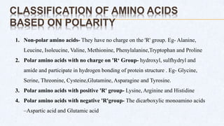
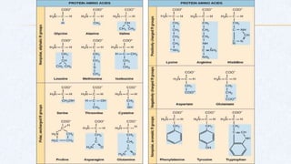
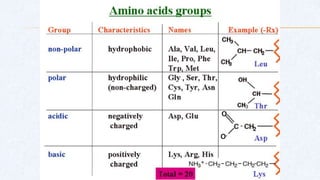
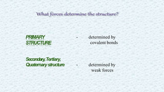
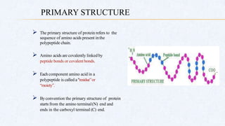
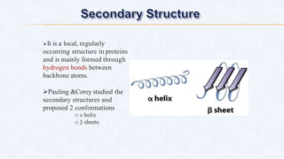
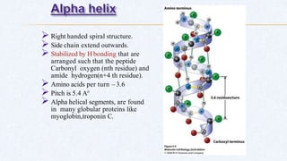
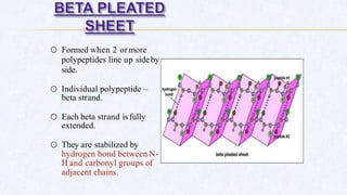
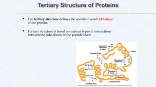
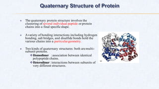
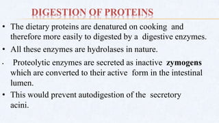
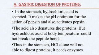
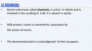
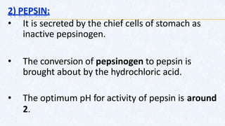
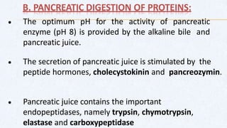
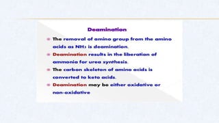
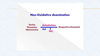
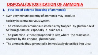
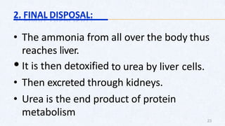
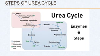
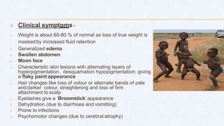
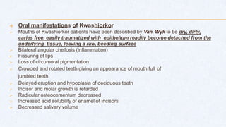
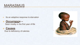
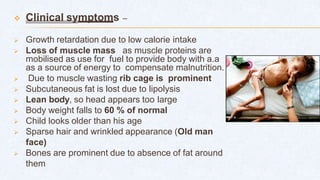
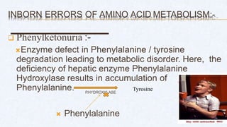
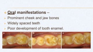
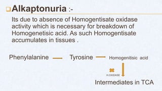
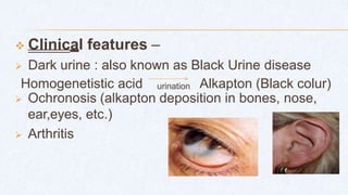
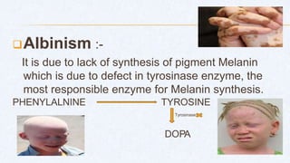
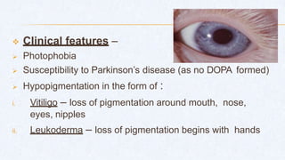
Proteins are the most abundant organic molecules of the living system.
They occur in every part of the cell and constitute about 50% of the cellular dry weight.
Proteins form the fundamental basis of structure and function of life.
Amino acids are the monomers that make up proteins
Proteins are naturally occurring polymers made up of amino acids and linked together by peptide bonds.
Proteins are the most abundant organic molecules in the living system.
The term "protein" is derived from the Greek word proteios, meaning holding the first place.
These are nitrogenous organic compounds that have large molecules weight of one or more long chains of amino acids.
Proteins are made from 20 ɑ-amino acids. (chains of amino acids)
A single unit of amino acid is known as a monomer. When many monomers combine together, they form polymers.
Absorption of proteins ppt
composition of protein ppt
digestion of protein ppt
Absorption of protein ppt
absorption of amino acid ppt
function of protein ppt
amino acid ppt
role enzyme ppt
this ppt covers about amino acids, classification, protein ,classifications, structure, denaturation, structure & fnctional relationship with applied aspects.
New Directions in Targeted Therapeutic Approaches for Older Adults With Mantl...i3 Health
i3 Health is pleased to make the speaker slides from this activity available for use as a non-accredited self-study or teaching resource.
This slide deck presented by Dr. Kami Maddocks, Professor-Clinical in the Division of Hematology and
Associate Division Director for Ambulatory Operations
The Ohio State University Comprehensive Cancer Center, will provide insight into new directions in targeted therapeutic approaches for older adults with mantle cell lymphoma.
STATEMENT OF NEED
Mantle cell lymphoma (MCL) is a rare, aggressive B-cell non-Hodgkin lymphoma (NHL) accounting for 5% to 7% of all lymphomas. Its prognosis ranges from indolent disease that does not require treatment for years to very aggressive disease, which is associated with poor survival (Silkenstedt et al, 2021). Typically, MCL is diagnosed at advanced stage and in older patients who cannot tolerate intensive therapy (NCCN, 2022). Although recent advances have slightly increased remission rates, recurrence and relapse remain very common, leading to a median overall survival between 3 and 6 years (LLS, 2021). Though there are several effective options, progress is still needed towards establishing an accepted frontline approach for MCL (Castellino et al, 2022). Treatment selection and management of MCL are complicated by the heterogeneity of prognosis, advanced age and comorbidities of patients, and lack of an established standard approach for treatment, making it vital that clinicians be familiar with the latest research and advances in this area. In this activity chaired by Michael Wang, MD, Professor in the Department of Lymphoma & Myeloma at MD Anderson Cancer Center, expert faculty will discuss prognostic factors informing treatment, the promising results of recent trials in new therapeutic approaches, and the implications of treatment resistance in therapeutic selection for MCL.
Target Audience
Hematology/oncology fellows, attending faculty, and other health care professionals involved in the treatment of patients with mantle cell lymphoma (MCL).
Learning Objectives
1.) Identify clinical and biological prognostic factors that can guide treatment decision making for older adults with MCL
2.) Evaluate emerging data on targeted therapeutic approaches for treatment-naive and relapsed/refractory MCL and their applicability to older adults
3.) Assess mechanisms of resistance to targeted therapies for MCL and their implications for treatment selection
Report Back from SGO 2024: What’s the Latest in Cervical Cancer?bkling
Are you curious about what’s new in cervical cancer research or unsure what the findings mean? Join Dr. Emily Ko, a gynecologic oncologist at Penn Medicine, to learn about the latest updates from the Society of Gynecologic Oncology (SGO) 2024 Annual Meeting on Women’s Cancer. Dr. Ko will discuss what the research presented at the conference means for you and answer your questions about the new developments.
Explore natural remedies for syphilis treatment in Singapore. Discover alternative therapies, herbal remedies, and lifestyle changes that may complement conventional treatments. Learn about holistic approaches to managing syphilis symptoms and supporting overall health.
TEST BANK for Operations Management, 14th Edition by William J. Stevenson, Ve...kevinkariuki227
TEST BANK for Operations Management, 14th Edition by William J. Stevenson, Verified Chapters 1 - 19, Complete Newest Version.pdf
Ethanol (CH3CH2OH), or beverage alcohol, is a two-carbon alcohol
that is rapidly distributed in the body and brain. Ethanol alters many
neurochemical systems and has rewarding and addictive properties. It
is the oldest recreational drug and likely contributes to more morbidity,
mortality, and public health costs than all illicit drugs combined. The
5th edition of the Diagnostic and Statistical Manual of Mental Disorders
(DSM-5) integrates alcohol abuse and alcohol dependence into a single
disorder called alcohol use disorder (AUD), with mild, moderate,
and severe subclassifications (American Psychiatric Association, 2013).
In the DSM-5, all types of substance abuse and dependence have been
combined into a single substance use disorder (SUD) on a continuum
from mild to severe. A diagnosis of AUD requires that at least two of
the 11 DSM-5 behaviors be present within a 12-month period (mild
AUD: 2–3 criteria; moderate AUD: 4–5 criteria; severe AUD: 6–11 criteria).
The four main behavioral effects of AUD are impaired control over
drinking, negative social consequences, risky use, and altered physiological
effects (tolerance, withdrawal). This chapter presents an overview
of the prevalence and harmful consequences of AUD in the U.S.,
the systemic nature of the disease, neurocircuitry and stages of AUD,
comorbidities, fetal alcohol spectrum disorders, genetic risk factors, and
pharmacotherapies for AUD.
Prix Galien International 2024 Forum ProgramLevi Shapiro
June 20, 2024, Prix Galien International and Jerusalem Ethics Forum in ROME. Detailed agenda including panels:
- ADVANCES IN CARDIOLOGY: A NEW PARADIGM IS COMING
- WOMEN’S HEALTH: FERTILITY PRESERVATION
- WHAT’S NEW IN THE TREATMENT OF INFECTIOUS,
ONCOLOGICAL AND INFLAMMATORY SKIN DISEASES?
- ARTIFICIAL INTELLIGENCE AND ETHICS
- GENE THERAPY
- BEYOND BORDERS: GLOBAL INITIATIVES FOR DEMOCRATIZING LIFE SCIENCE TECHNOLOGIES AND PROMOTING ACCESS TO HEALTHCARE
- ETHICAL CHALLENGES IN LIFE SCIENCES
- Prix Galien International Awards Ceremony
Pulmonary Thromboembolism - etilogy, types, medical- Surgical and nursing man...VarunMahajani
Disruption of blood supply to lung alveoli due to blockage of one or more pulmonary blood vessels is called as Pulmonary thromboembolism. In this presentation we will discuss its causes, types and its management in depth.
Lung Cancer: Artificial Intelligence, Synergetics, Complex System Analysis, S...Oleg Kshivets
RESULTS: Overall life span (LS) was 2252.1±1742.5 days and cumulative 5-year survival (5YS) reached 73.2%, 10 years – 64.8%, 20 years – 42.5%. 513 LCP lived more than 5 years (LS=3124.6±1525.6 days), 148 LCP – more than 10 years (LS=5054.4±1504.1 days).199 LCP died because of LC (LS=562.7±374.5 days). 5YS of LCP after bi/lobectomies was significantly superior in comparison with LCP after pneumonectomies (78.1% vs.63.7%, P=0.00001 by log-rank test). AT significantly improved 5YS (66.3% vs. 34.8%) (P=0.00000 by log-rank test) only for LCP with N1-2. Cox modeling displayed that 5YS of LCP significantly depended on: phase transition (PT) early-invasive LC in terms of synergetics, PT N0—N12, cell ratio factors (ratio between cancer cells- CC and blood cells subpopulations), G1-3, histology, glucose, AT, blood cell circuit, prothrombin index, heparin tolerance, recalcification time (P=0.000-0.038). Neural networks, genetic algorithm selection and bootstrap simulation revealed relationships between 5YS and PT early-invasive LC (rank=1), PT N0—N12 (rank=2), thrombocytes/CC (3), erythrocytes/CC (4), eosinophils/CC (5), healthy cells/CC (6), lymphocytes/CC (7), segmented neutrophils/CC (8), stick neutrophils/CC (9), monocytes/CC (10); leucocytes/CC (11). Correct prediction of 5YS was 100% by neural networks computing (area under ROC curve=1.0; error=0.0).
CONCLUSIONS: 5YS of LCP after radical procedures significantly depended on: 1) PT early-invasive cancer; 2) PT N0--N12; 3) cell ratio factors; 4) blood cell circuit; 5) biochemical factors; 6) hemostasis system; 7) AT; 8) LC characteristics; 9) LC cell dynamics; 10) surgery type: lobectomy/pneumonectomy; 11) anthropometric data. Optimal diagnosis and treatment strategies for LC are: 1) screening and early detection of LC; 2) availability of experienced thoracic surgeons because of complexity of radical procedures; 3) aggressive en block surgery and adequate lymph node dissection for completeness; 4) precise prediction; 5) adjuvant chemoimmunoradiotherapy for LCP with unfavorable prognosis.
Couples presenting to the infertility clinic- Do they really have infertility...Sujoy Dasgupta
Dr Sujoy Dasgupta presented the study on "Couples presenting to the infertility clinic- Do they really have infertility? – The unexplored stories of non-consummation" in the 13th Congress of the Asia Pacific Initiative on Reproduction (ASPIRE 2024) at Manila on 24 May, 2024.
2. CONTENT
1. Introduction
2. History
3. Functions of proteins
4. Composition
5. Structure of Amino acids
6. Classification of Amino acids
7. Structure of protein
8. Digestion of protein
9. General Metabolism of protein
10. Urea cycle
11. Metabolic Disturbances of Proteins
12. Inborn errors in metabolism
13. References
3. INTRODUCTION
Proteins are the most abundant organic molecules of
the living system.
They occur in every part of the cell and constitute
about 50% of the cellular dry weight.
Proteins form the fundamental basis of structure and
function of life.
Amino acids are the monomers that make up proteins
4. HISTORY
The term protein is derived from a Greek word
“proteios” meaning “holding the first place”
Berzelius( Swedish chemist) suggested the name
proteins to the group of organic compounds that are
utmost important to life.
Mulder (Dutch chemist) in 1838 used the term proteins
for the high molecular weight nitrogen- rich and most
abundant substances present in animals and plants
5. FUNCTIONS OF PROTEINS
Static /Structural
•Responsible for structure and strength of
body.
• Collagen and elastin in bone
•Matrix, vascular system and other organs
•a-keratin present in epidermal tissues
Dynamic
•These include proteins acting as enzyme
hormones
•Blood clotting factors
•Immunoglobulins
•Membrane receptors, storage proteins
• Muscle contraction, respiration etc
•Regarded as the working horses of cell
6. COMPOSITION OF PROTEINS
5 major elements:
Carbon-50 - 55%
Hydrogen-6 - 7.3%
Oxygen-19 - 24%
Nitrogen-13 - 19%
Sulphur- 0 – 4%
Estimation of nitrogen in the
laboratory mostly
by Kjeldahl's method is also used
to find out the
amount of protein in biological
fluids and foods
7. AMINO ACIDS
Amino acids are the monomers that make up
proteins.
Specifically, a protein is made up of one or more
linear chains of amino acids, each of which is called
a polypeptide.
There are 20 types of amino acids commonly found
in proteins.
8. STANDARD AMINO ACIDS
Amino acids are a group of
organic compounds containing
two functional groups amino
and carboxyl.
• amino group (-NH2) is basic
while
•carboxyl group (-COOH) is
acidic in nature
9. • Each amino acid has 4 different groups attatched to α-
carbon ( which is Catom next to COOH).
• These 4 groups are :amino group, COOH group ,
Hydrogen atom and side chain(R).
10. CLASSIFICATION OF AMINO ACIDS
A. Amino acid classification based on the Structure
B. Classification of amino acids based on Polarity
C. Nutritional classification of amino acids
11. AMINO ACID CLASSIFICATION BASED ON
THE STRUCTURE
1. Amino acids with aliphatic side
chains
• These are mono-amino mono-
carboxylic acids.
• Glycine
• Alanine
• Valine
• Leucine
• Isoleucine.
2. Hydroxyl group containing amino
acids :
•Serine
•Threonine
•Tyrosine
•(Tyrosine-being aromatic in nature-is
usually considered under aromatic
amino acids)
12. 3. Sulfur containing amino acids :
• Cysteine - sulfhydryl group
• Methionine -thioether group
4. Acidic amino acids and their amides
•Aspartic acid
• Glutamic acids
•Asparagine
• Glutamine
dicarboxylic mono
amino acids
are their
respective
amide
derivatives.
13. 5. Basic amino acids :
The three amino acids:
• Lysine
•Arginine (with guanidino group)
• Histidine (with imidazole ring)
are dibasic monocarboxylic acids.
6. Aromatic amino acids :
•Phenylalanine
•Tyrosine
•Tryptophan (with indole ring are
aromatic amino acids)
• Besides these, Histidine may also be
considered under this category.
7. lmino acids:
•Proline containing pyrrolidine ring is a unique amino acid.
•lt has an imino group (=NH), instead of an amino group (-
NH2) found in other amino acids.
14. CLASSIFICATION OF AMINO ACIDS
BASED ON POLARITY
1. Non-polar amino acids- They have no charge on the 'R' group. Eg- Alanine,
Leucine, Isoleucine, Valine, Methionine, Phenylalanine,Tryptophan and Proline
2. Polar amino acids with no charge on 'R‘ Group- hydroxyl, sulfhydryl and
amide and participate in hydrogen bonding of protein structure . Eg- Glycine,
Serine, Threonine, Cysteine,Glutamine, Asparagine and Tyrosine.
3. Polar amino acids with positive 'R' group- Lysine, Arginine and Histidine
4. Polar amino acids with negative 'R'group- The dicarboxylic monoamino acids
–Aspartic acid and Glutamic acid
15.
16.
17. STRUCTURE OF PROTEIN
Proteins are polymers of L-A-amino
acids
Structure of protein is complex which
can be divided into 4 levels of
orgnisations:
1. Primary structure
2. Secondary structure
3. Tertiary structure
4. Quaternary structure
19. The primary structure of protein refers to the
sequence of amino acids present inthe
polypeptide chain.
Amino acids are covalently linked by
peptide bonds or covalentbonds.
Eachcomponent amino acid in a
polypeptide is called a"residue”or
“moiety”.
Byconvention the primary structure of protein
starts from the amino terminal(N) end and
ends in the carboxyl terminal (C) end.
PRIMARY STRUCTURE
20. It is a local, regularly
occurring structure in proteins
and is mainly formed through
hydrogen bonds between
backbone atoms.
Pauling &Corey studied the
secondary structures and
proposed 2 conformations
o α helix
o β sheets.
21. Right handed spiral structure.
Side chain extend outwards.
Stabilized by H bonding that are
arranged such that the peptide
Carbonyl oxygen (nth residue) and
amide hydrogen(n+4 th residue).
Amino acids per turn – 3.6
Pitch is 5.4 A°
Alpha helical segments, are found
in many globular proteins like
myoglobin,troponin C.
22. o Formed when 2 ormore
polypeptides line up sideby
side.
o Individual polypeptide –
beta strand.
o Each beta strand is fully
extended.
o They are stabilized by
hydrogen bond between N-
H and carbonyl groups of
adjacent chains.
23. The tertiary structure defines the specific overall 3-D shape
of the protein.
Tertiary structure is based on various types of interactions
between the side-chains of the peptide chain
24. The quaternary protein structure involves the
clustering of several individual peptide or protein
chains into a final specificshape.
Avariety of bonding interactions including hydrogen
bonding, salt bridges, and disulfide bonds hold the
various chains into a particulargeometry.
Two kinds of quaternary structures: both aremulti-
subunit proteins.
Homodimer :association between identical
polypeptide chains.
Heterodimer :interactions between subunits of
very different structures.
26. DIGESTION OF PROTEINS
• The dietary proteins are denatured on cooking and
therefore more easily to digested by a digestive enzymes.
• All these enzymes are hydrolases in nature.
• Proteolytic enzymes are secreted as inactive zymogens
which are converted to their active form in the intestinal
lumen.
• This would prevent autodigestion of the secretory
acini.
27. THE PROTEOLYTIC ENZYMES INCLUDE:
• Endopeptidases:
They act on peptide bonds inside the protein molecule, so that the
protein becomes successively smaller andsmaller units. This
group includes pepsin, trypsin, chymotrypsin, and elastase.
• Exopeptidases:
This group acts at the peptide bond only at the end regionof the
chain.
• This includes carboxypeptidase acting on the peptide only at the
carboxyl terminal end on the chain
• A
minopeptidase, which acts on the peptide bond only at the
amino terminal end of the chain.
28. A. GASTRIC DIGESTION OF PROTEINS:
• In the stomach, hydrochloric acid is
secreted. It makes the pH optimum for the
action of pepsin and also activates pepsin.
•The acid also denatures the proteins. But
hydrochloric acid at body temperature could
not break the peptide bonds.
•Thus in the stomach, HCl alone will not
able to digest proteins; it needs enzymes.
29. 1) RENNIN:
• Rennin otherwise called chymosin, is active in infants and is
involved in the curdling of milk. It is absent in adults.
• Milk protein, casein is converted to paracasein by
the action ofrennin.
• The denatured protein is easilydigested further by pepsin.
30. 2) PEPSIN:
• It is secreted by the chief cells of stomach as
inactive pepsinogen.
• The conversion of pepsinogen to pepsin is
brought about by the hydrochloric acid.
• The optimum pH for activity of pepsin is around
2.
31. B. PANCREATIC DIGESTION OF PROTEINS:
• The optimum pH for the activity of pancreatic
enzyme (pH 8) is provided by the alkaline bile and
pancreatic juice.
• The secretion of pancreatic juice is stimulated by the
peptide hormones, cholecystokinin and pancreozymin.
• Pancreatic juice contains the important
endopeptidases, namely trypsin, chymotrypsin,
elastase and carboxypeptidase
32. 1) TRYPSIN:
• Trypsinogen is activated by enterokinase present on the intestinal
microvillus membranes. Once activated, the trypsin activates
other enzyme molecules.
•Trypsin catalyzes hydrolysis of the bonds formed by carboxyl
groups of Arg and Lys.
•Acute pancreatitis: Premature activation of
trypsinogen inside the pancreas itself will result in the
autodigestion of pancreatic cells. The result is acute
pancreatitis. It is a life-threatening condition
33. 2) CHYMOTRYPSIN:
• Trypsin will act on chymotrypsinogen, so that the active
site is formed. Thus, selective proteolysis produces the
catalytic site.
3) Carboxypeptidases:
• Trypsin and chymotrypsin degrade the proteins into small
peptides; these are further hydrolyzed into dipeptides
and tripeptides by carboxypeptidases present in the
pancreatic juice. They are metallo-enzymes requiring
zinc.
34. C. INTESTINAL DIGESTION OF PROTEINS:
Complete digestion of the small peptides to the
level of amino acids is brought about by enzymes
present in intestinal juice (succus entericus).
•The luminal surface of intestinal epithelial cells
contains Amino- peptidases, which release the N-
terminal amino acids successively.
36. General metabolism of amino acids:
• Dietary proteins and body proteins are broken down to amino
acids. This is called catabolic reactions.
• In transamination reaction, amino group of amino acid is removed
to produce the carbon skeleton (keto acid). The amino group is
excreted as urea.
• The carbon skeleton is used for synthesis of non- essential amino
acids.
• It is also used for gluconeogenesis or for complete oxidation.
• Amino acids are used for synthesis of body proteins; this is anabolic
reaction.
37. FORMATION OF AMMONIA
• The first step in the catabolism of amino
acids is to remove the amino group as
ammonia.
• Ammonia is highly toxic especially to the
nervous system.
• Detoxification of ammonia is by conversion to
urea and excretion through urine.
38. A. TRANSAMINATION
• Transamination is the exchange of amino group
between amino acid and another keto acid,
forming a new alpha amino acid.
• The enzyme catalyzing the reaction as a group
known as transaminases (amino transferases).
• These enzymes have pyridoxal phosphate as
prosthetic group.
• The reaction is readily reversible.
40. BIOLOGICAL SIGNIFICANCE OF TRANSAMINATION
1. First step of catabolism:
Ammonia is removed, and rest of the amino acid
is entering into catabolic pathway.
2. Synthesis of non-essential amino acids:
By means of transamination, all non-essential
amino acids could be synthesized by the body from
keto acids available for other sources 18
41. B. TRANS-DEAMINATION
• It means transamination followed by oxidative deamination.
• All amino acids are first transaminated to
glutamate, which is then finally deaminated.
• Glutamate dehydrogenase reaction is the final reaction which
removes the amino group of all amino acids.
20
AA+ketoacid1 Ketoacid2+ Glutamate Amino
grp+ketoacid3
transamination Trans-deamination
47. DISPOSAL/DETOXIFICATION OF AMMONIA
1. First line of defense (Trapping of ammonia):
• Even very minute quantity of ammonia may produce
toxicity in central nervous system.
• The intracellular ammonia is immediately trapped by glutamic acid
to form glutamine, especially in brain cells.
• The glutamine is then transported to liver,where the reaction is
reversed by the enzyme glutaminase.
• The ammonia thus generated is immediately detoxified into urea.
22
48. 2. FINAL DISPOSAL:
• The ammonia from all over the body thus
reaches liver.
•It is then detoxified to urea by liver cells.
• Then excreted through kidneys.
• Urea is the end product of protein
metabolism
23
50. UREA CYCLE
• The cycle is known as Krebs-Henseleit urea
cycle.
• As ornithine is the first member of the reaction
sequences, it is called as Ornithine cycle.
• The two nitrogen atoms of urea are derived from
two different sources, one from ammonia and
the other directly from aspartic acid.
24
51. STEPS OF UREA CYCLE
1. Formation of Carbamoyl Phosphate.
2. Formation of Citrulline.
3. Formation of Argininosuccinate.
4. Formation of Arginine.
5. Formation of Urea.
26
54. REGULATION OF THE UREA CYCLE
• During starvation, the activity of urea cycle
enzymes is elevated to meet the increased
rate of protein catabolism.
• The major regulatory steps is catalyzed by
CPS-I (Carbamoyl phosphate synthetase-I)
where the positive effectror is N-acetyl
glutamate (NAG).
29
55. DISORDERERS OF UREA CYCLE
• Deficiency of any of the urea cycle enzymes
would result in hyperammonemia.
• If block occur in one of the earlier steps, the
condition is more severe, since ammonia itself
accumulates.
• If deficiency occur in later enzymes, this result
in accumulation of other intermediates which
are less toxic and hence symptoms are less. 30
56. • The accumulation of ammonia in
blood (normally less than 50
mg/dl) and body fluids results in
toxic symptoms.
• Brain is very sensitive to ammonia.
• Child may be put on a low protein diet
and frequent small feeds are given.
• Since Citrulline is present in
significant quantities in milk,
breast milk is to be avoided in
Citrullinemia.
31
57. UREA LEVEL IN BLOOD AND URINE
• In clinical practice, blood urea level is taken as an indicator of
renal function.
• The normal urea level in plasma is from 20 to 40 mg/dl.
• Blood urea level is increased where renal function is inadequate.
• Urinary excretion of urea is 15 to 30 g/day (6-15 g
nitrogen/day).
• Urea constitutes 80% of urinary organic solids.
32
58. METABOLIC DISTURBANCES OF PROTEINS
I. Protein Energy Malnutrition (PEM) :-
•PEM is a spectrum of diseases whose
essential feature is a deficiency of protein
at one end as in Kwashiorkor and ;
•total inanition(starvation of infant due to
severe and prolonged restriction of all food
at other end as in Marasmus.
•In the middle of spectrum,there is
Marasmic Kwashiorkor in which there are
clinical features of both disorders.
59. Causes of PEM :-
Dietary deficiency
Serious illness
Infections in babies
Low socio-economic status
Problems in mother leading to inadequate
production of milk
Dietary factors like inadequate breast feeding, ignorance
of weaning
61. KWASHIORKOR
Its a maladaptive response to starvation due to lack
of physiological adaptation to unbalanced deficiency
where body utilizes proteins and conserves fat
Occurrence –
Seen predominantly between 1-5 years of age
Causes –
Due to insufficient intake of proteins, as the diet of
weaning child mainly consists of carbohydrates
62. Clinical symptoms -
Weight is about 60-80 % of normal as loss of true weight is
maskedby increased fluid retention
Generalized edema
Swollen abdomen
Moon face
Characteristic skin lesions with alternating layers of
hyperpigmentation, desquamation hypopigmentation; giving
a flaky paint appearance
Hair changes like loss of colour or alternate bands of pale
and darker colour, straightening and loss of firm
attachment to scalp
Eyelashes give a ‘Broomstick’ appearance
Dehydration (due to diarrhoea and vomitting)
Prone to infections
Psychomotor changes (due to cerebral atrophy)
63.
64. Oral manifestations of Kwashiorkor
Mouths of Kwashiorkor patients have been described by Van Wyk to be dry, dirty,
caries free, easily traumatized with epithelium readily become detached from the
underlying tissue, leaving a raw, beeding surface
Bilateral angular cheilosis (inflammation)
Fissuring of lips
Loss of circumoral pigmentation
Crowded and rotated teeth giving an appearance of mouth full of
jumbled teeth
Delayed eruption and hypoplasia of deciduous teeth
Incisor and molar growth is retarded
Radicular osteocementum decreased
Increased acid solubility of enamel of incisors
Decreased salivary volume
65. MARASMUS
d
Its an adaptive response to starvation
Occurrence –
Seen mostly in the first year of life
Causes –
Due to deficiency of calories
66. Clinical symptoms –
Growth retardation due to low calorie intake
Loss of muscle mass as muscle proteins are
mobilised as use for fuel to provide body with a.a
as a source of energy to compensate malnutrition.
Due to muscle wasting rib cage is prominent
Subcutaneous fat is lost due to lipolysis
Lean body, so head appears too large
Body weight falls to 60 % of normal
Child looks older than his age
Sparse hair and wrinkled appearance (Old man
face)
Bones are prominent due to absence of fat around
them
67.
68. DIFFERENTIATION BETWEEN KWASHIORKOR AND
MARASMUS
KWASHIORKOR
1. Malnutrition that occurs due to
insufficient intake of proteins
2. Large belly, diarrhoea, pigmented skin,
hair changes
3. Children of 1-5 yrs age
4. Weaned from mother’s milk to a diet
low in protein
5. Edema-Present (pitting type)
6. 60-80 % of normal body wt.
7. Decreased plasma albumin
8. Treated by High protein foods
MARASMUS
1. Malnutrition that occurs due to
starvation (i.e deficiency of proteins,
carbs. and fats in diet
2. Muscle wasting, skin foldings,
prominent rib cage , shrunken
abdomen
3. Children under 1 yr
4. Failed breastfeeding (inadequate
calorie intake)
5. Absent, rather wasting
6. Less than 60 % of normal
7. Normal / slightly decreased plasma
albumin
8. Treated by a Well banced diet
70. AMYLOIDOSIS
Disorder characterized by extracellular deposits
of fibrillar proteins which is a result of
aggregation of misfolded proteins.
Misfolded proteins being unstable start self
associating forming oligomers and fibrils that
are deposited in tissues.
71. Clinical manifestations :-
Weakness,fatigue,weightloss
Renal and hepatic diseases
Proteinuria
Cardiac arrythemia
Changes in skin color
Clay-colored stools
Feeling of fullness
Joint pain
Low red blood cell count (anemia)
Shortness of breath
Swelling of the tongue
Tingling and numbness in legs and feet
Weak hand grip
73. A. TONGUE AMYLOIDOSIS :-
Feature of systemic amyloidosis
Macroglossia with nodular deposits
Lateral ridging of enlarged tongue due to teeth
indentation
Firm tongue with yellow nodules on lateral surface
Interference with taste and hyposalivation may result from
amyloid deposition in salivary glands
Xerostomia and tongue pain
Sub mandibular swelling due to tongue enlargement can lead to
respiaratory obstruction.
74. B. PALATAL AMYLOIDOSIS :-
Rare condition
Feature of localized amyloidosis
Nodular deposits found on palate ,nasal septum
and maxillary sinus.
75. INBORN ERRORS OF AMINO ACID
METABOLISM
DISORDERS OF PHENYLALANINE AND TYROSINE
PHENYLKETONURIA
ALKAPTONURIA
ALBINISM
HARTNUP DISEASE
76. INBORN ERRORS OF AMINO ACID METABOLISM:-
Phenylketonuria :-
Enzyme defect in Phenylalanine / tyrosine
degradation leading to metabolic disorder. Here, the
deficiency of hepatic enzyme Phenylalanine
Hydroxylase results in accumulation of
Phenylalanine.
Phenylalanine
P
.HYDROXYLASE
Tyrosine
77. Oral manifestations –
Prominent cheek and jaw bones
Widely spaced teeth
Poor development of tooth enamel.
Patients are susceptible to tooth wear(erosion)
78. Alkaptonuria :-
Its due to absence of Homogentisate oxidase
activity which is necessary for breakdown of
Homogenetisic acid. As such Homogentisate
accumulates in tissues .
Phenylalanine Tyrosine Homogenitisic acid
H.OXIDASE
Intermediates in TCA
79. Clinical features –
Dark urine : also known as Black Urine disease
urination
Homogenetistic acid Alkapton (Black colur)
Ochronosis (alkapton deposition in bones, nose,
ear,eyes, etc.)
Arthritis
80. Albinism :-
It is due to lack of synthesis of pigment Melanin
which is due to defect in tyrosinase enzyme, the
most responsible enzyme for Melanin synthesis.
PHENYLALNINE TYROSINE
Tyrosinase
DOPA
81. Clinical features –
Photophobia
Susceptibility to Parkinson’s disease (as no DOPA formed)
Hypopigmentation in the form of :
i. Vitiligo – loss of pigmentation around mouth, nose,
eyes, nipples
ii. Leukoderma – loss of pigmentation begins with hands
82. REFERENCES
U.satyanarayan textbook of biochemistry (3rd
edition)
Shafer’s Textbook of oral pathology (7th
Robbin’s book of basic pathology (9th
edition)
Davidson’s book of medicine (22nd edition)
Neville’s textbook of oral and maxillofacial
pathology (2nd edition)