SlideShare a Scribd company logo
1 of 55
CHEMISTRY OF AMINO ACIDS
&
PROTEIN
MEGHANA C
Dept Of Biochemistry
ESICMC & Hospital Gulbargha
AMINO ACIDS
Amino acids, classifications based on side chain properties,
nutritional, requirement, based on metabolic fate, standard & non-
standard amino acids.
PROTEINS definition, classification based on chemical nature & solubility,
functions & nurtitional value
STRUCTURAL
ORGANIZATION OF
PROTEINS
primary, secondary tertiary & quarternary structure.
BIOLOGICALLY IMPORTANT
PEPTIDES
Glutathione, Thyrotropin releasing hormone, Enkeplins,
Vasopressin, Oxytocin, Gastrin, Antibiotics
STRUCTURE-FUNCTION
RELATIONSHIP OF PROTEIN
Fibrous protein, Globular protein
DENATURATION OF
PROTEIN
definition, causes, properties of a denatured protein, significance.
CONTENTS
What is Amino Acids?
Amino acids are basic structural building
blocks of protein.
Amino acids are the molecules having one
Amino group, one Carboxyl group, one
Hydrogen atom & one specific group R group
to the central Carbon atom
R group varies in structure, size, electric
charge & influence the solubility of amino acid
in water
The key element of amino acids are C,N,O,H.
Amino group Hydrogen Carboxyl group
PRIMARY STRUCTURE OF AMINO ACID
R- Group
variant
Amino Acids: Numbering
CH – CH – CH – CH – C – COOˉ
2 2
H
NH
I
I
3
1
2
3
4
5
6
I
2
NH
δ β α
γ
ε
LYSINE
In this amino acid α carbon atom is attached to 4 different groups, Amino
group, carboxyl group,hydrogen & side chain.
Numbering of carbon starts from carbon of carboxyl group, followed by α
carbon, β carbon, γ carbon, δ carbon, ε carbon.
To the ε carbon atom 2nd amino group is attached.
TYPES OF AMINO
ACIDS
STANDARD
AMINO ACIDS
NON
STANDARD
AMINO ACIDS
20
• Although there are 300 amino acids in nature. Standard
Amino Acids are 20 in numbers
• All protein are made up of just 20 amino acids in variant
combination
• During protein biosynthesis these 20 AA are incorporated
into protein & they are called STANDARD AMINO ACIDS
21. Selenocystine (active site of enzymes)
22. Pyrrolysine (bacterial species)
Non Standard Amino Acids does not incorporate into structure
of protein at the time of protein biosynthesis but still they play
several vital roles in the body.
D-AA
NP AA
AA-D
21. Selenocystine
Alanine ala
Arginine arg
Asparagine asn
Aspartic acid asp
Cysteine cys
Glutamine gln
Glutamic acid glu
Glycine gly
Histidine his
Isoleucine ile
Leucine leu
Lysine lys
Methionine met
Phenylalanine phe
Proline pro
Serine ser
Threonine thr
Tryptophan trp
Tyrosine tyr
Valine val
20 AMINOACIDS
CLASSIFICATION OF AMINO ACIDS
Based on structure &
Chemical Nature
Aliphatic side chain (Gly, Val, Ala)
Side chain with OH Group (Ser)
Side chain with ‘’S”
Side chain with acidic group
Side chain with basic group
Aromatic amino acids
Imino acids
Based On Polarity
Hydrophilic amino acids (Polar)
Hydrophobic amino acids (Non-Polar)
Based On Metabolic Fate
Based On Nutritional
Requirement
Ketogenic
Glucogenic
Both
Essential amino acids
Non-Essential amino acids
Semi-Essential amino acids
GLYCINE
Not attached to 4 different groups
Gly
Ala
Val
Leu
Iso
Branched chain amino
acids
Aliphatic Amino Acids
Side chain with Hydroxy Group
Serine
Thyrosine
theonine
SIDE CHAIN WITH SULPHUR GROOUP
Aspartic Acid
Side chain with acidic group
Side chain with basic group
(Dibasic monocarboxylic acid)
2 amino with 1 carboxyl group
Arg
Lys
His
Lysine
Aromatic AA, Phenylalanine
Imino Acid
Proline
It does not contain AA
It contains Imino Acid
Proline
Peptide Bonds, Peptides & Biologically Important peptides
(From the Greek word means "digested“) are short polymers of amino acid (monomers) linked by peptide
bonds, the covalent chemical bonds formed between two molecules when the carboxyl group of one molecule
reacts with the amino group of the other molecule.
The joining of amino acids in the process of making biochemical molecules like proteins is done by bonds
which are referred to as peptide bonds. This may be illustrated with the two simplest amino acids, glycine and
alanine.
PEPTIDES
When two or more amino acids are joined together by a peptide bond the resultant compound is peptide. A
peptide containing more than 10 amino acid is called polypeptide
PROTEINS ARE LARGE POLYPEPTIDES CONTAINING USUALLY MORE THAN 50 AMINO ACIDS
POLYPEPTIDE
A polypeptide is a long, continuous, and un branched peptide.
Proteins consist of one or more polypeptides arranged in a biologically
functional way and are often bound to cofactors, or other proteins.
Long peptides such as amyloid beta can be considered proteins, whereas
small proteins such as insulin can be considered peptides.
Peptide bond formation
Amino acids are linked together by condensation reaction between carboxylic and amino
groups from two different amino acids (with elimination of water).
The amide bond formed is called peptide bond.
The product is called a peptide, and named according to the number of amino acids
involved: e.g. dipeptide (2), tripeptide (3), decapeptide (10).
Big peptides (> 50 amino acids) are called polypeptides.
Peptides are denoted by the number of amino acid residues as di-, tri-, tetrapeptides and
the term “oligopeptides” is used for those with 10 or less amino acid residues.
Peptide bonds are formed by a
condensation reaction of carboxylic
group of an amino acid and amino
group of another amino acid with
removal of water molecule.
Peptide Bond Formation
Some biologically important peptides
GLUTATHIONE:
Tripeptide of glutamic acid, cysteine and glycine (GSH) functions:
1. Prevents oxidation of sulfhydryl group of proteins
2. Maintains RBC membrane structure and integrity
3. Prevents hemoglobin from getting oxidised
4. Helps in amino acid absorption
5. Helps in detoxification of xenobiotics
6. Help to scavenge free radicals.
THYROTROPIN RELEASING HORMONE (TRH):
Tripeptide secreted by hypothalamus which stimulates pituitary gland to release thyrotropic hormone
OXYTOCIN:
Nonapeptide hormone secreted by posterior pituitary, helps in contraction of uterus.
VASOPRESSIN (ADH):
Nonapeptide secreted by posterior pituitary which helps to retain water in kidneys and increase of blood pressure.
ANGIOTENSINS:
Decapeptide which has hypertensive effect.
BRADIKININ:
Nona & decapeptide and act as vasodilators.
ENKEPHALIN:
Penta-peptide found in brain. It inhibits sense of pain.
GASTROINTESTINAL HORMONES:
Gastrin, Secretin etc
ASPARTAME:
Dipeptide of aspartic acid and phenylalanine. It is 200 times sweeter than sucrose and used as a low calorie artificial
sweetener.
PROTEIN
• Proteins are polypeptides usually consisting of 50-2000 aminoacids
• However the largest protein known is a muscle protein known as TITIN
which has 2700 amino acids
• Protein are the most abundant molecules which make up about 75% of
dry weight of the body.
• Protein performs various function in the body. For example, Hormones,
enzymes, storage protein, transport protein, structural protein,
protective protein etc..
• Improper protein functions causes very large number of disease
Example, Lactosuria, Galactosemia, Sickle cell anemia.
STRUCTURE OF PROTEIN
Proteins are usually large molecules with a complex three
dimensional structure. The overall structure of a protein is described
at four levels. They are;
1.Primary structure (ASSEMBLING)
2.Secondary structure (FOLDING)
3.Tertiary structure (PACKING)
4.Quaternary structure (INTERACTION)
PEPTIDE BONDS
DISULPHIDE BOND
RAMCHANDRAN PLOT
BRANCHED PROTEIN
PSEUDO PEPTIDES
STRUCTURE OF PROTEIN
PRIMARY STRUCTURE
The primary structure refers to the sequence of amino acid rsidues
present in a protei. If the primary structure of the protein is known, we
know the following:
1.Total number of amino acid residues
2.N & C Terminal amino acid
3.Order or sequence of amino acids
4.Type or composition of amino acids
The bonds responsible for primary structure are covalent and
permanent bonds. They are:
1.Peptide bond
2.Disulphide bond
PRIMARY STRUCTURE
Peptide bond
The peptide bond is the most important bond in the proteins.
It is usually formed between the –COOH group of one aminno acid and –NH2
group of the adjacent amino acid.
Peptide bond is alternated by Cα---N on one side & C--- Cα Bond on the other
side.
Rotation is possible only around the C--- Cα & Cα---N bond but not around the
N—C bond.
Due to this restriction of movement, it is called partial double bond.
Due to the partial double bond character only the limited polypeptide chain
folding is allowed.
The angle between the Cα---N is called phi (Φ) angle
The angle between the Cα---C is called psi (Ψ) angle
The angle between the Cα---N is called phi (Φ) angle
The angle between the Cα---C is called psi (Ψ) angle
Due to the restriction, these angles can assume only limited
rotation
RAMACHANDRAN PLOT
Study of combination of phi & psi
angles which allow and disallow the
stable confirmation by the graphical
representation is called
RAMACHANDRAN PLOT
G.N. Ranachandran (1922-2001)
was a great Indian scientist who did
a markable work on structure of
protein.
DISULPHIDE BOND
Disulphide bonds are formed between two sulphide groups of two
cysteine residues present in different regions of the same polypeptide
chain (intra chain bonds) or between the two chains (inter chain bonds)
that are crosslinked.
CYSTEINE
BRANCHED CHAIN PROTEIN
As mentioned earlier, most proteins are linear polymers. However
many are branched in which branch points show disulphide bonds.
Also the different polypeptide chains are joined by disulphide bonds
For example, Insulin
Insulin has two chains
A Chain – 21 amino acids
B Chain – 31 amino acids
• There is an intra chain disulphide bond in A – Chain, between 6th & 11th residues.
• There are two interchain di-sulphide bonds are also seen
1. Between the cysteine at 7th position in both chain &
2. Between 20th cysteine in A chain & 19th cysteine in B Chain.
PSEUDO PEPTIDES
Usually most peptide bonds are formed between α---NH3 & α---COOH,
but sometimes non α-COOH is involved in peptide bond formation as in the case of
GLUTATHIONE where ˠ carboxylic group of glutamate forms peptide bond with amino
group of cysteine. Such a bond is called Pseudopeptide bond.
CO — NH ― C ― CO ― NH ― CH
COOH
I
CH
I
SH
α
γ
β
Cysteine
Glutamate
α
α
2
2
2
2
H
I
Glycine
Diagram showing peptide & pseudopeptide bond in
Glutathione
SECONDARY STRUCTURE
• Secondary structure refers
to regular, local structure of the
protein backbone, stabilised by
intramolecular and sometimes
intermolecular hydrogen bonding
of amide groups.
• There are two common types of
secondary structure. Alfa helix &
beta sheets.
• The most prevalent is the alpha
helix.
BETA – PLEATED
SHEETS
ALFA - HELIX
SECONDARY STRUCTURE
SECONDARY STRUCTURE
ALFA-HELIX
An alpha helix (or α-helix) is a sequence
of amino acids in a protein that are
twisted into a coil (a helix).
The alpha helix is the most common
structural arrangement in the secondary
structure of proteins.
SECONDARY STRUCTURE
(BETA PLEATED SHEETS)
This structure occurs when two
segments of a polypeptide chain
overlap one another and form a row of
hydrogen bonds with each other. This
can happen in a parallel arrangement,
Or in anti-parallel arrangement:
Antiparallel: In antiparallel beta
sheets, the neighbouring two
polypeptide strands run in the
opposite direction.
Parallel In parallel beta sheets,
all of the N-termini of polypeptide
strands are oriented in the same
direction.
LOOPS OR COILS & BETA BENDS
Major secondary structure confirmations are alfa
helix & beta pleated sheets. Proteins also contains
other secondary structures like loops & beta bends.
Loops or coils
Regiion between alfa helix & beta sheets they are
hairpin like structure, they are often the binding site
for antigen as in antibody.
Beta bends
These are seen at the end of beta antiparallel
sheets.
SUPER SECONDARY STRUCTURE MOTIFS
• In large globular proteins, alfa helix & beta pleated sheets assemble together in
different ways to form super secondary structural patterns or motifs.
• These motifs further interacts in tertiary & quarternary structures.
Some common super secondary structural motifs are :-
1. β-α-β loop
2. Β-hairpin motif
3. Greek key motif
The Greek-key motif
The β-hairpin motif
β-α-β motif
TERTIARY STRUCTURE
 The tertiary structure refers to the overall three
dimensional arrangement or folding of the
whole polypeptide chain. The amino acid
residues placed far apart in primary structures
or those present in different secondary structure
domains interact to form the tertiary structure.
 Tertiary structures are produced and maintained
by large number of weak bonds, such as
hydrogen bonds, electrostatics bonds and
Vander wall interactions. Many of times
intrachain covalent disulphide bond further
stabilize these structures.
QUARTERNARY STRUCTURE
1. Proteins are said to have quaternary
structure when they consist of more
than one i.e. two or more polypeptide
chains.
2. Each polypeptide chain is called
subunit or monomer and the protein
is called oligomeric or polymeric.
3. The monomeric units may be
identical (homopolymer) or different
(heteropolymer) .
4. The monomeric units are held together by non-covalent interaction
like hydrogen bonds, etc. between them. Disulphide bonds are also
involved many times.
5. A very good example is hemoglobin which consists of four subunits
of two types, which are two α and two β polypeptides chains
(molecular formula α2β2) . It is thus a quaternary protein.
6. Myoglobin has only one chain which is similar to β hemoglobin
chain. Hence, it has no quaternary structure. It has only primary,
secondary and tertiary level of structure.
STRUCTURE - FUNCTION RELATIONSHIP OF PROTEIN
Biological activity of protein is depends on It's full three dimensional
structure including the complete quaternary structure of an protein
disruption of three dimensional structure or the quaternary structure often
leads to loss of biological activity.
For example:-
1. Enzymes may become inactive.
2. Hormones may not produce biological action on the target tissue etc.
This is demonstrated by the denaturation of protein.
PROTEIN STRUCTURE DETERMINES ITS FORM AND FUNCTIONS
From structural point of view proteins are broadly divided into two types:-
1. Fibrous proteins (Collagen, Elastin)
2. Globular proteins (Enzymes, Hormones)
I. FIBROUS PROTEIN
1. Fibrous proteins, (e.g. Collagen, Elastin etc.) Have polypeptide chain
arranged in long strands or sheets.
2. They usually show a single type of secondary structure.
3. Due to this structure, they provide strength, shape, support and
external protection.
COLLAGEN
1. Collagen is a fibrous protein present in extracellular matrix.
2. Along with other components, it has a great tensile strength and is
inelastic.
3. It is one of the major protein of the body. (About 2.5% of total proteins)
and is present in most tissues and organs where plays a major role in
their shape, size and remodeling properties.
II. GLOBULAR PROTEINS
1. Globular proteins usually, are enzymes, hormones, growth factors,
regulatory proteins, etc.
2. Polypeptide chains of these proteins fold into spherical or globular shape
3. They show several types of secondary structures and super secondary
structural motif
DENATURATION
Denaturation is a process in which proteins lose the quaternary structure,
tertiary structure, and secondary structure (disruption of native protein
structure, higher level of protein organization is dismantled but the
primary structure is intact). which is present in their native state, by
application of some external stress or compound such as a strong acid or
base, a concentrated inorganic salt, an organic solvent (e.g., alcohol or
chloroform), agitation and radiation or heat. As a result protein looses its
biological function
If proteins in a living cell are denatured, this results in disruption of cell
activity and possibly cell death. Protein denaturation is also a
consequence of cell death.
The loss of solubility as a result of denaturation is called coagulation.
Denaturing agents ( cause of protein denaturation)
Physical agents like;
1. Heat
2. Mechanical Shaking
3. X-rays
4. UV-rays
Chemical agents include;
1. Strong acids
2. Strong base
3. Salts of heavy metals
4. Salicylate
5. Urea
Features of denaturation/ properties of denaturation
1. Protein looses its three dimensional form upon denaturation as a
result it looses its biological activity.
2. Primary structure is intact since peptide bond are not hydrolysed.
3. Denaturation is usually irreversable.
4. But as the peptide bonds are intact , sometimes when the
denaturing agent is removed, the process can be removed bringing
back the original structure of protein. This is known as re-
naturation.
5. Denaturation makes protein insoluble in the solvent in which it was
originally soluble.
Lyophilization
• It is the process by which water is evoporated at extremely low
temperatures in vaccum.
• Lyophilized proteins can be preserved for year together.
Coagulation
• When protein is heated irreersible denaturation results in
coagulation of protein.
• A coagulum is a thick, is a thick semi solid precipitate of protein.
• Albumin is easily coagulated as compared to globulin.
• Heat coagulation test is routinely used to detect the presence of
albumin in urine to detect kidney disease.
SIGNIFICANCE
In ischemic conditions (Myocardial Infarction) inadequate blood supply (low
oxygen to tissues) forces cell to depend on anaerobic glycolysis with increased
production of lactic acid. The intracellular denaturation causes denaturation of
protein resulting in coagulation necrosis.
DISEASE RELATED TO PROTEIN
FOLDING
A misfolded protein may result in neurological
diseases.
1. Prion disease (spongiform encephalopathies)
 Kuru in humans
 Mad cow disease in cattles
 Scrapie in sheep are the popular among
prion disease.
• These disease are caused by a protein known
as prion.
• The prion protein is present in the normal
persons. Its exact function is not known.
• A mutation causing misfolding of prion protein
leads to the abnormal accumulation of
misfolded prion protein resulting in damage of
neurons.
2. Alzheimer’s disease
A misfolded version of beta
protein in humans brains
accumulate in fibrous
deposits or plagues resulting
in dementia, progressive loss
of memory etc,. It is usually
seen in elderly patients.
AMINO ACID & PROTEIN CHEMISTRY.pptx

More Related Content

Similar to AMINO ACID & PROTEIN CHEMISTRY.pptx

Protein metabolism
Protein metabolismProtein metabolism
Protein metabolismkalakala1
 
Introduction to Proteins and Aminoacids with Clinical significance
Introduction to Proteins and Aminoacids with Clinical significanceIntroduction to Proteins and Aminoacids with Clinical significance
Introduction to Proteins and Aminoacids with Clinical significanceAhmad228
 
Proteins and Amino Acids
Proteins and Amino AcidsProteins and Amino Acids
Proteins and Amino AcidsJuliet Antiaye
 
Bioc 523
Bioc 523Bioc 523
Bioc 523mpark4
 
Classification and properties of protein
Classification and properties of proteinClassification and properties of protein
Classification and properties of proteinMark Philip Besana
 
Biol161 03
Biol161 03Biol161 03
Biol161 03gfb1
 
Amino acids and protein
Amino acids and proteinAmino acids and protein
Amino acids and proteinrupesh giri
 
Proteins chemistry project.pptx chemistry practical
Proteins chemistry project.pptx chemistry practicalProteins chemistry project.pptx chemistry practical
Proteins chemistry project.pptx chemistry practicalDevSharma303884
 
Introduction_to_proteins_and_amino_acids.pdf
Introduction_to_proteins_and_amino_acids.pdfIntroduction_to_proteins_and_amino_acids.pdf
Introduction_to_proteins_and_amino_acids.pdfFranceleiPatriceDant
 
Structure, functions and folding problems of protein
Structure, functions and folding problems of proteinStructure, functions and folding problems of protein
Structure, functions and folding problems of proteinRawat DA Greatt
 
aamino acids and proteinsHHHHHHHHHHHHHHHHHH.pptx
aamino acids and proteinsHHHHHHHHHHHHHHHHHH.pptxaamino acids and proteinsHHHHHHHHHHHHHHHHHH.pptx
aamino acids and proteinsHHHHHHHHHHHHHHHHHH.pptxamanuelmoges96
 
Biochemistry (amino acids and proteins-1) (8).pptx
Biochemistry (amino acids and proteins-1) (8).pptxBiochemistry (amino acids and proteins-1) (8).pptx
Biochemistry (amino acids and proteins-1) (8).pptxDereseBishaw
 
Biochemistry and-human-nutrition
Biochemistry and-human-nutritionBiochemistry and-human-nutrition
Biochemistry and-human-nutritionsunildawer
 
Chemistry of Protein.pdf
Chemistry of Protein.pdfChemistry of Protein.pdf
Chemistry of Protein.pdfAttiqueJavaid1
 

Similar to AMINO ACID & PROTEIN CHEMISTRY.pptx (20)

Protein metabolism
Protein metabolismProtein metabolism
Protein metabolism
 
Introduction to Proteins and Aminoacids with Clinical significance
Introduction to Proteins and Aminoacids with Clinical significanceIntroduction to Proteins and Aminoacids with Clinical significance
Introduction to Proteins and Aminoacids with Clinical significance
 
Proteins and Amino Acids
Proteins and Amino AcidsProteins and Amino Acids
Proteins and Amino Acids
 
Proteins
ProteinsProteins
Proteins
 
Bioc 523
Bioc 523Bioc 523
Bioc 523
 
Proteins
ProteinsProteins
Proteins
 
Classification and properties of protein
Classification and properties of proteinClassification and properties of protein
Classification and properties of protein
 
Biol161 03
Biol161 03Biol161 03
Biol161 03
 
Food proteins (2)
Food proteins (2)Food proteins (2)
Food proteins (2)
 
Amino acids and protein
Amino acids and proteinAmino acids and protein
Amino acids and protein
 
Proteins chemistry project.pptx chemistry practical
Proteins chemistry project.pptx chemistry practicalProteins chemistry project.pptx chemistry practical
Proteins chemistry project.pptx chemistry practical
 
Introduction_to_proteins_and_amino_acids.pdf
Introduction_to_proteins_and_amino_acids.pdfIntroduction_to_proteins_and_amino_acids.pdf
Introduction_to_proteins_and_amino_acids.pdf
 
Amino Acids-1.pptx
Amino Acids-1.pptxAmino Acids-1.pptx
Amino Acids-1.pptx
 
Structure, functions and folding problems of protein
Structure, functions and folding problems of proteinStructure, functions and folding problems of protein
Structure, functions and folding problems of protein
 
Protein-An overview
Protein-An overviewProtein-An overview
Protein-An overview
 
aamino acids and proteinsHHHHHHHHHHHHHHHHHH.pptx
aamino acids and proteinsHHHHHHHHHHHHHHHHHH.pptxaamino acids and proteinsHHHHHHHHHHHHHHHHHH.pptx
aamino acids and proteinsHHHHHHHHHHHHHHHHHH.pptx
 
Proteins
ProteinsProteins
Proteins
 
Biochemistry (amino acids and proteins-1) (8).pptx
Biochemistry (amino acids and proteins-1) (8).pptxBiochemistry (amino acids and proteins-1) (8).pptx
Biochemistry (amino acids and proteins-1) (8).pptx
 
Biochemistry and-human-nutrition
Biochemistry and-human-nutritionBiochemistry and-human-nutrition
Biochemistry and-human-nutrition
 
Chemistry of Protein.pdf
Chemistry of Protein.pdfChemistry of Protein.pdf
Chemistry of Protein.pdf
 

More from MEGHANA C

PLASMA PROTEIN (2).pptx
PLASMA PROTEIN (2).pptxPLASMA PROTEIN (2).pptx
PLASMA PROTEIN (2).pptxMEGHANA C
 
Pharmacology
PharmacologyPharmacology
PharmacologyMEGHANA C
 
Cerebro vascular accident
Cerebro vascular accidentCerebro vascular accident
Cerebro vascular accidentMEGHANA C
 
Cerebro vascular accident
Cerebro vascular accidentCerebro vascular accident
Cerebro vascular accidentMEGHANA C
 
Pre anesthetic medications by Meghana C
Pre anesthetic medications by Meghana CPre anesthetic medications by Meghana C
Pre anesthetic medications by Meghana CMEGHANA C
 
Intravenous Anestetics by Meghana C
Intravenous Anestetics by Meghana CIntravenous Anestetics by Meghana C
Intravenous Anestetics by Meghana CMEGHANA C
 
Radiological Examinations
Radiological ExaminationsRadiological Examinations
Radiological ExaminationsMEGHANA C
 
Nasogastric tube (ryles tube intubation) by meghana c
Nasogastric tube (ryles tube intubation) by meghana cNasogastric tube (ryles tube intubation) by meghana c
Nasogastric tube (ryles tube intubation) by meghana cMEGHANA C
 
Radioimmunoassay (modified copy)
Radioimmunoassay (modified copy)Radioimmunoassay (modified copy)
Radioimmunoassay (modified copy)MEGHANA C
 

More from MEGHANA C (9)

PLASMA PROTEIN (2).pptx
PLASMA PROTEIN (2).pptxPLASMA PROTEIN (2).pptx
PLASMA PROTEIN (2).pptx
 
Pharmacology
PharmacologyPharmacology
Pharmacology
 
Cerebro vascular accident
Cerebro vascular accidentCerebro vascular accident
Cerebro vascular accident
 
Cerebro vascular accident
Cerebro vascular accidentCerebro vascular accident
Cerebro vascular accident
 
Pre anesthetic medications by Meghana C
Pre anesthetic medications by Meghana CPre anesthetic medications by Meghana C
Pre anesthetic medications by Meghana C
 
Intravenous Anestetics by Meghana C
Intravenous Anestetics by Meghana CIntravenous Anestetics by Meghana C
Intravenous Anestetics by Meghana C
 
Radiological Examinations
Radiological ExaminationsRadiological Examinations
Radiological Examinations
 
Nasogastric tube (ryles tube intubation) by meghana c
Nasogastric tube (ryles tube intubation) by meghana cNasogastric tube (ryles tube intubation) by meghana c
Nasogastric tube (ryles tube intubation) by meghana c
 
Radioimmunoassay (modified copy)
Radioimmunoassay (modified copy)Radioimmunoassay (modified copy)
Radioimmunoassay (modified copy)
 

Recently uploaded

fourth grading exam for kindergarten in writing
fourth grading exam for kindergarten in writingfourth grading exam for kindergarten in writing
fourth grading exam for kindergarten in writingTeacherCyreneCayanan
 
Presentation by Andreas Schleicher Tackling the School Absenteeism Crisis 30 ...
Presentation by Andreas Schleicher Tackling the School Absenteeism Crisis 30 ...Presentation by Andreas Schleicher Tackling the School Absenteeism Crisis 30 ...
Presentation by Andreas Schleicher Tackling the School Absenteeism Crisis 30 ...EduSkills OECD
 
Accessible design: Minimum effort, maximum impact
Accessible design: Minimum effort, maximum impactAccessible design: Minimum effort, maximum impact
Accessible design: Minimum effort, maximum impactdawncurless
 
Sanyam Choudhary Chemistry practical.pdf
Sanyam Choudhary Chemistry practical.pdfSanyam Choudhary Chemistry practical.pdf
Sanyam Choudhary Chemistry practical.pdfsanyamsingh5019
 
Introduction to Nonprofit Accounting: The Basics
Introduction to Nonprofit Accounting: The BasicsIntroduction to Nonprofit Accounting: The Basics
Introduction to Nonprofit Accounting: The BasicsTechSoup
 
Software Engineering Methodologies (overview)
Software Engineering Methodologies (overview)Software Engineering Methodologies (overview)
Software Engineering Methodologies (overview)eniolaolutunde
 
Nutritional Needs Presentation - HLTH 104
Nutritional Needs Presentation - HLTH 104Nutritional Needs Presentation - HLTH 104
Nutritional Needs Presentation - HLTH 104misteraugie
 
Web & Social Media Analytics Previous Year Question Paper.pdf
Web & Social Media Analytics Previous Year Question Paper.pdfWeb & Social Media Analytics Previous Year Question Paper.pdf
Web & Social Media Analytics Previous Year Question Paper.pdfJayanti Pande
 
BAG TECHNIQUE Bag technique-a tool making use of public health bag through wh...
BAG TECHNIQUE Bag technique-a tool making use of public health bag through wh...BAG TECHNIQUE Bag technique-a tool making use of public health bag through wh...
BAG TECHNIQUE Bag technique-a tool making use of public health bag through wh...Sapna Thakur
 
Explore beautiful and ugly buildings. Mathematics helps us create beautiful d...
Explore beautiful and ugly buildings. Mathematics helps us create beautiful d...Explore beautiful and ugly buildings. Mathematics helps us create beautiful d...
Explore beautiful and ugly buildings. Mathematics helps us create beautiful d...christianmathematics
 
Student login on Anyboli platform.helpin
Student login on Anyboli platform.helpinStudent login on Anyboli platform.helpin
Student login on Anyboli platform.helpinRaunakKeshri1
 
Holdier Curriculum Vitae (April 2024).pdf
Holdier Curriculum Vitae (April 2024).pdfHoldier Curriculum Vitae (April 2024).pdf
Holdier Curriculum Vitae (April 2024).pdfagholdier
 
Russian Escort Service in Delhi 11k Hotel Foreigner Russian Call Girls in Delhi
Russian Escort Service in Delhi 11k Hotel Foreigner Russian Call Girls in DelhiRussian Escort Service in Delhi 11k Hotel Foreigner Russian Call Girls in Delhi
Russian Escort Service in Delhi 11k Hotel Foreigner Russian Call Girls in Delhikauryashika82
 
Measures of Dispersion and Variability: Range, QD, AD and SD
Measures of Dispersion and Variability: Range, QD, AD and SDMeasures of Dispersion and Variability: Range, QD, AD and SD
Measures of Dispersion and Variability: Range, QD, AD and SDThiyagu K
 
Interactive Powerpoint_How to Master effective communication
Interactive Powerpoint_How to Master effective communicationInteractive Powerpoint_How to Master effective communication
Interactive Powerpoint_How to Master effective communicationnomboosow
 
The basics of sentences session 2pptx copy.pptx
The basics of sentences session 2pptx copy.pptxThe basics of sentences session 2pptx copy.pptx
The basics of sentences session 2pptx copy.pptxheathfieldcps1
 
Unit-IV- Pharma. Marketing Channels.pptx
Unit-IV- Pharma. Marketing Channels.pptxUnit-IV- Pharma. Marketing Channels.pptx
Unit-IV- Pharma. Marketing Channels.pptxVishalSingh1417
 
Kisan Call Centre - To harness potential of ICT in Agriculture by answer farm...
Kisan Call Centre - To harness potential of ICT in Agriculture by answer farm...Kisan Call Centre - To harness potential of ICT in Agriculture by answer farm...
Kisan Call Centre - To harness potential of ICT in Agriculture by answer farm...Krashi Coaching
 

Recently uploaded (20)

fourth grading exam for kindergarten in writing
fourth grading exam for kindergarten in writingfourth grading exam for kindergarten in writing
fourth grading exam for kindergarten in writing
 
Presentation by Andreas Schleicher Tackling the School Absenteeism Crisis 30 ...
Presentation by Andreas Schleicher Tackling the School Absenteeism Crisis 30 ...Presentation by Andreas Schleicher Tackling the School Absenteeism Crisis 30 ...
Presentation by Andreas Schleicher Tackling the School Absenteeism Crisis 30 ...
 
INDIA QUIZ 2024 RLAC DELHI UNIVERSITY.pptx
INDIA QUIZ 2024 RLAC DELHI UNIVERSITY.pptxINDIA QUIZ 2024 RLAC DELHI UNIVERSITY.pptx
INDIA QUIZ 2024 RLAC DELHI UNIVERSITY.pptx
 
Accessible design: Minimum effort, maximum impact
Accessible design: Minimum effort, maximum impactAccessible design: Minimum effort, maximum impact
Accessible design: Minimum effort, maximum impact
 
Sanyam Choudhary Chemistry practical.pdf
Sanyam Choudhary Chemistry practical.pdfSanyam Choudhary Chemistry practical.pdf
Sanyam Choudhary Chemistry practical.pdf
 
Introduction to Nonprofit Accounting: The Basics
Introduction to Nonprofit Accounting: The BasicsIntroduction to Nonprofit Accounting: The Basics
Introduction to Nonprofit Accounting: The Basics
 
Software Engineering Methodologies (overview)
Software Engineering Methodologies (overview)Software Engineering Methodologies (overview)
Software Engineering Methodologies (overview)
 
Nutritional Needs Presentation - HLTH 104
Nutritional Needs Presentation - HLTH 104Nutritional Needs Presentation - HLTH 104
Nutritional Needs Presentation - HLTH 104
 
Web & Social Media Analytics Previous Year Question Paper.pdf
Web & Social Media Analytics Previous Year Question Paper.pdfWeb & Social Media Analytics Previous Year Question Paper.pdf
Web & Social Media Analytics Previous Year Question Paper.pdf
 
Mattingly "AI & Prompt Design: Structured Data, Assistants, & RAG"
Mattingly "AI & Prompt Design: Structured Data, Assistants, & RAG"Mattingly "AI & Prompt Design: Structured Data, Assistants, & RAG"
Mattingly "AI & Prompt Design: Structured Data, Assistants, & RAG"
 
BAG TECHNIQUE Bag technique-a tool making use of public health bag through wh...
BAG TECHNIQUE Bag technique-a tool making use of public health bag through wh...BAG TECHNIQUE Bag technique-a tool making use of public health bag through wh...
BAG TECHNIQUE Bag technique-a tool making use of public health bag through wh...
 
Explore beautiful and ugly buildings. Mathematics helps us create beautiful d...
Explore beautiful and ugly buildings. Mathematics helps us create beautiful d...Explore beautiful and ugly buildings. Mathematics helps us create beautiful d...
Explore beautiful and ugly buildings. Mathematics helps us create beautiful d...
 
Student login on Anyboli platform.helpin
Student login on Anyboli platform.helpinStudent login on Anyboli platform.helpin
Student login on Anyboli platform.helpin
 
Holdier Curriculum Vitae (April 2024).pdf
Holdier Curriculum Vitae (April 2024).pdfHoldier Curriculum Vitae (April 2024).pdf
Holdier Curriculum Vitae (April 2024).pdf
 
Russian Escort Service in Delhi 11k Hotel Foreigner Russian Call Girls in Delhi
Russian Escort Service in Delhi 11k Hotel Foreigner Russian Call Girls in DelhiRussian Escort Service in Delhi 11k Hotel Foreigner Russian Call Girls in Delhi
Russian Escort Service in Delhi 11k Hotel Foreigner Russian Call Girls in Delhi
 
Measures of Dispersion and Variability: Range, QD, AD and SD
Measures of Dispersion and Variability: Range, QD, AD and SDMeasures of Dispersion and Variability: Range, QD, AD and SD
Measures of Dispersion and Variability: Range, QD, AD and SD
 
Interactive Powerpoint_How to Master effective communication
Interactive Powerpoint_How to Master effective communicationInteractive Powerpoint_How to Master effective communication
Interactive Powerpoint_How to Master effective communication
 
The basics of sentences session 2pptx copy.pptx
The basics of sentences session 2pptx copy.pptxThe basics of sentences session 2pptx copy.pptx
The basics of sentences session 2pptx copy.pptx
 
Unit-IV- Pharma. Marketing Channels.pptx
Unit-IV- Pharma. Marketing Channels.pptxUnit-IV- Pharma. Marketing Channels.pptx
Unit-IV- Pharma. Marketing Channels.pptx
 
Kisan Call Centre - To harness potential of ICT in Agriculture by answer farm...
Kisan Call Centre - To harness potential of ICT in Agriculture by answer farm...Kisan Call Centre - To harness potential of ICT in Agriculture by answer farm...
Kisan Call Centre - To harness potential of ICT in Agriculture by answer farm...
 

AMINO ACID & PROTEIN CHEMISTRY.pptx

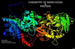
  • 1. CHEMISTRY OF AMINO ACIDS & PROTEIN MEGHANA C Dept Of Biochemistry ESICMC & Hospital Gulbargha
  • 2. AMINO ACIDS Amino acids, classifications based on side chain properties, nutritional, requirement, based on metabolic fate, standard & non- standard amino acids. PROTEINS definition, classification based on chemical nature & solubility, functions & nurtitional value STRUCTURAL ORGANIZATION OF PROTEINS primary, secondary tertiary & quarternary structure. BIOLOGICALLY IMPORTANT PEPTIDES Glutathione, Thyrotropin releasing hormone, Enkeplins, Vasopressin, Oxytocin, Gastrin, Antibiotics STRUCTURE-FUNCTION RELATIONSHIP OF PROTEIN Fibrous protein, Globular protein DENATURATION OF PROTEIN definition, causes, properties of a denatured protein, significance. CONTENTS
  • 3. What is Amino Acids? Amino acids are basic structural building blocks of protein. Amino acids are the molecules having one Amino group, one Carboxyl group, one Hydrogen atom & one specific group R group to the central Carbon atom R group varies in structure, size, electric charge & influence the solubility of amino acid in water The key element of amino acids are C,N,O,H.
  • 4. Amino group Hydrogen Carboxyl group PRIMARY STRUCTURE OF AMINO ACID R- Group variant
  • 5. Amino Acids: Numbering CH – CH – CH – CH – C – COOˉ 2 2 H NH I I 3 1 2 3 4 5 6 I 2 NH δ β α γ ε LYSINE In this amino acid α carbon atom is attached to 4 different groups, Amino group, carboxyl group,hydrogen & side chain. Numbering of carbon starts from carbon of carboxyl group, followed by α carbon, β carbon, γ carbon, δ carbon, ε carbon. To the ε carbon atom 2nd amino group is attached. TYPES OF AMINO ACIDS STANDARD AMINO ACIDS NON STANDARD AMINO ACIDS 20 • Although there are 300 amino acids in nature. Standard Amino Acids are 20 in numbers • All protein are made up of just 20 amino acids in variant combination • During protein biosynthesis these 20 AA are incorporated into protein & they are called STANDARD AMINO ACIDS 21. Selenocystine (active site of enzymes) 22. Pyrrolysine (bacterial species) Non Standard Amino Acids does not incorporate into structure of protein at the time of protein biosynthesis but still they play several vital roles in the body. D-AA NP AA AA-D
  • 6.
  • 7. 21. Selenocystine Alanine ala Arginine arg Asparagine asn Aspartic acid asp Cysteine cys Glutamine gln Glutamic acid glu Glycine gly Histidine his Isoleucine ile Leucine leu Lysine lys Methionine met Phenylalanine phe Proline pro Serine ser Threonine thr Tryptophan trp Tyrosine tyr Valine val 20 AMINOACIDS
  • 8. CLASSIFICATION OF AMINO ACIDS Based on structure & Chemical Nature Aliphatic side chain (Gly, Val, Ala) Side chain with OH Group (Ser) Side chain with ‘’S” Side chain with acidic group Side chain with basic group Aromatic amino acids Imino acids Based On Polarity Hydrophilic amino acids (Polar) Hydrophobic amino acids (Non-Polar) Based On Metabolic Fate Based On Nutritional Requirement Ketogenic Glucogenic Both Essential amino acids Non-Essential amino acids Semi-Essential amino acids
  • 9. GLYCINE Not attached to 4 different groups Gly Ala Val Leu Iso Branched chain amino acids Aliphatic Amino Acids Side chain with Hydroxy Group Serine Thyrosine theonine
  • 10. SIDE CHAIN WITH SULPHUR GROOUP Aspartic Acid Side chain with acidic group
  • 11. Side chain with basic group (Dibasic monocarboxylic acid) 2 amino with 1 carboxyl group Arg Lys His Lysine Aromatic AA, Phenylalanine Imino Acid Proline It does not contain AA It contains Imino Acid Proline
  • 12. Peptide Bonds, Peptides & Biologically Important peptides (From the Greek word means "digested“) are short polymers of amino acid (monomers) linked by peptide bonds, the covalent chemical bonds formed between two molecules when the carboxyl group of one molecule reacts with the amino group of the other molecule. The joining of amino acids in the process of making biochemical molecules like proteins is done by bonds which are referred to as peptide bonds. This may be illustrated with the two simplest amino acids, glycine and alanine. PEPTIDES When two or more amino acids are joined together by a peptide bond the resultant compound is peptide. A peptide containing more than 10 amino acid is called polypeptide PROTEINS ARE LARGE POLYPEPTIDES CONTAINING USUALLY MORE THAN 50 AMINO ACIDS
  • 13. POLYPEPTIDE A polypeptide is a long, continuous, and un branched peptide. Proteins consist of one or more polypeptides arranged in a biologically functional way and are often bound to cofactors, or other proteins. Long peptides such as amyloid beta can be considered proteins, whereas small proteins such as insulin can be considered peptides.
  • 14. Peptide bond formation Amino acids are linked together by condensation reaction between carboxylic and amino groups from two different amino acids (with elimination of water). The amide bond formed is called peptide bond. The product is called a peptide, and named according to the number of amino acids involved: e.g. dipeptide (2), tripeptide (3), decapeptide (10). Big peptides (> 50 amino acids) are called polypeptides. Peptides are denoted by the number of amino acid residues as di-, tri-, tetrapeptides and the term “oligopeptides” is used for those with 10 or less amino acid residues.
  • 15. Peptide bonds are formed by a condensation reaction of carboxylic group of an amino acid and amino group of another amino acid with removal of water molecule. Peptide Bond Formation
  • 16. Some biologically important peptides GLUTATHIONE: Tripeptide of glutamic acid, cysteine and glycine (GSH) functions: 1. Prevents oxidation of sulfhydryl group of proteins 2. Maintains RBC membrane structure and integrity 3. Prevents hemoglobin from getting oxidised 4. Helps in amino acid absorption 5. Helps in detoxification of xenobiotics 6. Help to scavenge free radicals. THYROTROPIN RELEASING HORMONE (TRH): Tripeptide secreted by hypothalamus which stimulates pituitary gland to release thyrotropic hormone OXYTOCIN: Nonapeptide hormone secreted by posterior pituitary, helps in contraction of uterus. VASOPRESSIN (ADH): Nonapeptide secreted by posterior pituitary which helps to retain water in kidneys and increase of blood pressure.
  • 17. ANGIOTENSINS: Decapeptide which has hypertensive effect. BRADIKININ: Nona & decapeptide and act as vasodilators. ENKEPHALIN: Penta-peptide found in brain. It inhibits sense of pain. GASTROINTESTINAL HORMONES: Gastrin, Secretin etc ASPARTAME: Dipeptide of aspartic acid and phenylalanine. It is 200 times sweeter than sucrose and used as a low calorie artificial sweetener.
  • 18. PROTEIN • Proteins are polypeptides usually consisting of 50-2000 aminoacids • However the largest protein known is a muscle protein known as TITIN which has 2700 amino acids • Protein are the most abundant molecules which make up about 75% of dry weight of the body. • Protein performs various function in the body. For example, Hormones, enzymes, storage protein, transport protein, structural protein, protective protein etc.. • Improper protein functions causes very large number of disease Example, Lactosuria, Galactosemia, Sickle cell anemia.
  • 19.
  • 20. STRUCTURE OF PROTEIN Proteins are usually large molecules with a complex three dimensional structure. The overall structure of a protein is described at four levels. They are; 1.Primary structure (ASSEMBLING) 2.Secondary structure (FOLDING) 3.Tertiary structure (PACKING) 4.Quaternary structure (INTERACTION) PEPTIDE BONDS DISULPHIDE BOND RAMCHANDRAN PLOT BRANCHED PROTEIN PSEUDO PEPTIDES
  • 22. PRIMARY STRUCTURE The primary structure refers to the sequence of amino acid rsidues present in a protei. If the primary structure of the protein is known, we know the following: 1.Total number of amino acid residues 2.N & C Terminal amino acid 3.Order or sequence of amino acids 4.Type or composition of amino acids The bonds responsible for primary structure are covalent and permanent bonds. They are: 1.Peptide bond 2.Disulphide bond
  • 24. Peptide bond The peptide bond is the most important bond in the proteins. It is usually formed between the –COOH group of one aminno acid and –NH2 group of the adjacent amino acid. Peptide bond is alternated by Cα---N on one side & C--- Cα Bond on the other side. Rotation is possible only around the C--- Cα & Cα---N bond but not around the N—C bond. Due to this restriction of movement, it is called partial double bond. Due to the partial double bond character only the limited polypeptide chain folding is allowed. The angle between the Cα---N is called phi (Φ) angle The angle between the Cα---C is called psi (Ψ) angle
  • 25. The angle between the Cα---N is called phi (Φ) angle The angle between the Cα---C is called psi (Ψ) angle Due to the restriction, these angles can assume only limited rotation
  • 26. RAMACHANDRAN PLOT Study of combination of phi & psi angles which allow and disallow the stable confirmation by the graphical representation is called RAMACHANDRAN PLOT G.N. Ranachandran (1922-2001) was a great Indian scientist who did a markable work on structure of protein.
  • 27. DISULPHIDE BOND Disulphide bonds are formed between two sulphide groups of two cysteine residues present in different regions of the same polypeptide chain (intra chain bonds) or between the two chains (inter chain bonds) that are crosslinked. CYSTEINE
  • 28. BRANCHED CHAIN PROTEIN As mentioned earlier, most proteins are linear polymers. However many are branched in which branch points show disulphide bonds. Also the different polypeptide chains are joined by disulphide bonds
  • 29. For example, Insulin Insulin has two chains A Chain – 21 amino acids B Chain – 31 amino acids • There is an intra chain disulphide bond in A – Chain, between 6th & 11th residues. • There are two interchain di-sulphide bonds are also seen 1. Between the cysteine at 7th position in both chain & 2. Between 20th cysteine in A chain & 19th cysteine in B Chain.
  • 30. PSEUDO PEPTIDES Usually most peptide bonds are formed between α---NH3 & α---COOH, but sometimes non α-COOH is involved in peptide bond formation as in the case of GLUTATHIONE where ˠ carboxylic group of glutamate forms peptide bond with amino group of cysteine. Such a bond is called Pseudopeptide bond.
  • 31. CO — NH ― C ― CO ― NH ― CH COOH I CH I SH α γ β Cysteine Glutamate α α 2 2 2 2 H I Glycine Diagram showing peptide & pseudopeptide bond in Glutathione
  • 32. SECONDARY STRUCTURE • Secondary structure refers to regular, local structure of the protein backbone, stabilised by intramolecular and sometimes intermolecular hydrogen bonding of amide groups. • There are two common types of secondary structure. Alfa helix & beta sheets. • The most prevalent is the alpha helix. BETA – PLEATED SHEETS ALFA - HELIX
  • 34. SECONDARY STRUCTURE ALFA-HELIX An alpha helix (or α-helix) is a sequence of amino acids in a protein that are twisted into a coil (a helix). The alpha helix is the most common structural arrangement in the secondary structure of proteins.
  • 35. SECONDARY STRUCTURE (BETA PLEATED SHEETS) This structure occurs when two segments of a polypeptide chain overlap one another and form a row of hydrogen bonds with each other. This can happen in a parallel arrangement, Or in anti-parallel arrangement:
  • 36. Antiparallel: In antiparallel beta sheets, the neighbouring two polypeptide strands run in the opposite direction. Parallel In parallel beta sheets, all of the N-termini of polypeptide strands are oriented in the same direction.
  • 37. LOOPS OR COILS & BETA BENDS Major secondary structure confirmations are alfa helix & beta pleated sheets. Proteins also contains other secondary structures like loops & beta bends. Loops or coils Regiion between alfa helix & beta sheets they are hairpin like structure, they are often the binding site for antigen as in antibody. Beta bends These are seen at the end of beta antiparallel sheets.
  • 38. SUPER SECONDARY STRUCTURE MOTIFS • In large globular proteins, alfa helix & beta pleated sheets assemble together in different ways to form super secondary structural patterns or motifs. • These motifs further interacts in tertiary & quarternary structures. Some common super secondary structural motifs are :- 1. β-α-β loop 2. Β-hairpin motif 3. Greek key motif The Greek-key motif The β-hairpin motif β-α-β motif
  • 39. TERTIARY STRUCTURE  The tertiary structure refers to the overall three dimensional arrangement or folding of the whole polypeptide chain. The amino acid residues placed far apart in primary structures or those present in different secondary structure domains interact to form the tertiary structure.  Tertiary structures are produced and maintained by large number of weak bonds, such as hydrogen bonds, electrostatics bonds and Vander wall interactions. Many of times intrachain covalent disulphide bond further stabilize these structures.
  • 40. QUARTERNARY STRUCTURE 1. Proteins are said to have quaternary structure when they consist of more than one i.e. two or more polypeptide chains. 2. Each polypeptide chain is called subunit or monomer and the protein is called oligomeric or polymeric. 3. The monomeric units may be identical (homopolymer) or different (heteropolymer) .
  • 41. 4. The monomeric units are held together by non-covalent interaction like hydrogen bonds, etc. between them. Disulphide bonds are also involved many times. 5. A very good example is hemoglobin which consists of four subunits of two types, which are two α and two β polypeptides chains (molecular formula α2β2) . It is thus a quaternary protein. 6. Myoglobin has only one chain which is similar to β hemoglobin chain. Hence, it has no quaternary structure. It has only primary, secondary and tertiary level of structure.
  • 42.
  • 43. STRUCTURE - FUNCTION RELATIONSHIP OF PROTEIN Biological activity of protein is depends on It's full three dimensional structure including the complete quaternary structure of an protein disruption of three dimensional structure or the quaternary structure often leads to loss of biological activity. For example:- 1. Enzymes may become inactive. 2. Hormones may not produce biological action on the target tissue etc. This is demonstrated by the denaturation of protein.
  • 44. PROTEIN STRUCTURE DETERMINES ITS FORM AND FUNCTIONS From structural point of view proteins are broadly divided into two types:- 1. Fibrous proteins (Collagen, Elastin) 2. Globular proteins (Enzymes, Hormones)
  • 45. I. FIBROUS PROTEIN 1. Fibrous proteins, (e.g. Collagen, Elastin etc.) Have polypeptide chain arranged in long strands or sheets. 2. They usually show a single type of secondary structure. 3. Due to this structure, they provide strength, shape, support and external protection. COLLAGEN 1. Collagen is a fibrous protein present in extracellular matrix. 2. Along with other components, it has a great tensile strength and is inelastic. 3. It is one of the major protein of the body. (About 2.5% of total proteins) and is present in most tissues and organs where plays a major role in their shape, size and remodeling properties.
  • 46. II. GLOBULAR PROTEINS 1. Globular proteins usually, are enzymes, hormones, growth factors, regulatory proteins, etc. 2. Polypeptide chains of these proteins fold into spherical or globular shape 3. They show several types of secondary structures and super secondary structural motif
  • 47. DENATURATION Denaturation is a process in which proteins lose the quaternary structure, tertiary structure, and secondary structure (disruption of native protein structure, higher level of protein organization is dismantled but the primary structure is intact). which is present in their native state, by application of some external stress or compound such as a strong acid or base, a concentrated inorganic salt, an organic solvent (e.g., alcohol or chloroform), agitation and radiation or heat. As a result protein looses its biological function If proteins in a living cell are denatured, this results in disruption of cell activity and possibly cell death. Protein denaturation is also a consequence of cell death. The loss of solubility as a result of denaturation is called coagulation.
  • 48. Denaturing agents ( cause of protein denaturation) Physical agents like; 1. Heat 2. Mechanical Shaking 3. X-rays 4. UV-rays Chemical agents include; 1. Strong acids 2. Strong base 3. Salts of heavy metals 4. Salicylate 5. Urea
  • 49. Features of denaturation/ properties of denaturation 1. Protein looses its three dimensional form upon denaturation as a result it looses its biological activity. 2. Primary structure is intact since peptide bond are not hydrolysed. 3. Denaturation is usually irreversable. 4. But as the peptide bonds are intact , sometimes when the denaturing agent is removed, the process can be removed bringing back the original structure of protein. This is known as re- naturation. 5. Denaturation makes protein insoluble in the solvent in which it was originally soluble.
  • 50. Lyophilization • It is the process by which water is evoporated at extremely low temperatures in vaccum. • Lyophilized proteins can be preserved for year together. Coagulation • When protein is heated irreersible denaturation results in coagulation of protein. • A coagulum is a thick, is a thick semi solid precipitate of protein. • Albumin is easily coagulated as compared to globulin. • Heat coagulation test is routinely used to detect the presence of albumin in urine to detect kidney disease.
  • 51. SIGNIFICANCE In ischemic conditions (Myocardial Infarction) inadequate blood supply (low oxygen to tissues) forces cell to depend on anaerobic glycolysis with increased production of lactic acid. The intracellular denaturation causes denaturation of protein resulting in coagulation necrosis.
  • 52. DISEASE RELATED TO PROTEIN FOLDING A misfolded protein may result in neurological diseases. 1. Prion disease (spongiform encephalopathies)  Kuru in humans  Mad cow disease in cattles  Scrapie in sheep are the popular among prion disease. • These disease are caused by a protein known as prion. • The prion protein is present in the normal persons. Its exact function is not known. • A mutation causing misfolding of prion protein leads to the abnormal accumulation of misfolded prion protein resulting in damage of neurons.
  • 53. 2. Alzheimer’s disease A misfolded version of beta protein in humans brains accumulate in fibrous deposits or plagues resulting in dementia, progressive loss of memory etc,. It is usually seen in elderly patients.