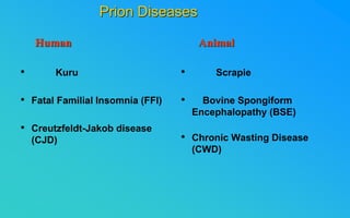
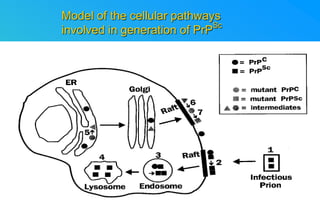
PRIONS
THE
INFECTIOUS PROTEINS
Paras Yadav*, Jaspreet Singh Arora*, Sachinandan De*, Tirtha Kumar
Datta*, Surender Lal Goswami*, Aarti Bhardwaj$, Shalini Jain# and
Hariom Yadav#
*Animal Biotechnology, #Animal Biochemistry Division, National Dairy Research
Institute, Karnal-132001, Haryana, India
$Meerut Institute of Engeenering and Technology, Meerut, U.P., India
• Stanley B. Prusiner coined the term proin from Proteinaceous infective particle
and changed to prion to sound it rhythmic.
• Prion diseases were caused by misfolded proteins.
• Elucidated the gene and mechanism by which wild type protein bring about the
clinical disease.
INTRODUCTION
• Kuru
• Fatal Familial Insomnia (FFI)
• Creutzfeldt-Jakob disease
(CJD)
• Scrapie
• Bovine Spongiform
Encephalopathy (BSE)
• Chronic Wasting Disease
(CWD)
Prion Diseases
Human Animal
Classification of prion diseases
• Infectious/Exogenous
– e.g., Kuru, BSE (mad cow disease), Scrapie
– Spread by
• Consumption of infected material.
• Transfusion.
• Sporadic
• Familial/Hereditary
– Due to autosomal dominant mutation of PrP.
Differences between cellular and scrapie proteins
PrPC PrPSC
Solubility
Soluble Non soluble
Structure
Alpha-helical Beta-sheeted
Multimerisation state
Monomeric Multimeric
Infectivity
Non infectious Infectious
Susceptibility to Proteinase K
Susceptible Resistant
Steps in the biosynthesis of PrPc
Post-translational processing of PrP
S S
A
C B
ER Golgi
Endosome
Cellular trafficking and cleavage of PrP
Mechanism of Internalization of PrPC
Hypothetical model for a PrPc
receptor
Model for the function of
LRP- LR as the receptor for PrP
HSPG
Dependent
binding
domain
Direct binding
domain (aa
161-179)
LRP/LR
PrP
BDI
(aa 144-179)
Heparan sulfate chain
(HS)
Sulfated domains
HSPG
Proteogl
ycan
moiety
GPI
BDII (aa 53-93)
Cell Culture Systems for Prion Propagation
Sequence of prion protein
PrPC + Cu (Copper)

Antioxidant activity

Resistance to oxidative stress

Prevent neuronal dysfunction
(Brown et al., 2002)
• Antioxidative
Functions of Prion protein
• Other functions
Models for the conversion of PrPc
to PrPsc
Time taken for Transformation of mutant PrP
c
to a PrP
Sc
state
Detergent
insoluble
PIPLC-
resistant
Synthesis of
Mutant PrP
c
Protease-
resistant
<10 min
BFA
180
C
0.5-1 hr
1-6 hr
Endoplasmic Reticulum
Plasma membrane/
Endocytic Pathway
Effect of conformation of PrP on Pro K
Model of the cellular pathways
involved in generation of PrPSc
Proposed model of PrPc
aggregation and induction of CtmPrP
Pathogenesis
Mechanism of PrPsc
induced apoptosis
 They generate a C-terminal fragment(C2) which has molecular
weight of 27-30 kDs.
 Increase in intracellular levels of Calcium increase
production of terminal fragments .
 Calpastatin prevents production of C2.
 Inhibitors of lysosomal proteases has no effect on C2
production.
Telling et al.,2004
What are Calpains?
 It was proposed that prion-associated toxicity involves altered
trafficking of PrP
c
.
 Inhibition of ubiquitin-proteasome system(UPS).
 Deposition of aggresomes of PrP
Sc
in nerve cells.
 Induction of Apoptosis with activation of Caspase 3 and
Caspase 8.
 Complete molecular basis for neuronal death is not known.
 Aggregates of over expressed PrP
c
does not cause cell death.
Tabrizi et al., 2005
Role of Caspases
The AGAAAAGA Palindrome in Prion Generation
Factors Responsible for Prion Propagation
Norstrom & Mastrianni, 2005
Factors Responsible for Prion Propagation cont…
• PrP
c
association with lipid rafts in the early secretory pathway.
• Creutzfeldt–Jakob disease (CJD) in Libyan Jews, linked to the E200K
mutation in PRNP (E200KCJD).
E.g. Hsp70, GroEL
Model for chaperone-supervised PrP conversion
Phospholipase A2 Inhibitors prevent prion replication.
Platelet-activating Factor Antagonists also inhibits prion replication.
Bate et al.,2004
 Drugs which share a N-benzylidene-benzohydrazide core structure.
 Trimethylamine N-oxide (TMAO), can prevent formation of PrP
Sc
.
Factors that prevent Prion Replication
Bertsch et al.,2005
Bennion, 2004
Gross and Microscopic Changes
Grossly there is Cortical atrophy and ventricular dilatation
may also be present.
Gross changes
Scrapie BSE
CJD
Kuru
Microscopic changes
• Gliosis within plaques.
• Loss of oligodendrocytes within plaques.
• Axons usually remain intact in plaques.
• Both CD4+ and CD8+ lymphocytes are present in active
lesions.
(Kretzschmar et al.,1996, Wilesmith et al., 1997).
Other microscopic changes
Diagnosis can be made by:
1. Clinical signs and Symptoms.
2. Detection of Scrapie
Associated fibrils.
3. Detection of Abnormal Prion
protein (PrPsc) by Western blotting.
4. Two dimensional Gel Electrophoresis.
5. Imunodiagnosis of Prion disease.
6. Bioassay in Mice.
Diagnosis
Scrapie Associated
fibrils.
PrPC
PrPSC
PrPC
Mechanism of plaque formation
PrPsc fibrils
Plaque
Molecular hallmark of the disorder is the accumulation of abnormal prion
protein(PrP
Sc
).
Physiological functions of cellular prion protein (PrP
c
) is not clear.
Identity of intracellular compartment where PrPc
to PrPSc
occurs is not
established.
Prion peptide of 106-126 residues is found to be neurotoxic.
Studies on prion protein will open the avenues for treatment of other
neurodegenerative disorders.
Conclusion
30511 prions.ppt

30511 prions.ppt

  • 1.
    PRIONS THE INFECTIOUS PROTEINS Paras Yadav*,Jaspreet Singh Arora*, Sachinandan De*, Tirtha Kumar Datta*, Surender Lal Goswami*, Aarti Bhardwaj$, Shalini Jain# and Hariom Yadav# *Animal Biotechnology, #Animal Biochemistry Division, National Dairy Research Institute, Karnal-132001, Haryana, India $Meerut Institute of Engeenering and Technology, Meerut, U.P., India
  • 2.
    • Stanley B.Prusiner coined the term proin from Proteinaceous infective particle and changed to prion to sound it rhythmic. • Prion diseases were caused by misfolded proteins. • Elucidated the gene and mechanism by which wild type protein bring about the clinical disease. INTRODUCTION
  • 3.
    • Kuru • FatalFamilial Insomnia (FFI) • Creutzfeldt-Jakob disease (CJD) • Scrapie • Bovine Spongiform Encephalopathy (BSE) • Chronic Wasting Disease (CWD) Prion Diseases Human Animal
  • 4.
    Classification of priondiseases • Infectious/Exogenous – e.g., Kuru, BSE (mad cow disease), Scrapie – Spread by • Consumption of infected material. • Transfusion. • Sporadic • Familial/Hereditary – Due to autosomal dominant mutation of PrP.
  • 5.
    Differences between cellularand scrapie proteins PrPC PrPSC Solubility Soluble Non soluble Structure Alpha-helical Beta-sheeted Multimerisation state Monomeric Multimeric Infectivity Non infectious Infectious Susceptibility to Proteinase K Susceptible Resistant
  • 6.
    Steps in thebiosynthesis of PrPc
  • 7.
  • 8.
  • 9.
  • 10.
    Hypothetical model fora PrPc receptor
  • 11.
    Model for thefunction of LRP- LR as the receptor for PrP HSPG Dependent binding domain Direct binding domain (aa 161-179) LRP/LR PrP BDI (aa 144-179) Heparan sulfate chain (HS) Sulfated domains HSPG Proteogl ycan moiety GPI BDII (aa 53-93)
  • 12.
    Cell Culture Systemsfor Prion Propagation
  • 13.
  • 14.
    PrPC + Cu(Copper)  Antioxidant activity  Resistance to oxidative stress  Prevent neuronal dysfunction (Brown et al., 2002) • Antioxidative Functions of Prion protein • Other functions
  • 15.
    Models for theconversion of PrPc to PrPsc
  • 16.
    Time taken forTransformation of mutant PrP c to a PrP Sc state Detergent insoluble PIPLC- resistant Synthesis of Mutant PrP c Protease- resistant <10 min BFA 180 C 0.5-1 hr 1-6 hr Endoplasmic Reticulum Plasma membrane/ Endocytic Pathway
  • 17.
    Effect of conformationof PrP on Pro K
  • 18.
    Model of thecellular pathways involved in generation of PrPSc
  • 19.
    Proposed model ofPrPc aggregation and induction of CtmPrP
  • 20.
  • 21.
  • 22.
     They generatea C-terminal fragment(C2) which has molecular weight of 27-30 kDs.  Increase in intracellular levels of Calcium increase production of terminal fragments .  Calpastatin prevents production of C2.  Inhibitors of lysosomal proteases has no effect on C2 production. Telling et al.,2004 What are Calpains?
  • 23.
     It wasproposed that prion-associated toxicity involves altered trafficking of PrP c .  Inhibition of ubiquitin-proteasome system(UPS).  Deposition of aggresomes of PrP Sc in nerve cells.  Induction of Apoptosis with activation of Caspase 3 and Caspase 8.  Complete molecular basis for neuronal death is not known.  Aggregates of over expressed PrP c does not cause cell death. Tabrizi et al., 2005 Role of Caspases
  • 24.
    The AGAAAAGA Palindromein Prion Generation Factors Responsible for Prion Propagation Norstrom & Mastrianni, 2005
  • 25.
    Factors Responsible forPrion Propagation cont… • PrP c association with lipid rafts in the early secretory pathway. • Creutzfeldt–Jakob disease (CJD) in Libyan Jews, linked to the E200K mutation in PRNP (E200KCJD).
  • 26.
    E.g. Hsp70, GroEL Modelfor chaperone-supervised PrP conversion
  • 27.
    Phospholipase A2 Inhibitorsprevent prion replication. Platelet-activating Factor Antagonists also inhibits prion replication. Bate et al.,2004  Drugs which share a N-benzylidene-benzohydrazide core structure.  Trimethylamine N-oxide (TMAO), can prevent formation of PrP Sc . Factors that prevent Prion Replication Bertsch et al.,2005 Bennion, 2004
  • 28.
    Gross and MicroscopicChanges Grossly there is Cortical atrophy and ventricular dilatation may also be present. Gross changes
  • 29.
  • 30.
    • Gliosis withinplaques. • Loss of oligodendrocytes within plaques. • Axons usually remain intact in plaques. • Both CD4+ and CD8+ lymphocytes are present in active lesions. (Kretzschmar et al.,1996, Wilesmith et al., 1997). Other microscopic changes
  • 31.
    Diagnosis can bemade by: 1. Clinical signs and Symptoms. 2. Detection of Scrapie Associated fibrils. 3. Detection of Abnormal Prion protein (PrPsc) by Western blotting. 4. Two dimensional Gel Electrophoresis. 5. Imunodiagnosis of Prion disease. 6. Bioassay in Mice. Diagnosis Scrapie Associated fibrils.
  • 32.
  • 33.
  • 34.
  • 35.
    Molecular hallmark ofthe disorder is the accumulation of abnormal prion protein(PrP Sc ). Physiological functions of cellular prion protein (PrP c ) is not clear. Identity of intracellular compartment where PrPc to PrPSc occurs is not established. Prion peptide of 106-126 residues is found to be neurotoxic. Studies on prion protein will open the avenues for treatment of other neurodegenerative disorders. Conclusion