Central Giant Cell Granuloma :
WHO has defined it as an intraosseous lesion consisting of cellular and fibrous tissue that contains multiple foci of hemorrhage, aggregation of multinucleated giant cells and occasionally trabaculae of woven bone
Etiology JAFFE (1953): considered this lesion to be a local reparative reaction of bone, possibly to intramedullary hemorrhage or trauma, hence the term reparative giant cell granuloma was accepted.
Charles A Waldron & W G Shafer (1966) suggested trauma be an important etiological factor in the initiation of the CGCG of the jaws
Thoma K H (1986) suggested that the lesion may be due to capillary injury caused by defective wall due to some type of trauma
J V Soames and J C Southam (1997) suggested that it could be a reaction to some form of hemodynamic disturbance in bone marrow, perhaps associated with trauma and hemorrhage REGEZI AND SCIUBBA(1999) :
Suggested that
Response to previous traumatic or inflammatory episodes.
This lesion is charecterised by proliferation of fibroblasts and multinucleated giant cells, in a densely packed stromaThe CGCG is a benign process that occurs almost exclusively within the jaw bones
CLINICAL PRESENTATION
Found predominantly in children and young adult
It has a female predilection (2:1)
Most commonly affected site is the anterior portion of the jaws, with an increased frequency of occurrence in the mandible
Majority of the CGCG of jaws are painless, expansion of bone is detected on routine examination
Few cases may be associated with pain, paresthesia or perforation of a cortical bone plate, occasionally resulting in the ulceration of the mucosal surface by the underlying lesion
Radiographic featuresCentral giant cell lesions present as radiolucent defects. Which may be unilocular or multilocular.
The defect is usually well delineated
The lesion may vary from a 5×5mm incidental radiographic findings to a destructive lesion greater than 10cm in size.
radiographic findings
A small unilocular lesion may be confused with periapical granuloma or cysts.
multilocular giant cell lesions cannot be radiographically distinguished from ameloblastomas or other multilocular lesions. Based on clinical and radiological features CGCG may be divided into two categories
- Non-aggressive lesion
- Aggressive lesion
The non aggressive lesion makes up most cases and exhibit few or no symptoms, they demonstrate slow growth and do not show cortical perforation or root resorption of teeth involved in the lesion. The aggressive lesions are characterized by pain, rapid growth, cortical perforation and root resorption and show marked tendency to recur when compared with non aggressive typeSoft spongy, brownish to reddish friable tissue of various size.
A specimen is usually coated with fresh or coagulated blood. Giant cell lesions of the jaws show a variety of features. Common to all is the presence of few to many multinucleated
Dentin dysplasia (DD) is a rare hereditary disturbance is inherited as an autosomal dominant trait.
unknown etiology that affects approximately 1 :100,000.
In 1972, Witkop classified it into type I and type II which affect both dentitions.DD Type I
Radicular dentin dysplasia
Characterized by:-
1.Both dentitions are affected.
2.Normal appearing crowns
3.No or only rudimentary root development (rootless teeth)
4.Incomplete or total obliteration of the pulp chamber.
5.Teeth may exhibit extreme mobility and exfoliate prematurely.DD type II
coronal dentin dysplasia
Characterized by:-
1.partial pulpal obliteration.
2.Thistle-tube-or flame-shaped coronal pulp chambers
3. Thread-like root canals
4. Usually the absence of periapical radiolucencies.
5. In this type of anomaly, teeth roots are of normal shape and contour.The enamel and the immediately subjacent dentin appear normal.
Deeper layers of dentin show an atypical tubular pattern with an amorphous, atubular area, and irregular organization.
Normal dentinal tubule formation appears to have been blocked so that new dentine forms around obstacles and takes on the characteristic appearances described as “lava flowing around boulders”The radiograph revealed features of dentine dysplasia type I with normal appearance of crown but no root development Autosomal Dominant Disorder:
Manifested in heterozygous states
At least one parent of index case is usually affected
Both males and females are affected.
Clinical feature can be modified by variation in penetrance and expressivity. Some individual inherit the mutant gene but are phenotpically normal. This is reffered to as “incomplete penetrance”.
In many condition the age of onset is delayed.
Inheritance Pattern:
Typical pattern is a heterozygous affected parent with a homozygous unaffected parent.
Every child has one chance in two of having the disease
Both sexes are affected equally..Autosomal Recessive Disorder
Largest category of Mendelian disorder
Usually does not affect the parent of the affected individual, but sibling may show the disease.
Complete penetrance is common.
Onset is frequently early in life.
Usually affect enzymatic proteins.
Pattern Of Inheritance:
Typical pattern is two heterozygous unaffected (carrier) parent.
The triat does not usually affect the parent, but siblings may show the disease
Siblings have one chance in four of being affected
Both sexes affected equally.
Necrotising periodontal diseases, Necrotising periodontal diseases as a manifestation of systemic diseases.
By Dr. Ritam Kundu, MDS PGT, Dr. R. Ahmed Dental College & Hospital, Kolkata, India.
the most common viral infections that affects the maxillofacial area
sources( burket's oral medicine 11th ed,oral and maxillofacial pathology neville 2e )
Title: Sense of Smell
Presenter: Dr. Faiza, Assistant Professor of Physiology
Qualifications:
MBBS (Best Graduate, AIMC Lahore)
FCPS Physiology
ICMT, CHPE, DHPE (STMU)
MPH (GC University, Faisalabad)
MBA (Virtual University of Pakistan)
Learning Objectives:
Describe the primary categories of smells and the concept of odor blindness.
Explain the structure and location of the olfactory membrane and mucosa, including the types and roles of cells involved in olfaction.
Describe the pathway and mechanisms of olfactory signal transmission from the olfactory receptors to the brain.
Illustrate the biochemical cascade triggered by odorant binding to olfactory receptors, including the role of G-proteins and second messengers in generating an action potential.
Identify different types of olfactory disorders such as anosmia, hyposmia, hyperosmia, and dysosmia, including their potential causes.
Key Topics:
Olfactory Genes:
3% of the human genome accounts for olfactory genes.
400 genes for odorant receptors.
Olfactory Membrane:
Located in the superior part of the nasal cavity.
Medially: Folds downward along the superior septum.
Laterally: Folds over the superior turbinate and upper surface of the middle turbinate.
Total surface area: 5-10 square centimeters.
Olfactory Mucosa:
Olfactory Cells: Bipolar nerve cells derived from the CNS (100 million), with 4-25 olfactory cilia per cell.
Sustentacular Cells: Produce mucus and maintain ionic and molecular environment.
Basal Cells: Replace worn-out olfactory cells with an average lifespan of 1-2 months.
Bowman’s Gland: Secretes mucus.
Stimulation of Olfactory Cells:
Odorant dissolves in mucus and attaches to receptors on olfactory cilia.
Involves a cascade effect through G-proteins and second messengers, leading to depolarization and action potential generation in the olfactory nerve.
Quality of a Good Odorant:
Small (3-20 Carbon atoms), volatile, water-soluble, and lipid-soluble.
Facilitated by odorant-binding proteins in mucus.
Membrane Potential and Action Potential:
Resting membrane potential: -55mV.
Action potential frequency in the olfactory nerve increases with odorant strength.
Adaptation Towards the Sense of Smell:
Rapid adaptation within the first second, with further slow adaptation.
Psychological adaptation greater than receptor adaptation, involving feedback inhibition from the central nervous system.
Primary Sensations of Smell:
Camphoraceous, Musky, Floral, Pepperminty, Ethereal, Pungent, Putrid.
Odor Detection Threshold:
Examples: Hydrogen sulfide (0.0005 ppm), Methyl-mercaptan (0.002 ppm).
Some toxic substances are odorless at lethal concentrations.
Characteristics of Smell:
Odor blindness for single substances due to lack of appropriate receptor protein.
Behavioral and emotional influences of smell.
Transmission of Olfactory Signals:
From olfactory cells to glomeruli in the olfactory bulb, involving lateral inhibition.
Primitive, less old, and new olfactory systems with different path
These simplified slides by Dr. Sidra Arshad present an overview of the non-respiratory functions of the respiratory tract.
Learning objectives:
1. Enlist the non-respiratory functions of the respiratory tract
2. Briefly explain how these functions are carried out
3. Discuss the significance of dead space
4. Differentiate between minute ventilation and alveolar ventilation
5. Describe the cough and sneeze reflexes
Study Resources:
1. Chapter 39, Guyton and Hall Textbook of Medical Physiology, 14th edition
2. Chapter 34, Ganong’s Review of Medical Physiology, 26th edition
3. Chapter 17, Human Physiology by Lauralee Sherwood, 9th edition
4. Non-respiratory functions of the lungs https://academic.oup.com/bjaed/article/13/3/98/278874
Lung Cancer: Artificial Intelligence, Synergetics, Complex System Analysis, S...Oleg Kshivets
RESULTS: Overall life span (LS) was 2252.1±1742.5 days and cumulative 5-year survival (5YS) reached 73.2%, 10 years – 64.8%, 20 years – 42.5%. 513 LCP lived more than 5 years (LS=3124.6±1525.6 days), 148 LCP – more than 10 years (LS=5054.4±1504.1 days).199 LCP died because of LC (LS=562.7±374.5 days). 5YS of LCP after bi/lobectomies was significantly superior in comparison with LCP after pneumonectomies (78.1% vs.63.7%, P=0.00001 by log-rank test). AT significantly improved 5YS (66.3% vs. 34.8%) (P=0.00000 by log-rank test) only for LCP with N1-2. Cox modeling displayed that 5YS of LCP significantly depended on: phase transition (PT) early-invasive LC in terms of synergetics, PT N0—N12, cell ratio factors (ratio between cancer cells- CC and blood cells subpopulations), G1-3, histology, glucose, AT, blood cell circuit, prothrombin index, heparin tolerance, recalcification time (P=0.000-0.038). Neural networks, genetic algorithm selection and bootstrap simulation revealed relationships between 5YS and PT early-invasive LC (rank=1), PT N0—N12 (rank=2), thrombocytes/CC (3), erythrocytes/CC (4), eosinophils/CC (5), healthy cells/CC (6), lymphocytes/CC (7), segmented neutrophils/CC (8), stick neutrophils/CC (9), monocytes/CC (10); leucocytes/CC (11). Correct prediction of 5YS was 100% by neural networks computing (area under ROC curve=1.0; error=0.0).
CONCLUSIONS: 5YS of LCP after radical procedures significantly depended on: 1) PT early-invasive cancer; 2) PT N0--N12; 3) cell ratio factors; 4) blood cell circuit; 5) biochemical factors; 6) hemostasis system; 7) AT; 8) LC characteristics; 9) LC cell dynamics; 10) surgery type: lobectomy/pneumonectomy; 11) anthropometric data. Optimal diagnosis and treatment strategies for LC are: 1) screening and early detection of LC; 2) availability of experienced thoracic surgeons because of complexity of radical procedures; 3) aggressive en block surgery and adequate lymph node dissection for completeness; 4) precise prediction; 5) adjuvant chemoimmunoradiotherapy for LCP with unfavorable prognosis.
Flu Vaccine Alert in Bangalore Karnatakaaddon Scans
As flu season approaches, health officials in Bangalore, Karnataka, are urging residents to get their flu vaccinations. The seasonal flu, while common, can lead to severe health complications, particularly for vulnerable populations such as young children, the elderly, and those with underlying health conditions.
Dr. Vidisha Kumari, a leading epidemiologist in Bangalore, emphasizes the importance of getting vaccinated. "The flu vaccine is our best defense against the influenza virus. It not only protects individuals but also helps prevent the spread of the virus in our communities," he says.
This year, the flu season is expected to coincide with a potential increase in other respiratory illnesses. The Karnataka Health Department has launched an awareness campaign highlighting the significance of flu vaccinations. They have set up multiple vaccination centers across Bangalore, making it convenient for residents to receive their shots.
To encourage widespread vaccination, the government is also collaborating with local schools, workplaces, and community centers to facilitate vaccination drives. Special attention is being given to ensuring that the vaccine is accessible to all, including marginalized communities who may have limited access to healthcare.
Residents are reminded that the flu vaccine is safe and effective. Common side effects are mild and may include soreness at the injection site, mild fever, or muscle aches. These side effects are generally short-lived and far less severe than the flu itself.
Healthcare providers are also stressing the importance of continuing COVID-19 precautions. Wearing masks, practicing good hand hygiene, and maintaining social distancing are still crucial, especially in crowded places.
Protect yourself and your loved ones by getting vaccinated. Together, we can help keep Bangalore healthy and safe this flu season. For more information on vaccination centers and schedules, residents can visit the Karnataka Health Department’s official website or follow their social media pages.
Stay informed, stay safe, and get your flu shot today!
These lecture slides, by Dr Sidra Arshad, offer a quick overview of physiological basis of a normal electrocardiogram.
Learning objectives:
1. Define an electrocardiogram (ECG) and electrocardiography
2. Describe how dipoles generated by the heart produce the waveforms of the ECG
3. Describe the components of a normal electrocardiogram of a typical bipolar leads (limb II)
4. Differentiate between intervals and segments
5. Enlist some common indications for obtaining an ECG
Study Resources:
1. Chapter 11, Guyton and Hall Textbook of Medical Physiology, 14th edition
2. Chapter 9, Human Physiology - From Cells to Systems, Lauralee Sherwood, 9th edition
3. Chapter 29, Ganong’s Review of Medical Physiology, 26th edition
4. Electrocardiogram, StatPearls - https://www.ncbi.nlm.nih.gov/books/NBK549803/
5. ECG in Medical Practice by ABM Abdullah, 4th edition
6. ECG Basics, http://www.nataliescasebook.com/tag/e-c-g-basics
Anti ulcer drugs and their Advance pharmacology ||
Anti-ulcer drugs are medications used to prevent and treat ulcers in the stomach and upper part of the small intestine (duodenal ulcers). These ulcers are often caused by an imbalance between stomach acid and the mucosal lining, which protects the stomach lining.
||Scope: Overview of various classes of anti-ulcer drugs, their mechanisms of action, indications, side effects, and clinical considerations.
Title: Sense of Taste
Presenter: Dr. Faiza, Assistant Professor of Physiology
Qualifications:
MBBS (Best Graduate, AIMC Lahore)
FCPS Physiology
ICMT, CHPE, DHPE (STMU)
MPH (GC University, Faisalabad)
MBA (Virtual University of Pakistan)
Learning Objectives:
Describe the structure and function of taste buds.
Describe the relationship between the taste threshold and taste index of common substances.
Explain the chemical basis and signal transduction of taste perception for each type of primary taste sensation.
Recognize different abnormalities of taste perception and their causes.
Key Topics:
Significance of Taste Sensation:
Differentiation between pleasant and harmful food
Influence on behavior
Selection of food based on metabolic needs
Receptors of Taste:
Taste buds on the tongue
Influence of sense of smell, texture of food, and pain stimulation (e.g., by pepper)
Primary and Secondary Taste Sensations:
Primary taste sensations: Sweet, Sour, Salty, Bitter, Umami
Chemical basis and signal transduction mechanisms for each taste
Taste Threshold and Index:
Taste threshold values for Sweet (sucrose), Salty (NaCl), Sour (HCl), and Bitter (Quinine)
Taste index relationship: Inversely proportional to taste threshold
Taste Blindness:
Inability to taste certain substances, particularly thiourea compounds
Example: Phenylthiocarbamide
Structure and Function of Taste Buds:
Composition: Epithelial cells, Sustentacular/Supporting cells, Taste cells, Basal cells
Features: Taste pores, Taste hairs/microvilli, and Taste nerve fibers
Location of Taste Buds:
Found in papillae of the tongue (Fungiform, Circumvallate, Foliate)
Also present on the palate, tonsillar pillars, epiglottis, and proximal esophagus
Mechanism of Taste Stimulation:
Interaction of taste substances with receptors on microvilli
Signal transduction pathways for Umami, Sweet, Bitter, Sour, and Salty tastes
Taste Sensitivity and Adaptation:
Decrease in sensitivity with age
Rapid adaptation of taste sensation
Role of Saliva in Taste:
Dissolution of tastants to reach receptors
Washing away the stimulus
Taste Preferences and Aversions:
Mechanisms behind taste preference and aversion
Influence of receptors and neural pathways
Impact of Sensory Nerve Damage:
Degeneration of taste buds if the sensory nerve fiber is cut
Abnormalities of Taste Detection:
Conditions: Ageusia, Hypogeusia, Dysgeusia (parageusia)
Causes: Nerve damage, neurological disorders, infections, poor oral hygiene, adverse drug effects, deficiencies, aging, tobacco use, altered neurotransmitter levels
Neurotransmitters and Taste Threshold:
Effects of serotonin (5-HT) and norepinephrine (NE) on taste sensitivity
Supertasters:
25% of the population with heightened sensitivity to taste, especially bitterness
Increased number of fungiform papillae
Tom Selleck Health: A Comprehensive Look at the Iconic Actor’s Wellness Journeygreendigital
Tom Selleck, an enduring figure in Hollywood. has captivated audiences for decades with his rugged charm, iconic moustache. and memorable roles in television and film. From his breakout role as Thomas Magnum in Magnum P.I. to his current portrayal of Frank Reagan in Blue Bloods. Selleck's career has spanned over 50 years. But beyond his professional achievements. fans have often been curious about Tom Selleck Health. especially as he has aged in the public eye.
Follow us on: Pinterest
Introduction
Many have been interested in Tom Selleck health. not only because of his enduring presence on screen but also because of the challenges. and lifestyle choices he has faced and made over the years. This article delves into the various aspects of Tom Selleck health. exploring his fitness regimen, diet, mental health. and the challenges he has encountered as he ages. We'll look at how he maintains his well-being. the health issues he has faced, and his approach to ageing .
Early Life and Career
Childhood and Athletic Beginnings
Tom Selleck was born on January 29, 1945, in Detroit, Michigan, and grew up in Sherman Oaks, California. From an early age, he was involved in sports, particularly basketball. which played a significant role in his physical development. His athletic pursuits continued into college. where he attended the University of Southern California (USC) on a basketball scholarship. This early involvement in sports laid a strong foundation for his physical health and disciplined lifestyle.
Transition to Acting
Selleck's transition from an athlete to an actor came with its physical demands. His first significant role in "Magnum P.I." required him to perform various stunts and maintain a fit appearance. This role, which he played from 1980 to 1988. necessitated a rigorous fitness routine to meet the show's demands. setting the stage for his long-term commitment to health and wellness.
Fitness Regimen
Workout Routine
Tom Selleck health and fitness regimen has evolved. adapting to his changing roles and age. During his "Magnum, P.I." days. Selleck's workouts were intense and focused on building and maintaining muscle mass. His routine included weightlifting, cardiovascular exercises. and specific training for the stunts he performed on the show.
Selleck adjusted his fitness routine as he aged to suit his body's needs. Today, his workouts focus on maintaining flexibility, strength, and cardiovascular health. He incorporates low-impact exercises such as swimming, walking, and light weightlifting. This balanced approach helps him stay fit without putting undue strain on his joints and muscles.
Importance of Flexibility and Mobility
In recent years, Selleck has emphasized the importance of flexibility and mobility in his fitness regimen. Understanding the natural decline in muscle mass and joint flexibility with age. he includes stretching and yoga in his routine. These practices help prevent injuries, improve posture, and maintain mobilit
Factory Supply Best Quality Pmk Oil CAS 28578–16–7 PMK Powder in Stockrebeccabio
Factory Supply Best Quality Pmk Oil CAS 28578–16–7 PMK Powder in Stock
Telegram: bmksupplier
signal: +85264872720
threema: TUD4A6YC
You can contact me on Telegram or Threema
Communicate promptly and reply
Free of customs clearance, Double Clearance 100% pass delivery to USA, Canada, Spain, Germany, Netherland, Poland, Italy, Sweden, UK, Czech Republic, Australia, Mexico, Russia, Ukraine, Kazakhstan.Door to door service
Hot Selling Organic intermediates
TEST BANK for Operations Management, 14th Edition by William J. Stevenson, Ve...kevinkariuki227
TEST BANK for Operations Management, 14th Edition by William J. Stevenson, Verified Chapters 1 - 19, Complete Newest Version.pdf
TEST BANK for Operations Management, 14th Edition by William J. Stevenson, Ve...
PRIMARY HERPETIC GINGIVOSTOMATITIS
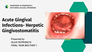
1. Presented by
OLGA MCFRANCIS
FINAL YEAR BDS PART 1
Acute Gingival
Infections- Herpetic
Gingivostomatitis
DEPARTMENT OF PERIODONTICS ,
GOVT DENTAL COLLEGE, TRIVANDRUM
2. CONTENTS
• Introduction
• Classification
• Primary Herpetic
Gingivostomatitis
• Mode of Transmission
• Pathogenesis
• General Features
• Clinical Features
• Histopathology
• Diagnosis
• Differential Diagnosis
• Conclusion
• Recent Studies
• Previous year Questions
• Bibliography
• Treatment
3. INTRODUCTION
Acute gingival infections often present with
painful intraoral or perioral lesions. Patients
presenting with acute infections require
urgent treatment to relieve symptoms and to
resolve the infection.This seminar reviews the
presenting history, signs, symptoms, clinical
features, diagnosis, treatment,etc of primary
herpetic gingivostomatitis.
5. Primary Herpetic Gingivostomatitis
• Herpes simplex, an acute infectious disease, is probably the most
common viral disease affecting man, with the exception of viral
respiratory infections.
• HSV is composed of double-stranded DNA, protein capsid,
tegument and lipid envelope,which contain glycoproteins derived
from the nuclear membrane of host cells.
• There are two immunologically different types of HSV: type 1 and
type 2
• HSV-1 predominantly affects the face, lips, oral cavity and upper
body skin
• HSV-2 usually affects the genitals and skin of the lower half of the
body.
6.
7. Mode of
transmission
• It can be due to droplet spread.
• There is no animal reservoir for
this virus.
• The incubation period appears
to range from 2 to 20 days, with
an average of 6 days before
development of lesions.
8. Pathogenesis
• The virus is attached to the cells at the inoculation site through
specific receptors .
• It replicates too many virions to the maximum number, and
discharges to neighbouring cells.
• Subsequently, it affects adjacent cells, spreads to distant
sensory nerve endings and autonomic axons, further to
adjacent related ganglia,and remains latent there.
• The lymph node is involved through viral proteins by mobile
dendritic cells and begins its primary immune response.
10. General Features
• Mostly in infants and children less than 6 years
old.
• Seen in adolescents and adults also.
• Male : Female equal
• Mostly Primary Infections are asymptomatic.
• As part of the primary infection, the virus ascends
through the sensory and autonomic nerves where
it persists as latent HSV inneuronal ganglia that
innervate the site.
11. • In approximately one-third of the
world’s population, secondary
manifestations result from various
stimuli, such as sunlight, trauma,
fever, and stress.
• These secondary manifestations
include herpes labialis,herpetic
stomatitis,herpes genitalis, ocular
herpes, and herpetic encephalitis.
12. • Secondary herpetic stomatitis can
occur on the palate, on the gingiva or
on the mucosa as a result of dental
treatment that traumatizes or
stimulates the latent virus in the ganglia
that innervate the area.
• It may be present as pain away from the
site of treatment 2–4 days later.
13. • Diffuse,erythematous, shiny involvement of the
gingiva and the adjacent oral mucosa, with
varying degrees of edema and gingival
bleeding.
• During its initial stage, it is characterized by the
presence of discrete, spherical gray vesicles,
which may occur on the gingiva, the labial and
buccal mucosae, the soft palate, the pharynx,
the sublingual mucosa, and the tongue
Clinical Features
Oral signs
14. • After 24 h, vesicles rupture and form painful small
ulcers with red, elevated, halo-like margins and
depressed yellowish or grayish-white central
portions.
• These occur either in widely separated areas or in
clusters where confluence occurs.
• Course of the disease is limited to 7–10 days.
• The diffuse gingival erythema and edema that
appear early during the course of the disease persist
for several days after the ulcerative lesions have
healed.
• Scarring does not occur in the areas of healed
ulcerations.
15.
16. Oral Symptoms
• It is accompanied by generalized “soreness” of
the oral cavity, which interferes with eating,
drinking, and oral hygiene.
• Ruptured vesicles are the focal sites of pain.
• They are sensitive to touch, thermal changes,
foods such as condiments and fruit juices, and
the action of coarse foods.
• In infants, it is marked by irritability and refusal
to take food.
17. Extraoral and systemic signs and
symptoms
• Cervical adenitis
• Fever as high as 101–105°F (38–40.6°C)
• Generalized malaise
18. • Primary herpetic gingivostomatitis is the
result of an acute infection by HSV, and it
has an acute onset.
Histor
y
19. • The herpetic vesicle is an intraepithelial blister filled with
fluid. The infected cells are swollen and have pale
eosinophilic cytoplasm and large vesicular nuclei,
described as 'ballooning degeneration', while others
characteristically contain intranuclear inclusions known as
Lipschutz bodies.
• These are eosinophilic, ovoid homogeneous structures
within the nucleus, which tend to displace the nucleolus
and nuclear chromatin peripherally.
• The displacement of chromatin often produces a peri-
inclusion halo.
Histopathology
21. Histopathology
• Cytoplasm of the infected cells forms giant
cells.
• The subjacent connective tissue is usually
infiltrated by inflammatory cells.
• When the vesicle ruptures, the surface of the
tissue is covered by exudates made up of
fibrin, polymorphonuclear leukocytes and
degenerated cells.
• The lesions heal by peripheral epithelial
proliferation.
22. • Diagnosed both clinically and by laboratory
procedures.
• Scrapings obtained from the base of the
lesions are stained with Wright's and Giemsa
stains.
• Pap stain shows balloon cells, multinucleated
giant cells and intranuclear inclusions.
• Although cytological procedures give a quick
result but it will not differentiate between HSV
and varicella zoster virus (VZV).
Diagnosis
Pap Smear
24. Diagnosis
• HSV can be demonstrated in the
laboratory by the isolation of
virus in tissue culture or by DNA
in the scrapings from lesions.
• The most sensitive and accurate
method for diagnosing these
lesions is the PCR technique.
27. Treatment
• Topical Lignocaine for pain relief.
• Acyclovir 15mg/kg 5 times daily for 5-7 days reduces
duration of disease, halts the progression of lesions and
reduces infectivity.
28. • According to Irish Journal of Medical Science( 1971) , an article on Adolescents
and Herpetic Gingivostomatitis: an Italian Overview published on 15 May 2021,
• In Italian adolescents, PHGS was diagnosed atleast 48 h after onset and an
antibiotic therapy was prescription in order to prevent secondary bacterial
infections.
• 15 patients had been treated with non alcoholic chlorhexidine rinses ( Group A)
• 29 patients with non alcoholic chlorhexidine rinses plus hyaluronic acid gel (
Group B),
• 30 patients with non alcoholic chlorhexidine rinses plus Mucosyte ( Group C).
• A significant improvement of the pain scoring and lesions severity was noted in
Group C .
• Among topical therapies , an association of Verbascoside and Sodium
Hyaluronhate seems to favor faster healing.
Recent Studies
30. Bibliography
• Newman and Carranza's CLINICAL
PERIODONTOLOGY - Third South Asian Edition
• Essentials of CLINICAL PERIODONTOLOGY AND
PERIODONTICS by Shantipriya Reddy - 6th Edition
• Shafer's Textbook of ORAL PATHOLOGY -Ninth
Edition