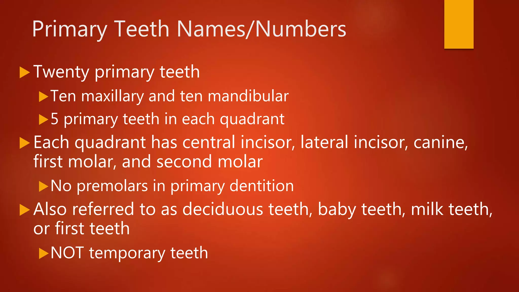
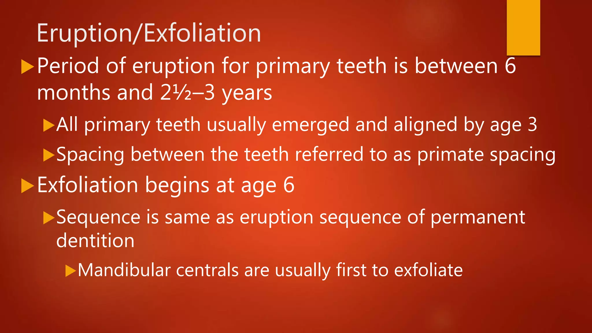
Primary Dentition &
Tooth Development
CHAPTER 13 & 14
Primary Dentition- Review
CHAPTER 13
Objectives
At the completion of this chapter, you will be
able to
Identify name, number, and eruption dates of
primary teeth
Describe function of primary teeth
Compare primary teeth to permanent teeth
Primary Teeth Names/Numbers
Twenty primary teeth
Ten maxillary and ten mandibular
5 primary teeth in each quadrant
Each quadrant has central incisor, lateral incisor, canine,
first molar, and second molar
No premolars in primary dentition
Also referred to as deciduous teeth, baby teeth, milk teeth,
or first teeth
NOT temporary teeth
Primary Teeth Names/
Numbers (cont’d.)
Eruption/Exfoliation
Period of eruption for primary teeth is between 6
months and 2½–3 years
All primary teeth usually emerged and aligned by age 3
Spacing between the teeth referred to as primate spacing
Exfoliation begins at age 6
Sequence is same as eruption sequence of permanent
dentition
Mandibular centrals are usually first to exfoliate
Comparison With Permanent Teeth
 Primary anterior teeth and secondary molars resemble their permanent
counterparts
 Primary teeth have more pronounced cervical ridge
 Roots of posterior teeth are more flared (divergent)
Allowing growth of the permanent teeth forming beneath them
Cervix appears to be more constricted than permanent teeth
 Primary crowns have less enamel than permanent teeth, and pulp
horns extend more occlusally
 Pulp is closer to the surface of primary crowns making it necessary to
reduce excessive friction when polishing.
Maxillary Incisors
Maxillary Central Incisors: E, F
Maxillary Lateral Incisors: D, G
Labial crown of incisor is smooth with a
straight incisal edge
No mamelons
Crown is wide with a cingulum and marginal
ridges on lingual
Maxillary Incisors (cont’d.)
Maxillary Canines
C, H
Broad cervical ridge on
canine causes cervix to
appear constricted
Short, pointed cusp with
long, slender root
Maxillary First Molar
B, I
Number of cusps varies from 2 to 4
No dividing groove on buccal surface
Three roots on all maxillary molars
Occlusal surface has central fossa and mesial
triangular fossa connected by central groove
Maxillary Second Molar
A, J
Anatomy is same as permanent maxillary first molar
Two buccal cusps divided by buccal groove
Two lingual cusps with a cusp of Carabelli on
mesiolingual
Three roots
Two buccal, one lingual
Maxillary First and Second Molars
Mandibular Incisors
Mandibular Central Incisors: O, P
First tooth to exfoliate
Mandibular Lateral Incisors: N, Q
Both labial and lingual surfaces are
smooth
Slight cingulum marginal ridges on
lingual
Mandibular Canines
M, R
Buccal surface has pronounced cervical ridge
Lingual surface
has cingulum
and lingual ridges
Mandibular First Molar
L, S
No definite anatomy, like maxillary first molar
Only primary tooth not anatomically identical to any
permanent tooth
Usually two buccal cusps divided by depression (rather
than a groove) and two lingual cusps
Two long, slender roots are divergent
Occlusal surface has central groove crossed by
buccal/lingual grooves
Mandibular Second Molar
K, T
Identical anatomy and groove pattern to
permanent mandibular first molar
Grooves divide three buccal cusps and two
lingual cusps
Two long, thin, divergent roots
Can be twice as long as crown
Mandibular First and Second Molars
Importance of Primary Teeth
 Both form and function of primary dentition is important
Each primary tooth has same function as permanent tooth that
follows
Maintains a solid position for permanent tooth replacement
 Damage to primary teeth could cause problems with
permanent dentition
 When both deciduous and permanent teeth are present in
the oral cavity its called mixed dentition.
Summary
 Primary dentition
10 maxillary and 10 mandibular teeth
A-T for universal system
No primary premolars
 Eruption is completed between 2½ and 3 years
Primary tooth resembles permanent version but smaller
 Posterior teeth have flared roots to fit permanent teeth
developing below
Tooth Development
CHAPTER 14
General Information
Development of a human begins with an
embryo, which has three layers
Ectoderm forms lining of oral cavity
As well as outer covering of the body
Mesoderm forms the skeletal/muscular
systems, cementum, dentin, and pulp
Endoderm forms the lining of internal organs
General Information
 During the third week of fetal life, when the embryo is only 3mm ling, the
face begins its development.
 At one end of the embryo there is an invagination of the ectoderm
forming the stomodeum, or primitive mouth, which becomes oral and
nasal cavities.
 The primitive mouth is lined with ectoderm and becomes the oral
epithelium.
 Beneath this is the mesenchyme, developed from mesoderm, which becomes
the underlying connective tissue.
 Between the fifth and sixth week in utero, the first signs of tooth
development is evident.
 Teeth are formed from the ectoderm and mesoderm in a complex
histologic process simplified into the following pattern.
Growth and Development
Formation of teeth
Progression begins during fifth or sixth week in utero
Process continues until 10 maxillary and 10 mandibular
teeth are formed
Permanent teeth begin forming around fourth or fifth
month of fetal life but do not calcify until birth
Proper diet during tooth formation is crucial for long-
term health
Growth and Development
Initiation: Bud Stage
 First stage of odontogenesis or origin of tooth, is referred
to as bud stage
During this stage, tooth begins formation from dental lamina
referred to as initiation
Dental lamina is growth from oral epithelium that gives rise to
tooth buds
10 growths on each arch later become primary teeth
Permanent teeth develop similarly
Proliferation: Cap Stage
Bud of tooth grows and changes shape during cap
stage
Organ is indented on lower side and appears much like a
cap, hence name
Primary embryonic ectoderm layer develops into the
oral epithelium
Epithelium matures into the enamel of the tooth
Proliferation: Cap Stage
Primary embryonic mesoderm layer develops into
connective tissue during
Proliferation when the cells multiply
Histodifferentiation when the cells develop into different
tissues
Morphodifferentiation, when the cells begin to outline the
future shape of the developing organ
Morpho/Histodifferentiation: Bell Stage
Further specialization of cells takes place in the bell
stage
Inner epithelium of enamel organ become ameloblasts,
enamel-forming cells
Dental papilla peripheral cells become odontoblasts,
dentin-forming
Dental sac form cementoblasts, cementum-forming cells
Enclosed area of tissue that later matures into the dentin and
pulp of the tooth.
Apposition-Maturation State
Odontogenesis reaches completion in this stage
Tissues of enamel, dentin, and cementum formed in layers and
fused together
 Calcium and other minerals are deposited in formed teeth in
this stage.
Process is called calcification and is last developmental
state prior to eruption
The final stage is attrition, the wearing away of the incisal
or occlusal surfaces
Apposition-Maturation State
 The root of the tooth does not develop fully prior to eruption.
 The phase when the tooth passes through the bone and the oral mucosa and into its place in the
oral cavity.
 Twenty of the permanent teeth are located below and distal to the primary teeth.
 Which ones aren’t?
 As permanent teeth erupt, they apply pressure to the apices of the roots of the
primary teeth.
 During this force, osteoclasts, bone resorbsion cells, dissolve the rooth of the primary tooth.
 This Resorption first takes place at the apex and continues up toward the crown
 When very little root structure is left, the tooth loosens because of lack of support… and?
 Children often assist the final stages of loosening the tooth by moving it back and forth until
they break the attaching fibers.
Eruption
Eruption is the movement of the tooth
from its position within the jaw to its
position in the oral cavity
The process is divided into active and
passive eruption
Eruption and Importance of Primaries
Primary teeth occupy and maintain space in the
dental arches for permanent teeth and act as guides
during the eruption process.
If the primary teeth are removed early, the spaces
may be diminished, causing crowding when the
permanent teeth erupt.
Active Eruption
Process where crown moves from within jaw into oral
cavity
Continues until tooth meets its antagonist in opposite jaw
1½ to 3 years for completion of deciduous root
3 years for completion of the permanent root
The entire process of permanent tooth development,
from initiation to completion, takes about 10 years.
Passive Eruption
Increase in length of clinical crown caused by
gingival recession is referred to as passive
eruption
Factors such as wear from use or trauma can
cause peridontial attrition/breakdown
Exposure of cementum
Wearing of the enamel
Gingival recession
Developmental Anomalies
Disturbances in development stage of teeth and
bones can cause abnormalities
Can be hereditary or nutritional factors
Excessive fluoride
Disturbances during the formation of the tooth can
cause faulty enamel
An extra tooth forms called a supernumerary tooth
Summary
Tooth development begins between 5-6 week in utero
and continues until entire tooth is formed
2–3 years for primary teeth
9–10 years for permanent teeth
Once tooth is formed, it cannot repair itself
Proper diet during tooth formation and preventive care
after eruption is critical

Primary Dentition and Tooth Development

  • 1.
    Primary Dentition & ToothDevelopment CHAPTER 13 & 14
  • 2.
  • 3.
    Objectives At the completionof this chapter, you will be able to Identify name, number, and eruption dates of primary teeth Describe function of primary teeth Compare primary teeth to permanent teeth
  • 4.
    Primary Teeth Names/Numbers Twentyprimary teeth Ten maxillary and ten mandibular 5 primary teeth in each quadrant Each quadrant has central incisor, lateral incisor, canine, first molar, and second molar No premolars in primary dentition Also referred to as deciduous teeth, baby teeth, milk teeth, or first teeth NOT temporary teeth
  • 5.
  • 6.
    Eruption/Exfoliation Period of eruptionfor primary teeth is between 6 months and 2½–3 years All primary teeth usually emerged and aligned by age 3 Spacing between the teeth referred to as primate spacing Exfoliation begins at age 6 Sequence is same as eruption sequence of permanent dentition Mandibular centrals are usually first to exfoliate
  • 7.
    Comparison With PermanentTeeth  Primary anterior teeth and secondary molars resemble their permanent counterparts  Primary teeth have more pronounced cervical ridge  Roots of posterior teeth are more flared (divergent) Allowing growth of the permanent teeth forming beneath them Cervix appears to be more constricted than permanent teeth  Primary crowns have less enamel than permanent teeth, and pulp horns extend more occlusally  Pulp is closer to the surface of primary crowns making it necessary to reduce excessive friction when polishing.
  • 8.
    Maxillary Incisors Maxillary CentralIncisors: E, F Maxillary Lateral Incisors: D, G Labial crown of incisor is smooth with a straight incisal edge No mamelons Crown is wide with a cingulum and marginal ridges on lingual
  • 9.
  • 10.
    Maxillary Canines C, H Broadcervical ridge on canine causes cervix to appear constricted Short, pointed cusp with long, slender root
  • 11.
    Maxillary First Molar B,I Number of cusps varies from 2 to 4 No dividing groove on buccal surface Three roots on all maxillary molars Occlusal surface has central fossa and mesial triangular fossa connected by central groove
  • 12.
    Maxillary Second Molar A,J Anatomy is same as permanent maxillary first molar Two buccal cusps divided by buccal groove Two lingual cusps with a cusp of Carabelli on mesiolingual Three roots Two buccal, one lingual
  • 13.
    Maxillary First andSecond Molars
  • 14.
    Mandibular Incisors Mandibular CentralIncisors: O, P First tooth to exfoliate Mandibular Lateral Incisors: N, Q Both labial and lingual surfaces are smooth Slight cingulum marginal ridges on lingual
  • 15.
    Mandibular Canines M, R Buccalsurface has pronounced cervical ridge Lingual surface has cingulum and lingual ridges
  • 16.
    Mandibular First Molar L,S No definite anatomy, like maxillary first molar Only primary tooth not anatomically identical to any permanent tooth Usually two buccal cusps divided by depression (rather than a groove) and two lingual cusps Two long, slender roots are divergent Occlusal surface has central groove crossed by buccal/lingual grooves
  • 17.
    Mandibular Second Molar K,T Identical anatomy and groove pattern to permanent mandibular first molar Grooves divide three buccal cusps and two lingual cusps Two long, thin, divergent roots Can be twice as long as crown
  • 18.
    Mandibular First andSecond Molars
  • 19.
    Importance of PrimaryTeeth  Both form and function of primary dentition is important Each primary tooth has same function as permanent tooth that follows Maintains a solid position for permanent tooth replacement  Damage to primary teeth could cause problems with permanent dentition  When both deciduous and permanent teeth are present in the oral cavity its called mixed dentition.
  • 20.
    Summary  Primary dentition 10maxillary and 10 mandibular teeth A-T for universal system No primary premolars  Eruption is completed between 2½ and 3 years Primary tooth resembles permanent version but smaller  Posterior teeth have flared roots to fit permanent teeth developing below
  • 21.
  • 22.
    General Information Development ofa human begins with an embryo, which has three layers Ectoderm forms lining of oral cavity As well as outer covering of the body Mesoderm forms the skeletal/muscular systems, cementum, dentin, and pulp Endoderm forms the lining of internal organs
  • 23.
    General Information  Duringthe third week of fetal life, when the embryo is only 3mm ling, the face begins its development.  At one end of the embryo there is an invagination of the ectoderm forming the stomodeum, or primitive mouth, which becomes oral and nasal cavities.  The primitive mouth is lined with ectoderm and becomes the oral epithelium.  Beneath this is the mesenchyme, developed from mesoderm, which becomes the underlying connective tissue.  Between the fifth and sixth week in utero, the first signs of tooth development is evident.  Teeth are formed from the ectoderm and mesoderm in a complex histologic process simplified into the following pattern.
  • 24.
    Growth and Development Formationof teeth Progression begins during fifth or sixth week in utero Process continues until 10 maxillary and 10 mandibular teeth are formed Permanent teeth begin forming around fourth or fifth month of fetal life but do not calcify until birth Proper diet during tooth formation is crucial for long- term health
  • 25.
  • 26.
    Initiation: Bud Stage First stage of odontogenesis or origin of tooth, is referred to as bud stage During this stage, tooth begins formation from dental lamina referred to as initiation Dental lamina is growth from oral epithelium that gives rise to tooth buds 10 growths on each arch later become primary teeth Permanent teeth develop similarly
  • 27.
    Proliferation: Cap Stage Budof tooth grows and changes shape during cap stage Organ is indented on lower side and appears much like a cap, hence name Primary embryonic ectoderm layer develops into the oral epithelium Epithelium matures into the enamel of the tooth
  • 28.
    Proliferation: Cap Stage Primaryembryonic mesoderm layer develops into connective tissue during Proliferation when the cells multiply Histodifferentiation when the cells develop into different tissues Morphodifferentiation, when the cells begin to outline the future shape of the developing organ
  • 29.
    Morpho/Histodifferentiation: Bell Stage Furtherspecialization of cells takes place in the bell stage Inner epithelium of enamel organ become ameloblasts, enamel-forming cells Dental papilla peripheral cells become odontoblasts, dentin-forming Dental sac form cementoblasts, cementum-forming cells Enclosed area of tissue that later matures into the dentin and pulp of the tooth.
  • 30.
    Apposition-Maturation State Odontogenesis reachescompletion in this stage Tissues of enamel, dentin, and cementum formed in layers and fused together  Calcium and other minerals are deposited in formed teeth in this stage. Process is called calcification and is last developmental state prior to eruption The final stage is attrition, the wearing away of the incisal or occlusal surfaces
  • 31.
    Apposition-Maturation State  Theroot of the tooth does not develop fully prior to eruption.  The phase when the tooth passes through the bone and the oral mucosa and into its place in the oral cavity.  Twenty of the permanent teeth are located below and distal to the primary teeth.  Which ones aren’t?  As permanent teeth erupt, they apply pressure to the apices of the roots of the primary teeth.  During this force, osteoclasts, bone resorbsion cells, dissolve the rooth of the primary tooth.  This Resorption first takes place at the apex and continues up toward the crown  When very little root structure is left, the tooth loosens because of lack of support… and?  Children often assist the final stages of loosening the tooth by moving it back and forth until they break the attaching fibers.
  • 32.
    Eruption Eruption is themovement of the tooth from its position within the jaw to its position in the oral cavity The process is divided into active and passive eruption
  • 33.
    Eruption and Importanceof Primaries Primary teeth occupy and maintain space in the dental arches for permanent teeth and act as guides during the eruption process. If the primary teeth are removed early, the spaces may be diminished, causing crowding when the permanent teeth erupt.
  • 34.
    Active Eruption Process wherecrown moves from within jaw into oral cavity Continues until tooth meets its antagonist in opposite jaw 1½ to 3 years for completion of deciduous root 3 years for completion of the permanent root The entire process of permanent tooth development, from initiation to completion, takes about 10 years.
  • 35.
    Passive Eruption Increase inlength of clinical crown caused by gingival recession is referred to as passive eruption Factors such as wear from use or trauma can cause peridontial attrition/breakdown Exposure of cementum Wearing of the enamel Gingival recession
  • 36.
    Developmental Anomalies Disturbances indevelopment stage of teeth and bones can cause abnormalities Can be hereditary or nutritional factors Excessive fluoride Disturbances during the formation of the tooth can cause faulty enamel An extra tooth forms called a supernumerary tooth
  • 37.
    Summary Tooth development beginsbetween 5-6 week in utero and continues until entire tooth is formed 2–3 years for primary teeth 9–10 years for permanent teeth Once tooth is formed, it cannot repair itself Proper diet during tooth formation and preventive care after eruption is critical