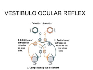
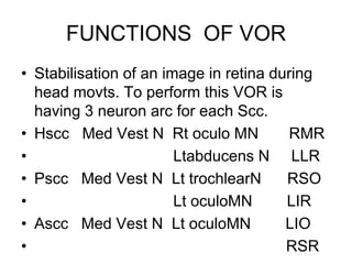
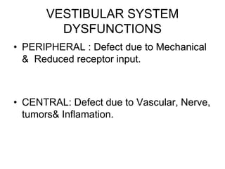
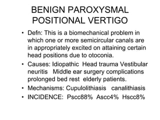
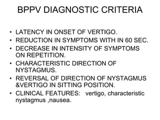
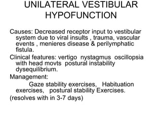
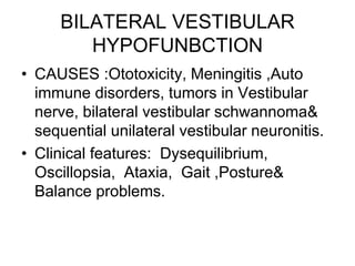
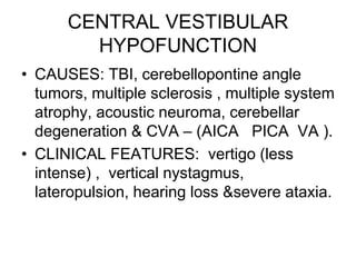
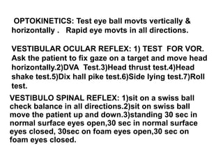
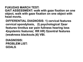
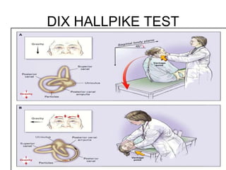
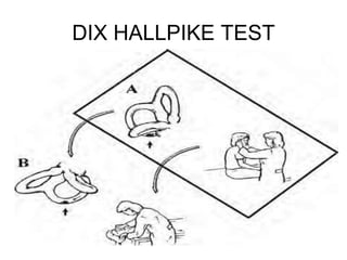
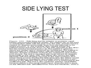
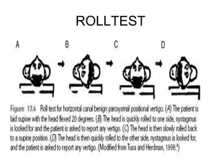
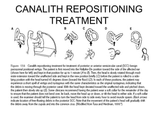
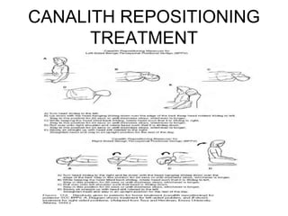
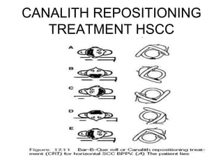
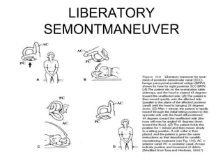
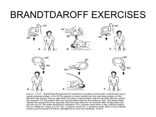
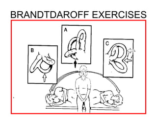
VESTIBULAR AND BALANCE
REHABILITATION
• M.UMASANKAR,BPT
• PHYSICAL
THERAPIST,
• MANIPALHOSPITAL,
• BANGALORE-17.
OVERVIEW
• VESTIBULAR SYSTEM ANATOMY.
• VESTIBULAR SYSTEM PHYSIOLOGY.
• VESTIBULAR SYSTEM DYSFUNCTION.
• VESTIBULAR SYSTEM EVALUATION.
• VESTIBULAR SYSTEM PHYSICAL
THERAPY MANAGEMENT &
REHABILITATION.
ANATOMY OF VESTIBULAR
SYSTEM
PERIPHERAL : vestibular
apparatus
CENTRAL : vestibular nerve
vestibular nuclei and its
connection
VESTIBULAR APPARATUS
• BONY LABYRINTH:3
sccs vestibule and
cochlea filled with
perilymph
•
MEMBRANOUS
LABYRINTH: 5 sensory
organs membranous
portion of 3sccs and 2
otolith organs filled
with endolymph
SEMI CIRCULAR CANALS
• 3sccs orthogonal to
each other.
HSCC: inclined30degree
upward&paralleltoeart
hPSCC:92 degree
inclined from plane of
hscc. it is in vertical
plane situated
backwards&outwards.
ASCC:90degree inclined
from plane of hscc
situatedForwards&out
wards. 3coplanar pairs.
SEMICIRCULAR CANALS
SENSE ORGANS
•
AMPULLA
CUPULA
CRISTA
AMPULLARIS
HAIR CELLS (stereo
cilium &kinocilium)
EXCITATION &INHIBITION HAIR
CELLS
• Due to head movts
Endolymphatic Pressure Difference
Occurs in cupula and it moves to
Stimulate the Hair cells.
•
Change in membrane potential of
hair cells Causes Deflection in
stereocilia.,
• Deflection of stereocilia towards
Kinocilia leads to Excitation
(Depolarization)
• Deflection of stereocilia away from
the kinocilia leads to inhibition
(hyperpolarization)
MOVEMENT OF HAIR CELLS
VESTIBULAR VASCULAR SUPPLY:
Anterior Vestibular artery Supplies 3 Sccs and Otoliths.
Common cochlear artery.
VESTIBULAR NERVE SUPPLY:
Superior branch supplies Ascc, Hscc and Utricle.
Inferior branch supplies Pscc and Saccule.
VESTBULAR NUCLEI: Superior,Medial,Inferior,Lateral and other
small nuclei.
AREA CONNECTED TO IT: Cerebellum and floculonodular
lobule.
Functions of Vestibular System
• Stabilisation of visual images on retina
during head motion for clear vision.
• Maintaining postural stability during head
movts.
• Providing information used for spatial
orientation.
PHYSIOLOGY OF VESTIBULAR
SYSTEM
• TONIC FIRING RATE.
• PUSH PULL MECHANISM.
• INHIBITORY CUTT OFF.
• VELOCITY STORAGE SYSTEM.
• VESTIBULO OCULAR REFLEX.
• VOR GAIN AND VOR PHASE.
• VESTIBULO SPINAL REFLEX.
• RETINAL SLIP.
VESTIBULO OCULAR REFLEX
FUNCTIONS OF VOR
• Stabilisation of an image in retina during
head movts. To perform this VOR is
having 3 neuron arc for each Scc.
• Hscc Med Vest N Rt oculo MN RMR
• Ltabducens N LLR
• Pscc Med Vest N Lt trochlearN RSO
• Lt oculoMN LIR
• Ascc Med Vest N Lt oculoMN LIO
• RSR
VESTIBULAR SYSTEM
DYSFUNCTIONS
• PERIPHERAL : Defect due to Mechanical
& Reduced receptor input.
• CENTRAL: Defect due to Vascular, Nerve,
tumors& Inflamation.
BENIGN PAROXYSMAL
POSITIONAL VERTIGO
• Defn: This is a biomechanical problem in
which one or more semicircular canals are
in appropriately excited on attaining certain
head positions due to otoconia.
• Causes: Idiopathic Head trauma Vestibular
neuritis Middle ear surgery complications
prolonged bed rest elderly patients.
• Mechanisms: Cupulolithiasis canalithiasis
• INCIDENCE: Pscc88% Ascc4% Hscc8%
BPPV DIAGNOSTIC CRITERIA
• LATENCY IN ONSET OF VERTIGO.
• REDUCTION IN SYMPTOMS WITH IN 60 SEC.
• DECREASE IN INTENSITY OF SYMPTOMS
ON REPETITION.
• CHARACTERISTIC DIRECTION OF
NYSTAGMUS.
• REVERSAL OF DIRECTION OF NYSTAGMUS
&VERTIGO IN SITTING POSITION.
• CLINICAL FEATURES: vertigo, characteristic
nystagmus ,nausea.
UNILATERAL VESTIBULAR
HYPOFUNCTION
Causes: Decreased receptor input to vestibular
system due to viral insults , trauma, vascular
events , menieres disease & perilymphatic
fistula.
Clinical features: vertigo nystagmus oscillopsia
with head movts postural instability
dysequilibrium.
Management:
Gaze stability exercises, Habituation
exercises, postural stability Exercises.
(resolves with in 3-7 days)
BILATERAL VESTIBULAR
HYPOFUNBCTION
• CAUSES :Ototoxicity, Meningitis ,Auto
immune disorders, tumors in Vestibular
nerve, bilateral vestibular schwannoma&
sequential unilateral vestibular neuronitis.
• Clinical features: Dysequilibrium,
Oscillopsia, Ataxia, Gait ,Posture&
Balance problems.
CENTRAL VESTIBULAR
HYPOFUNCTION
• CAUSES: TBI, cerebellopontine angle
tumors, multiple sclerosis , multiple system
atrophy, acoustic neuroma, cerebellar
degeneration & CVA – (AICA PICA VA ).
• CLINICAL FEATURES: vertigo (less
intense) , vertical nystagmus,
lateropulsion, hearing loss &severe ataxia.
ELDERLY PATIENTS
• CAUSES: Reduced visual, vestibular &
proprioceptive function.
• Symptoms: Inability to do dual task.
• Management: comprehensive
programme. (vestibular exercises,
proprioceptive training, Rom Exercises&
correction Exercises)
• Outcome will be only in indoor activities.
CENTRAL AND PERIPHERAL
VESTIBULAR HYPOFUNCTION
• CENTRAL:
• Vertigo is less intense.
• Permanent hearing
loss.
• Severe ataxia
• Vertical nystagmus.
(pendular nystagmus)
• Lesions above
vestibular nuclei
causes lateropulsion
,head tilt &visual
perceptual deficit.
• PERIPHERAL:
• Vertigo is severe.
• Temporary hearing
loss. (fullness of ear
tinnitus)
• Less or no ataxia.
• Characteristic
nystagmus (slow & fast
components present)
resolved by gaze
fixation mild ataxia.
INDICATIONS
CONTRAINDICATIONS
• BPPV.
• UVH.
• BVH.
• CVH.
• ELDERLY PATIENTS
• MOTION SICKNESS
• POST OP CP ANALE
TUMORS,MD,PLF.
RECENT NECK
TRAUMA&SURGERY.
ATLANTO AXIAL
INSTABILITY.
CERVICAL MYELOPATHY.
CAROTID SINUS
SYNCOPE.
SUDDEN HEARING LOSS.
DISCHARGE OF FLUID OR
CSF FROM EAR
UNCOOPERATIVE
PATIENTS
ARNOLD CHARI
MALFORMATION.
VESTIBULAR EVALUATION
• SUBJECTIVE:
• HISTORY: PAST MEDICAL(LONG TERM
ANTIBIOTICS) PRESENT(VESTIBULAR
SUPPRESANTS)
SURGICAL(LABYRINTHECTOMY)
• CHIEF COMPLAINTS: dizzy, environment
is spinning, reduced balance while
walking,nausea ,vomitting.
On examination
• HIGHER MENTAL FUNCTIONS.
• CRANIAL NERVE EXAMINATION.
• SENSORY&MOTOR EXAMINATIONS.
• SYMPTOMS: vertigo, dysequillibrium, light
headedness, oscillopsia, nystagmus, nausea,
vomitting, diplopia.(latency period of symptoms is
less and prolonged long time means cupulolithiasis)
• ONSET INTENSITY DURATION FREQUENCY
CONSTANT OR DUE TO HEAD MOVTS.
OPTOKINETICS: Test eye ball movts vertically &
horizontally . Rapid eye movts in all directions.
VESTIBULAR OCULAR REFLEX: 1) TEST FOR VOR.
Ask the patient to fix gaze on a target and move head
horizontally.2)DVA Test.3)Head thrust test.4)Head
shake test.5)Dix hall pike test.6)Side lying test.7)Roll
test.
VESTIBULO SPINAL REFLEX: 1)sit on a swiss ball
check balance in all directions.2)sit on swiss ball
move the patient up and down.3)standing 30 sec in
normal surface eyes open,30 sec in normal surface
eyes closed, 30sec on foam eyes open,30 sec on
foam eyes closed.
FUKUDAS MARCH TEST:
GAIT ASSESSMENT: walk with gaze fixation on one
object, walk with gaze fixation on one object with
head movts.
DIFFERENTIAL DIAGNOSIS: 1) cervical features
cervical spondylosis, 2) psychological 3)ear
features tinnitus ear pain fullness hearing loss
4)systemic features( RR HR) 5)central features
(weakness blackouts.)6) VBI.
DIAGNOSIS:
PROBLEM LIST:
GOALS:
DVA SNELLEN CHART
DIX HALLPIKE TEST
DIX HALLPIKE TEST
SIDE LYING TEST
ROLLTEST
TESTS AND MEASURES
• VISUAL ANALOGUE SCALE.
• MOTION SENSITIVITY QUOTIENT.
• FUNCTIONAL DISABILITY SCALE.
• DYNAMIC HANDICAP INVENTORY.
• VESTIBULAR DISORDERS ACTIVITIES
OF DAILY LIVING SCALE.
MOTION SENSITIVITY QUOTIENT
PHYSICAL THERAPY
MANGAEMENT FOR
VESTIBULAR
HYPOFUNCTION
• CANALITHIASIS:CRT
EPLEYSMANUVER.
• CUPULOLITHIASIS:LIBERATORY
SEMONT MANUVER.
• HOME PROGRAMME:BRANDT DAROFF
EXERCISES.
Epley’s maneuver - 1992
• based on canalolithiasis
• easy to perform
• short duration (5-7 min)
• Additional measures:
• vibration
• vestibular suppressant
• head in upright position for 48 hs
» Not necessary!!!
CANALITH REPOSITIONING
TREATMENT
CANALITH REPOSITIONING
TREATMENT
CANALITH REPOSITIONING
TREATMENT HSCC
LIBERATORY
SEMONTMANEUVER
BRANDTDAROFF EXERCISES
VESTIBULAR ADAPTATION
EXERCISES
• Defn: The use of exercises based on generating a
retinal slip error signal does appear to induce
recovery.
• Pre requisites: Normal DVINPUT opto kinetic
system cerebellum.
• Indications: Acute Vestibular hypo function
reduced vestibular gain.
• Methods to develop:
• Should incorporate eye movts &head movts.
• Should perform in different frequencies.
• Perform in various positions.
• Exercises in stable base.
Expose to variety of tasks and environments.
Exercises in walking.
Functional activities.
1-4 days: opto kinetic GAZE STABILITY
EXERCISES(20 -40 rep without symptoms indication
for progression)
Upto 2 weeks: VOR Exercises.
Upto 4 weeks: VSR Exercises.
Exercises in unstable surfaces (swiss ball,
foam,trampoline)
VESTIBULAR HABITUATION
EXERCISES
• Defn: Habituation refers to a reduction in
Symptoms through repetitive exposure to
the movement.
• Physiological mechanism: By repeating
the provoking positions the synapse no. is
reduced.
• Indications: chronic vestibular hypo
function, non specific vertigo, central
vertigo.
Management: Evaluation: using MSQ for
specific provoking movts(4movts).
select 4 movts which provoking symptoms
& habituate it.
Gradually add Exercises like (vestibular ball,
trampoline, rotator chair) (yoga& swimming)
Exercises should be specific to vocation&
environment.
5 rep 3 times a day for 4 weeks to 2
months.
Instruct the patient about increase in
symptoms
CAWTHORNE COOKSEY
EXERCISES
MRS. HARIPRIYA,B.P.T,
• MANAGER
P.M.R.DEPT,MANIPAL
HOSPITAL,
• BANGALORE.17.
• DR.JOHN SOLOMON
PHD,PROFESSOR
MCOAHS,MANIPAL
UNIVERSITY,MANIPAL.
• MBBS(KMC) MD
PHY.MED&REHAB(AIM
MS) NEURO
REHAB(NIMHANS)
CONSULTANT
PHYSIATRIST, H.O.D.
P.M.R. DEPT.MANIPAL
HOSPITAL
BANGALORE.17.
DR.MAHESWARAPPA B.M
SINCERE THANKS
MR.KARVANNAN,
MPT,
• MR.V.PREM,MPT,
• PROFESSOR,MCOAHS,
• BANGALORE.
• PHYSICAL
REHABILITATION(SUSAN O
SILLIVAIN).
• VESTIBULAR
REHABILITATION(SUSAN
HERDMAN).
LECTURER,
MCOAHS,
BANGALORE.
REFERENCES:
*GRAYS
ANATOMY.
*MEDICAL
PHYSIOLOGY
(GUYTON).
THANKS

Presentation01mhb vestibular rehab

  • 2.
    VESTIBULAR AND BALANCE REHABILITATION •M.UMASANKAR,BPT • PHYSICAL THERAPIST, • MANIPALHOSPITAL, • BANGALORE-17.
  • 3.
    OVERVIEW • VESTIBULAR SYSTEMANATOMY. • VESTIBULAR SYSTEM PHYSIOLOGY. • VESTIBULAR SYSTEM DYSFUNCTION. • VESTIBULAR SYSTEM EVALUATION. • VESTIBULAR SYSTEM PHYSICAL THERAPY MANAGEMENT & REHABILITATION.
  • 4.
    ANATOMY OF VESTIBULAR SYSTEM PERIPHERAL: vestibular apparatus CENTRAL : vestibular nerve vestibular nuclei and its connection
  • 5.
    VESTIBULAR APPARATUS • BONYLABYRINTH:3 sccs vestibule and cochlea filled with perilymph • MEMBRANOUS LABYRINTH: 5 sensory organs membranous portion of 3sccs and 2 otolith organs filled with endolymph
  • 6.
    SEMI CIRCULAR CANALS •3sccs orthogonal to each other. HSCC: inclined30degree upward&paralleltoeart hPSCC:92 degree inclined from plane of hscc. it is in vertical plane situated backwards&outwards. ASCC:90degree inclined from plane of hscc situatedForwards&out wards. 3coplanar pairs.
  • 7.
  • 8.
    EXCITATION &INHIBITION HAIR CELLS •Due to head movts Endolymphatic Pressure Difference Occurs in cupula and it moves to Stimulate the Hair cells. • Change in membrane potential of hair cells Causes Deflection in stereocilia., • Deflection of stereocilia towards Kinocilia leads to Excitation (Depolarization) • Deflection of stereocilia away from the kinocilia leads to inhibition (hyperpolarization)
  • 9.
  • 10.
    VESTIBULAR VASCULAR SUPPLY: AnteriorVestibular artery Supplies 3 Sccs and Otoliths. Common cochlear artery. VESTIBULAR NERVE SUPPLY: Superior branch supplies Ascc, Hscc and Utricle. Inferior branch supplies Pscc and Saccule. VESTBULAR NUCLEI: Superior,Medial,Inferior,Lateral and other small nuclei. AREA CONNECTED TO IT: Cerebellum and floculonodular lobule.
  • 11.
    Functions of VestibularSystem • Stabilisation of visual images on retina during head motion for clear vision. • Maintaining postural stability during head movts. • Providing information used for spatial orientation.
  • 12.
    PHYSIOLOGY OF VESTIBULAR SYSTEM •TONIC FIRING RATE. • PUSH PULL MECHANISM. • INHIBITORY CUTT OFF. • VELOCITY STORAGE SYSTEM. • VESTIBULO OCULAR REFLEX. • VOR GAIN AND VOR PHASE. • VESTIBULO SPINAL REFLEX. • RETINAL SLIP.
  • 13.
  • 14.
  • 15.
    FUNCTIONS OF VOR •Stabilisation of an image in retina during head movts. To perform this VOR is having 3 neuron arc for each Scc. • Hscc Med Vest N Rt oculo MN RMR • Ltabducens N LLR • Pscc Med Vest N Lt trochlearN RSO • Lt oculoMN LIR • Ascc Med Vest N Lt oculoMN LIO • RSR
  • 16.
    VESTIBULAR SYSTEM DYSFUNCTIONS • PERIPHERAL: Defect due to Mechanical & Reduced receptor input. • CENTRAL: Defect due to Vascular, Nerve, tumors& Inflamation.
  • 17.
    BENIGN PAROXYSMAL POSITIONAL VERTIGO •Defn: This is a biomechanical problem in which one or more semicircular canals are in appropriately excited on attaining certain head positions due to otoconia. • Causes: Idiopathic Head trauma Vestibular neuritis Middle ear surgery complications prolonged bed rest elderly patients. • Mechanisms: Cupulolithiasis canalithiasis • INCIDENCE: Pscc88% Ascc4% Hscc8%
  • 18.
    BPPV DIAGNOSTIC CRITERIA •LATENCY IN ONSET OF VERTIGO. • REDUCTION IN SYMPTOMS WITH IN 60 SEC. • DECREASE IN INTENSITY OF SYMPTOMS ON REPETITION. • CHARACTERISTIC DIRECTION OF NYSTAGMUS. • REVERSAL OF DIRECTION OF NYSTAGMUS &VERTIGO IN SITTING POSITION. • CLINICAL FEATURES: vertigo, characteristic nystagmus ,nausea.
  • 19.
    UNILATERAL VESTIBULAR HYPOFUNCTION Causes: Decreasedreceptor input to vestibular system due to viral insults , trauma, vascular events , menieres disease & perilymphatic fistula. Clinical features: vertigo nystagmus oscillopsia with head movts postural instability dysequilibrium. Management: Gaze stability exercises, Habituation exercises, postural stability Exercises. (resolves with in 3-7 days)
  • 20.
    BILATERAL VESTIBULAR HYPOFUNBCTION • CAUSES:Ototoxicity, Meningitis ,Auto immune disorders, tumors in Vestibular nerve, bilateral vestibular schwannoma& sequential unilateral vestibular neuronitis. • Clinical features: Dysequilibrium, Oscillopsia, Ataxia, Gait ,Posture& Balance problems.
  • 21.
    CENTRAL VESTIBULAR HYPOFUNCTION • CAUSES:TBI, cerebellopontine angle tumors, multiple sclerosis , multiple system atrophy, acoustic neuroma, cerebellar degeneration & CVA – (AICA PICA VA ). • CLINICAL FEATURES: vertigo (less intense) , vertical nystagmus, lateropulsion, hearing loss &severe ataxia.
  • 22.
    ELDERLY PATIENTS • CAUSES:Reduced visual, vestibular & proprioceptive function. • Symptoms: Inability to do dual task. • Management: comprehensive programme. (vestibular exercises, proprioceptive training, Rom Exercises& correction Exercises) • Outcome will be only in indoor activities.
  • 23.
    CENTRAL AND PERIPHERAL VESTIBULARHYPOFUNCTION • CENTRAL: • Vertigo is less intense. • Permanent hearing loss. • Severe ataxia • Vertical nystagmus. (pendular nystagmus) • Lesions above vestibular nuclei causes lateropulsion ,head tilt &visual perceptual deficit. • PERIPHERAL: • Vertigo is severe. • Temporary hearing loss. (fullness of ear tinnitus) • Less or no ataxia. • Characteristic nystagmus (slow & fast components present) resolved by gaze fixation mild ataxia.
  • 24.
    INDICATIONS CONTRAINDICATIONS • BPPV. • UVH. •BVH. • CVH. • ELDERLY PATIENTS • MOTION SICKNESS • POST OP CP ANALE TUMORS,MD,PLF. RECENT NECK TRAUMA&SURGERY. ATLANTO AXIAL INSTABILITY. CERVICAL MYELOPATHY. CAROTID SINUS SYNCOPE. SUDDEN HEARING LOSS. DISCHARGE OF FLUID OR CSF FROM EAR UNCOOPERATIVE PATIENTS ARNOLD CHARI MALFORMATION.
  • 25.
    VESTIBULAR EVALUATION • SUBJECTIVE: •HISTORY: PAST MEDICAL(LONG TERM ANTIBIOTICS) PRESENT(VESTIBULAR SUPPRESANTS) SURGICAL(LABYRINTHECTOMY) • CHIEF COMPLAINTS: dizzy, environment is spinning, reduced balance while walking,nausea ,vomitting.
  • 26.
    On examination • HIGHERMENTAL FUNCTIONS. • CRANIAL NERVE EXAMINATION. • SENSORY&MOTOR EXAMINATIONS. • SYMPTOMS: vertigo, dysequillibrium, light headedness, oscillopsia, nystagmus, nausea, vomitting, diplopia.(latency period of symptoms is less and prolonged long time means cupulolithiasis) • ONSET INTENSITY DURATION FREQUENCY CONSTANT OR DUE TO HEAD MOVTS.
  • 27.
    OPTOKINETICS: Test eyeball movts vertically & horizontally . Rapid eye movts in all directions. VESTIBULAR OCULAR REFLEX: 1) TEST FOR VOR. Ask the patient to fix gaze on a target and move head horizontally.2)DVA Test.3)Head thrust test.4)Head shake test.5)Dix hall pike test.6)Side lying test.7)Roll test. VESTIBULO SPINAL REFLEX: 1)sit on a swiss ball check balance in all directions.2)sit on swiss ball move the patient up and down.3)standing 30 sec in normal surface eyes open,30 sec in normal surface eyes closed, 30sec on foam eyes open,30 sec on foam eyes closed.
  • 28.
    FUKUDAS MARCH TEST: GAITASSESSMENT: walk with gaze fixation on one object, walk with gaze fixation on one object with head movts. DIFFERENTIAL DIAGNOSIS: 1) cervical features cervical spondylosis, 2) psychological 3)ear features tinnitus ear pain fullness hearing loss 4)systemic features( RR HR) 5)central features (weakness blackouts.)6) VBI. DIAGNOSIS: PROBLEM LIST: GOALS:
  • 29.
  • 30.
  • 31.
  • 32.
  • 33.
  • 34.
    TESTS AND MEASURES •VISUAL ANALOGUE SCALE. • MOTION SENSITIVITY QUOTIENT. • FUNCTIONAL DISABILITY SCALE. • DYNAMIC HANDICAP INVENTORY. • VESTIBULAR DISORDERS ACTIVITIES OF DAILY LIVING SCALE.
  • 35.
  • 36.
    PHYSICAL THERAPY MANGAEMENT FOR VESTIBULAR HYPOFUNCTION •CANALITHIASIS:CRT EPLEYSMANUVER. • CUPULOLITHIASIS:LIBERATORY SEMONT MANUVER. • HOME PROGRAMME:BRANDT DAROFF EXERCISES.
  • 37.
    Epley’s maneuver -1992 • based on canalolithiasis • easy to perform • short duration (5-7 min) • Additional measures: • vibration • vestibular suppressant • head in upright position for 48 hs » Not necessary!!!
  • 38.
  • 39.
  • 40.
  • 41.
  • 42.
  • 43.
  • 44.
    VESTIBULAR ADAPTATION EXERCISES • Defn:The use of exercises based on generating a retinal slip error signal does appear to induce recovery. • Pre requisites: Normal DVINPUT opto kinetic system cerebellum. • Indications: Acute Vestibular hypo function reduced vestibular gain. • Methods to develop: • Should incorporate eye movts &head movts. • Should perform in different frequencies. • Perform in various positions. • Exercises in stable base.
  • 45.
    Expose to varietyof tasks and environments. Exercises in walking. Functional activities. 1-4 days: opto kinetic GAZE STABILITY EXERCISES(20 -40 rep without symptoms indication for progression) Upto 2 weeks: VOR Exercises. Upto 4 weeks: VSR Exercises. Exercises in unstable surfaces (swiss ball, foam,trampoline)
  • 46.
    VESTIBULAR HABITUATION EXERCISES • Defn:Habituation refers to a reduction in Symptoms through repetitive exposure to the movement. • Physiological mechanism: By repeating the provoking positions the synapse no. is reduced. • Indications: chronic vestibular hypo function, non specific vertigo, central vertigo.
  • 47.
    Management: Evaluation: usingMSQ for specific provoking movts(4movts). select 4 movts which provoking symptoms & habituate it. Gradually add Exercises like (vestibular ball, trampoline, rotator chair) (yoga& swimming) Exercises should be specific to vocation& environment. 5 rep 3 times a day for 4 weeks to 2 months. Instruct the patient about increase in symptoms
  • 48.
  • 49.
    MRS. HARIPRIYA,B.P.T, • MANAGER P.M.R.DEPT,MANIPAL HOSPITAL, •BANGALORE.17. • DR.JOHN SOLOMON PHD,PROFESSOR MCOAHS,MANIPAL UNIVERSITY,MANIPAL. • MBBS(KMC) MD PHY.MED&REHAB(AIM MS) NEURO REHAB(NIMHANS) CONSULTANT PHYSIATRIST, H.O.D. P.M.R. DEPT.MANIPAL HOSPITAL BANGALORE.17. DR.MAHESWARAPPA B.M SINCERE THANKS
  • 50.
    MR.KARVANNAN, MPT, • MR.V.PREM,MPT, • PROFESSOR,MCOAHS, •BANGALORE. • PHYSICAL REHABILITATION(SUSAN O SILLIVAIN). • VESTIBULAR REHABILITATION(SUSAN HERDMAN). LECTURER, MCOAHS, BANGALORE. REFERENCES: *GRAYS ANATOMY. *MEDICAL PHYSIOLOGY (GUYTON).
  • 51.