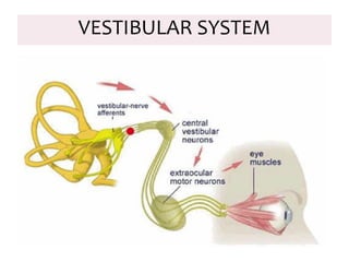
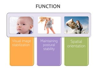
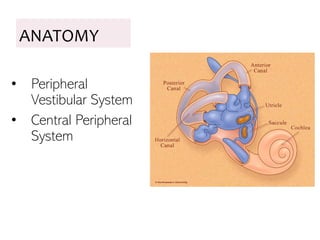
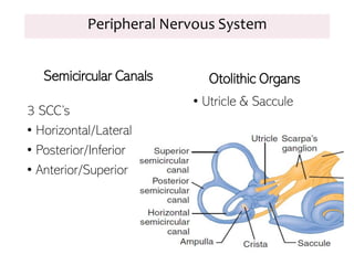
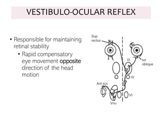
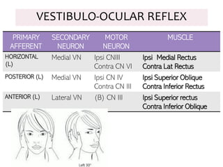
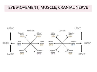
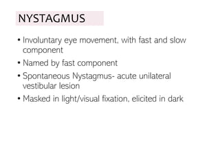
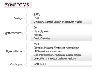
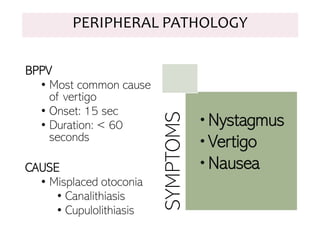
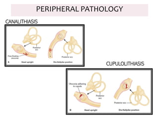
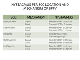
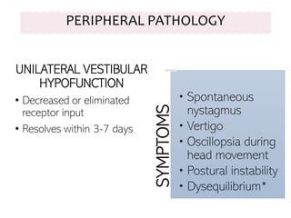
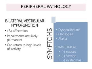
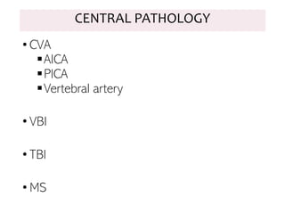
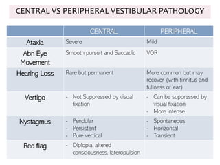
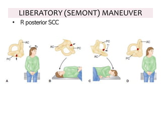
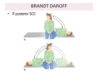
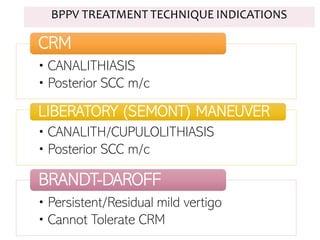
VESTIBULAR SYSTEM
Visual image
stabilization
Maintaining
postural
stability
Spatial
orientation
FUNCTION
ANATOMY
• Peripheral
Vestibular System
• Central Peripheral
System
Peripheral Nervous System
Semicircular Canals Otolithic Organs
• Utricle & Saccule
3 SCC’s
• Horizontal/Lateral
• Posterior/Inferior
• Anterior/Superior
OTOLITH ORGANS
“HUVS”
SEMICIRCULAR CANALS
PUSH-PULL DYNAMIC
CENTRAL VESTIBULAR SYSTEM
CORTEX
- Parietal and insular
lobes
• Thalamus
• Reticular System
• Cerebellum
VESTIBULO-OCULAR REFLEX
• Responsible for maintaining
retinal stability
• Rapid compensatory
eye movement opposite
direction of the head
motion
PRIMARY
AFFERENT
SECONDARY
NEURON
MOTOR
NEURON
MUSCLE
HORIZONTAL
(L)
Medial VN Ipsi CNIII
Contra CN VI
Ipsi Medial Rectus
Contra Lat Rectus
POSTERIOR (L) Medial VN Ipsi CN IV
Contra CN III
Ipsi Superior Oblique
Contra Inferior Rectus
ANTERIOR (L) Lateral VN (B) CN III Ipsi Superior rectus
Contra Inferior Oblique
VESTIBULO-OCULAR REFLEX
EYE MOVEMENT; MUSCLE; CRANIAL NERVE
RPSCC
RHSCC LHSCC
RASCC
LASCC
LPSCC
NYSTAGMUS
• Involuntary eye movement, with fast and slow
component
• Named by fast component
• Spontaneous Nystagmus- acute unilateral
vestibular lesion
• Masked in light/visual fixation, elicited in dark
SYMPTOMS
Vertigo
• BPPV
• UVH
• Unilateral Central Lesion (Vestibular Nuclei)
Lightheadedness
• OH
• Hypoglycemia
• Anxiety
• Panic Disorder
Dysequilibrium
• BVH
• Chronic Unilateral Vestibular hypofuction
• LE Somatosensation loss
• Upper brainsterm/Vestibular Cortex lesion
• cerebellar and motor pathway lesions
Oscillopsia • VOR deficit
VESTIBULAR SYSTEM
DYSFUNCTION
Peripheral Pathologies
PERIPHERAL PATHOLOGY
BPPV
• Most common cause
of vertigo
• Onset: 15 sec
• Duration: < 60
seconds
CAUSE
• Misplaced otoconia
• Canalithiasis
• Cupulolithiasis
SYMPTOMS
•Nystagmus
•Vertigo
•Nausea
CANALITHIASIS
CUPULOLITHIASIS
PERIPHERAL PATHOLOGY
NYSTAGMUS PER SCC LOCATION AND
MECHANISM OF BPPV
SCC MECHANISM NYSTAGMUS
Right posterior Cupulo
Canal
Persistent UBN c R torsion
Transient UBN c R torsion
Left Posterior Cupulo
Canal
Persistent UBN c L torsion
Transient UBN c L torsion
Horizontal Cupulo
Canal
Persistent ageotropic
Transient geotropic
Right Superior Cupulo
Canal
Persistent DBN c R torsion
Left Superior Cupulo
Canal
Persistent DBN c L torsion
UNILATERAL VESTIBULAR
HYPOFUNCTION
• Decreased or eliminated
receptor input
• Resolves within 3-7 days
SYMPTOMS
• Spontaneous
nystagmus
• Vertigo
• Oscillopsia during
head movement
• Postural instability
• Dysequilibrium*
PERIPHERAL PATHOLOGY
BILATERAL VESTIBULAR
HYPOFUNCTION
• (B) affectation
• Impairments are likely
permanent
• Can return to high levels
of activity
SYMPTOMS
• Dysequilibrium*
• Oscillopsia
• Ataxia
SYMMETRICAL
• (-) nausea
• (-) Vertigo
• (-) nystagmus
PERIPHERAL PATHOLOGY
• CVA
▪AICA
▪PICA
▪Vertebral artery
• VBI
• TBI
• MS
CENTRAL PATHOLOGY
CENTRAL VS PERIPHERAL VESTIBULAR PATHOLOGY
CENTRAL PERIPHERAL
Ataxia Severe Mild
Abn Eye
Movement
Smooth pursuit and Saccadic VOR
Hearing Loss Rare but permanent More common but may
recover (with tinnitus and
fullness of ear)
Vertigo - Not Suppressed by visual
fixation
- Can be suppressed by
visual fixation
- More intense
Nystagmus - Pendular
- Persistent
- Pure vertical
- Spontaneous
- Horizontal
- Transient
Red flag - Diplopia, altered
consciousness, lateropulsion
OCULAR TILT REACTION
ASSESSMENTS
HEAD IMPULSE TEST
HSCC
HEAD IMPULSE TEST
Vertical Semicircular Canal
HEAD SHAKING INDUCED NYSTAGMUS
• Tests unilateral
peripheral vestibular
defect
INSTRUCTION:
1. Patient close eyes
2. Flex head 30
3. Oscillate horizontally 20
cycles (2 repetitions per
second
DIX-HALLPIKE Test
• Patient sits on the
examination table
• Manually rotate head
45
• Quickly brought to
supine with neck 30
extended beyond
neutral
*ASCC & PSCC
• Patient sits on the
examination table
• Head rotated
horizontally 45
degrees
• Quickly bring patient
down to side opposite
to the side of head
rotation
DIX-HALLPIKE Test
ROLL TEST
*HSCC
Caloric test
• RESPONSE: COWS
• Limitations
• Only HSCC can be
assessed
• Stimulation
corresponds to a
frequency (0.025 Hz)
that is much lower
than the natural
frequencies of head
movement (1 to 20
Hz).
ROTATIONAL CHAIR TEST
• Rotate subject in the
dark/ Eyes closed using
a chair.
• Normal: Nystagmus
should be generated
• Compare VOR of each
ear/direction
• Considered the standard
test for bilateral vestibular
hypofunction
• Limitation:
-Only HSCC can be
assessed
DYNAMIC VISUAL ACUITY TEST
Measurement of visual
acuity during horizontal
movement of the head.
Speed: >100deg/sec
CLINICAL TEST FOR SENSORY INTERACTION OF
BALANCE
INTERVENTIONS
BPPV
CANALITH REPOSITIONING MANEUVER
• Anterior or posterior SCC
• For R horizontal SCC
CANALITH REPOSITIONING MANEUVER
LIBERATORY (SEMONT) MANEUVER
• R posterior SCC
BRANDT DAROFF
• R posterior SCC
BPPV TREATMENT TECHNIQUE INDICATIONS
• CANALITHIASIS
• Posterior SCC m/c
CRM
• CANALITH/CUPULOLITHIASIS
• Posterior SCC m/c
LIBERATORY (SEMONT) MANEUVER
• Persistent/Residual mild vertigo
• Cannot Tolerate CRM
BRANDT-DAROFF
INTERVENTIONS
Unilateral Vestibular Hypofunction
GAZE STABILITY EXERCISE
•Improve VOR
•Expose patient
to “RETINAL
SLIP”
HABITUATION EXERCISE
•Find provoking position and maintain for
30 seconds
•Do 3-5 reps, 2-3x/day
•Make activity diary
•2 weeks
BALANCE EXERCISE
•Develop balance strategy
•Challenging but safe enough to be done
independently
•INCORPORATE HEAD MOVEMENT
INTERVENTIONS
Bilateral Vestibular Hypofunction
•2x is not recommended
GAZE STABILITY TRAINING
•Prone to fall
BALANCE EXERCISE
VESTIBULAR ADAPTATION
MENIERE’S DISEASE
Tx: decreasing fluid build up or surgery
Symptoms:
Low frequency hearing
loss + episodic vertigo
Fullness of ear Tinnitus
Due to increase in endolymphatic fluid
PERILYMPHATIC FISTULA
• Cause: rupture of the oval or round windows.
• MOI: trauma (loud noise, excessive pressure,
head trauma)
• Sx: Vertigo and Hearing Loss
• Tx:
• Bed rest
• Surgical patches of fistula
• PT: residual sx or hypofunction post-op
Vestibular System (Ear) Lecture for students

Vestibular System (Ear) Lecture for students