The document summarizes the third stage of labor and delivery of the placenta. It describes:
- The third stage begins after delivery of the fetus and lasts until delivery of the placenta, usually 10-15 minutes.
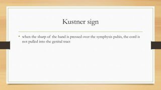
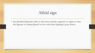
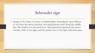
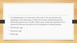
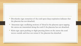
- There are two mechanisms of placental separation - central (Schultze) and edge (Duncan) separation. Signs that the placenta has separated from the uterus include the Kustner, Alfeld and Schroeder signs.
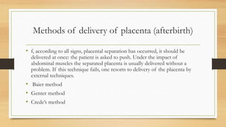
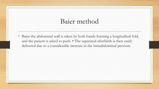
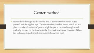
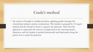
- Methods for delivering the placenta include having the mother push, or using external techniques like the Baier, Genter, or Crede's method which apply pressure to the uterus to express the placenta.