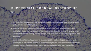
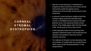
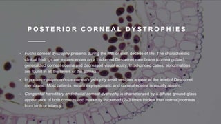
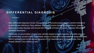
Local Housewife and effective ☎️ 8250192130 🍉🍓 Sexy Girls VIP Call Girls Chan...
David Rajasekaran Prasannakumar 1432.pptx
1. C O R N E A L
D Y S T R O P H Y
David Rajasekaran
Prasannakumar
1432
2. I N T R O D U C T I O N
• Corneal dystrophies are often progressive, eye disorders in which
abnormal material often accumulates in the clear outer layer of the eye.
• It is a group of rare hereditary disorders characterized by bilateral
abnormal deposition of substances in the transparent front part of the
eye called the cornea.
• A corneal dystrophy can be caused by an accumulation of extraneous
material in the cornea, including lipids and cholesterol crystals.
3. S I G N S & S Y M P T O M S
• Corneal dystrophy may not significantly affect vision in the early stages. However, it does require proper evaluation and treatment for restoration of optimal vision.
Corneal dystrophies usually manifest themselves during the first or second decade but sometimes later. It appears as grayish white lines, circles, or clouding of
the cornea.
• Corneal dystrophy can also have a crystalline appearance. They affect vision in widely differing ways. Some cause severe visual impairment, while a few cause no
vision problems and are diagnosed during a specialized eye examination by an opthalmologist. Other dystrophies may cause repeated episodes of pain without leading
to permanent loss of vision.
There are over 20 corneal dystrophies that affect all parts of the cornea. These diseases share many traits:
• They are usually inherited, Most progress gradually.
• They affect the right and left eyes equally.
• They are not caused by outside factors, such as injury or diet.
• Most usually begin in one of the five corneal layers and may later spread to nearby layers.
• Most do not affect other parts of the body, nor are they related to diseases affecting other parts of the eye or body.
• Most can occur in otherwise totally healthy people, male or female.
4. G E N E T I C S
• Different corneal dystrophies are caused by mutations in the CHST6,
KRT3, KRT12, PIP5K3, SLC4A11, TACSTD2, TGFBI, and UBIAD1 genes.
Mutations in TGFBI which encodes transforming growth factor beta
induced cause several forms of corneal dystrophies including granular
corneal dystrophy, lattice corneal dystrophy, epithelial basement
membrane dystrophy, Reis-Bucklers corneal dystrophy, and Thiel–Behnke
dystrophy.
• Corneal dystrophies may have a simple autosomal dominant, autosomal
recessive or rarely X-linked recessive Mendelian mode of inheritance
5. D I A G N O S I S
• Diagnosis can be established on clinical grounds and this may be enhanced with studies on
surgically excised corneal tissue and in some cases with molecular genetic analyses.
• As clinical manifestations widely vary with the different entities, corneal dystrophies should be
suspected when corneal transparency is lost or corneal opacities occur spontaneously,
particularly in both corneas, and especially in the presence of a positive family history or in the
offspring of consanguineous parents.
6. S U P E R F I C I A L C O R N E A L
D Y S T R O P H I E S
• Meesmann dystrophy is characterized by distinct tiny bubble-like, punctate opacities that
form in the central corneal epithelium and to a lesser extent in the peripheral cornea of
both eyes during infancy that persists throughout life.
• Symmetrical reticular opacities form in the superficial central cornea of both eyes at
about 4–5 years of age in Reis-Bücklers corneal dystrophy. Patient remains
asymptomatic until epithelial erosions precipitate acute episodes of ocular hyperemia,
pain, and photophobia.
• Visual acuity eventually becomes reduced during the second and third decades of life
following a progressive superficial haze and an irregular corneal surface.
7. S U P E R F I C I A L C O R N E A L D Y S T R O P H I E
S
• In Thiel–Behnke dystrophy, sub-epithelial corneal opacities form a honeycomb-shaped
pattern in the superficial cornea.
• Multiple prominent gelatinous mulberry-shaped nodules form beneath the corneal
epithelium during the first decade of life in gelatinous drop-like corneal dystrophy which
cause photophobia, tearing, corneal foreign body sensation and severe progressive loss
of vision.
• Lisch epithelial corneal dystrophy is characterized by feather shaped opacities and
microcysts in the corneal epithelium that are arranged in a band-shaped and sometimes
whorled pattern. Painless blurred vision sometimes begins after sixty years of life
8. C O R N E A L
S T R O M A L
D Y S T R O P H I E S
• Macular corneal dystrophy is manifested by a
progressive dense cloudiness of the entire corneal
stroma that usually first appears during
adolescence and eventually causing severe visual
impairment.
• In granular corneal dystrophy multiple small white
discrete irregular spots that resemble bread
crumbs or snowflakes become apparent beneath
Bowman zone in the superficial central corneal
stroma. They initially appear within the first decade
of life. Visual acuity is more or less normal.
• Lattice dystrophy starts as fine branching linear
opacities in Bowman's layer in the central area and
spreads to the periphery. Recurrent corneal
erosions may occur.
• The hallmark of Schnyder corneal dystrophy is the
accumulation of crystals within the corneal stroma
which cause corneal clouding typically in a ring-
shaped fashion.
9. P O S T E R I O R C O R N E A L D Y S T R O P H I E S
• Fuchs corneal dystrophy presents during the fifth or sixth decade of life. The characteristic
clinical findings are excrescences on a thickened Descemet membrane (cornea guttae),
generalized corneal edema and decreased visual acuity. In advanced cases, abnormalities
are found in all the layers of the cornea.
• In posterior polymorphous corneal dystrophy small vesicles appear at the level of Descemet
membrane. Most patients remain asymptomatic and corneal edema is usually absent.
• Congenital hereditary endothelial corneal dystrophy is characterized by a diffuse ground-glass
appearance of both corneas and markedly thickened (2–3 times thicker than normal) corneas
from birth or infancy.
10. D I F F E R E N T I A L D I A G N O S I S
• Main differential diagnosis include various causes of monoclonal gammopathy, lecithin-cholesterol-
acyltransferase deficiency, Fabry disease, cystinosis, tyrosine transaminase deficiency, systemic
lysosomal storage diseases, and several skin diseases (X-linked ichthyosis, keratosis follicularis
spinolosa decalvans).
• Historically, an accumulation of small gray variable shaped punctate opacities of variable shape in
the central deep corneal stroma immediately anterior to Descemet membrane were designated deep
filiform dystrophy and cornea farinata because of their resemblance to commas, circles, lines,
threads (filiform), flour (farina) or dots.
11. D I F F E R E N T I A L D I A G N O S I S
• These abnormalities are now known to accompany X-linked ichthyosis, steroid sulfatase
deficiency, caused by steroid sulfatase gene mutations and are currently usually not included
under the rubric of the corneal dystrophies.
• In the past, the designation vortex corneal dystrophy (corneal verticillata) was applied to a
corneal disorder characterized by the presence of innumerable tiny brown spots arranged in
curved whirlpool-like lines in the superficial cornea.
• An autosomal dominant mode of transmission was initially suspected, but later it was realized
that these individuals were affected hemizygous males and asymptomatic female carriers of an
X-linked systemic metabolic disease caused by a deficiency of α-galactosidase, known as
Fabry disease.
12. E P I T H E L I A L
&
S U B E P I T H E L I
A L
D Y S T R O P H I E
S
• Epithelial basement membrane
dystrophy
• Epithelial recurrent erosion dystrophies
(Franceschetti corneal
dystrophy, Dystrophia Smolandiensis,
and Dystrophia Helsinglandica)
• Subepithelial mucinous corneal
dystrophy
• Meesmann corneal dystrophy
• Lisch epithelial corneal dystrophy
• Gelatinous drop-like corneal dystrophy
(AR)
13. B O W M A N
L AY E R
D Y S T R O P H I E
S
• Reis–Bücklers corneal dystrophy
• Thiel–Behnke corneal dystrophy
• Stromal dystrophies-
• TGFB1 corneal dystrophies
• Lattice corneal dystrophy, type 1 variants
(III, IIIA, I/IIIA, IV) of lattice corneal
dystrophy
• Granular corneal dystrophy, type 1
• Granular corneal dystrophy, type 2
14. S T R O M A L D Y S T R O P H I E S
• Macular corneal dystrophy (AR)
• Schnyder crystalline corneal dystrophy
• Congenital stromal corneal dystrophy
• Fleck corneal dystrophy
• Posterior amorphous corneal dystrophy
• Central cloudy dystrophy of François
• Pre-Descemet corneal dystrophy
15. E N D O T H E L I A
L
D Y S T R O P H I E
S
• Fuchs' dystrophy
• Posterior polymorphous corneal dystrophy
• Congenital hereditary endothelial dystrophy
(AR)
• X-linked endothelial corneal dystrophy
16. T R E AT M E N T
• Early stages may be asymptomatic and may not require any intervention. Initial treatment may include hypertonic
eyedrops and ointment to reduce the corneal edema and may offer symptomatic improvement prior to surgical
intervention.
• Suboptimal vision caused by corneal dystrophy may be helped with scleral contact lenses but eventually usually requires
surgical intervention in the form of corneal transplantation. Penetrating keratoplasty, a common type of corneal
transplantation, is commonly performed for extensive corneal dystrophy.
• With penetrating keratoplasty (corneal transplant), the long-term results are good to excellent. Recent surgical
improvements have been made which have increased the success rate for this procedure. However, recurrence of the
disease in the donor graft may happen. Superficial corneal dystrophies do not need a penetrating keratoplasty as the
deeper corneal tissue is unaffected, therefore a lamellar keratoplasty may be used instead.
• Phototherapeutic keratectomy (PTK) can be used to excise or ablate the abnormal corneal tissue. Patients with superficial
corneal opacities are suitable candidates for this procedure.