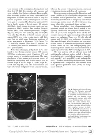
This study aimed to predict the risk of malignancy in women with adnexal masses using preoperative factors. The researchers analyzed 395 patients and found:
1) Tumor morphology on ultrasound, elevated serum CA 125 levels, presence of ascites, and older age were associated with higher risk of malignancy.
2) Patients with solid or complex masses and CA 125 > 35 U/mL had a positive predictive value of 84.7% for malignancy.
3) Purely cystic masses had a 100% negative predictive value for ruling out malignancy.
4) The combination of complex/solid mass and elevated CA 125 best defined patients at high risk of ovarian cancer.
Austin Journal of Clinical & Diagnostic Research is a multidisciplinary, Rapid-Publication journal. Austin Journal of Clinical & Diagnostic Research Journal is open to scientists from all countries. The mission of the journal is to promote topics of Clinical & Diagnostic research, as well as stimulate international cooperation in these areas. The journal will consider articles from every legitimate specialty.
Austin Publishing Group's mission to facilitate immediate access to scientific data through an Open Access platform is greatly supported by invaluable contributions from the strong editorial and advisory boards.
Austin Publishing Group is moving ahead with a vision to develop an optimized knowledge sharing platform and an enlightening interactive network for researchers all over the world through its scientific publications and meetings.
The KRAS-Variant and miRNA Expression in RTOG Endometrial Cancer Clinical Tri...UCLA
The KRAS-variant may be a genetic marker of risk for type 2 endometrial cancers. In addition, tumor miRNA expression appears to be associated with patient age, lymphovascular invasion and the KRAS-variant, supporting the hypothesis that altered tumor biology can be measured by miRNA expression, and that the KRAS-variant likely impacts endometrial tumor biology.
PRIMARY SQUAMOUS CELL CANCER OF BREAST: A CASE REPORTKETAN VAGHOLKAR
Primary squamous cell cancer (SqCC) of the breast is a rather rare disease. These tumors are known to be
quite aggressive in nature and are usually found to be treatment-resistant. Currently, there is no standard treatment
guideline for the management of primary SqCC of the breast. In this case report, we present a case of primary SqCC of
the breast in 60-year old postmenopausal women presenting as pigmented lesion over the right breast (no lump). Initial
skin biopsy (core) done by dermatologist revealed squamous cell cancer in situ (Bowen’s disease); however surgical
resection of the lesion and subsequent histopathological examination revealed primary SqCC (no secondary sites were
found elsewhere in the body).
Clinicopathologic Features and Survival Analysis of Non-metastatic Breast Can...Hugo Raul Castro Salguero
Background: Breast cancer (BC) is a leading cause of cancer related death
worldwide. Unfortunately, data concerning clinicopathologic features of this
malignancy in non-developed countries is scarce. This study aims to characterize a
cohort of Guatemalan female patients with non-metastatic BC and to determine
risk factors for overall survival (OS).
Cost-Effectiveness of Contralateral Prophylactic
Mastectomy Versus Routine Surveillance in Patients
With Unilateral Breast Cancer
Benjamin Zendejas, James P. Moriarty, Jamie O’Byrne, Amy C. Degnim, David R. Farley, and Judy C. Boughey
Cancer de mama
Clinica Ruber
Dr Juan Carlos Meneu
The KRAS-Variant Is Associated with Risk of Developing Double Primary Breast ...UCLA
A germline microRNA binding site-disrupting variant, the KRAS-variant (rs61764370), is associated with an increased risk of developing several cancers. Because this variant is most strongly associated with ovarian cancer risk in patients from hereditary breast and ovarian families (HBOC), and with the risk of premenopausal triple negative breast cancer, we evaluated the association of the KRAS-variant with women with personal histories of both breast and ovarian cancer, referred to as double primary patients.
Dr. Maurie Markman, President of Science and Medicine at Cancer Treatment Centers of America, shares his expertise on the latest developments in immunotherapy for ovarian cancer.
Anaesthesia considerations and Implications during Oncologic and Non-Oncologi...Apollo Hospitals
Cancer has been the leading cause of mortality in both developed and developing countries. With the advancement in chemotherapeutic agents, the quality and lifespan of patients with advanced malignancies has improved. These patients often come to hospitals for various types of elective and emergency surgeries. The attending anaesthesiologist faces a daunting task while managing these patients as there can be gross physiological derangements in most of the organ systems. A careful and thorough preoperative assessment, optimisation of physiological milieu, vigilant intraoperative monitoring, anticipation of potential complications and postoperative pain control is essential for reducing perioperative mortality and morbidity in these patients.
Original StudyType of Breast Cancer Diagnosis, Screening,a.docxvannagoforth
Original Study
Type of Breast Cancer Diagnosis, Screening,
and Survival
Carla Cedolini,1 Serena Bertozzi,1 Ambrogio P. Londero,2 Sergio Bernardi,3,4
Luca Seriau,1 Serena Concina,1 Federico Cattin,1 Andrea Risaliti1
Abstract
Organized, invitational breast cancer screening in our population succeeded in detecting early-stage tumors,
which have been consequently treated more frequently with breast and axillary conservative surgery, com-
plementary breast irradiation, and eventual hormonal therapy. The diagnosis of invasive cancer with screening
in our population resulted in a survival gain at 5 years from the diagnosis.
Introduction: Breast cancer screening is known to reduce mortality. In the present study, we analyzed the prevalence
of breast cancers detected through screening, before and after introduction of an organized screening, and we
evaluated the overall survival of these patients in comparison with women with an extrascreening imaging-detected
breast cancer or those with palpable breast cancers. Materials and Methods: We collected data about all women
who underwent a breast operation for cancer in our department between 2001 and 2008, focusing on type of tumor
diagnosis, tumor characteristics, therapies administered, and patient outcome in terms of overall survival, and re-
currences. Data was analyzed by R (version 2.15.2), and P < .05 was considered significant. Results: Among the 2070
cases of invasive breast cancer we considered, 157 were detected by regional mammographic screening (group A),
843 by extrascreening breast imaging (group B: 507 by mammography and 336 by ultrasound), and 1070 by extra-
screening breast objective examination (group C). The 5-year overall survival in groups A, B, and C were, respectively,
99% (95% CI, 98%-100%), 98% (95% CI, 97%-99%), and 91% (95% CI, 90%-93%), with a significant difference
between the first 2 groups and the third (P < .05) and a trend between groups A and B (P ¼ .081). Conclusion: The
diagnosis of invasive breast cancer with screening in our population resulted in a survival gain at 5 years from the
diagnosis, but a longer follow-up is necessary to confirm this data.
Clinical Breast Cancer, Vol. 14, No. 4, 235-40 ª 2014 Elsevier Inc. All rights reserved.
Keywords: Breast cancer, Breast cancer screening, Invasive breast cancer, Mammographic screening, Overall survival
Introduction
Because of the detection of early-stage tumors, breast cancer
screening reduced breast cancer mortality in Europe by 25%-31%
in patients who were invited for screening and by 38%-48% in
those who were actually screened during the last decade of the
twentieth century and the first decade of the twenty-first.1 In our
region of Italy, an organized breast cancer screening was firstly intro-
duced in 2005, but despite the high compliance of invited women
1Clinic of Surgery
2Clinic of Obstetrics and Gynecology
University of Udine, Udine, Italy
3Department of Surgery, Ospedale Civile di Latisana, Udine, Italy
4 ...
Management of ovarian masses e Clinical situations & recommendations Apollo Hospitals
Adenexal mass is a common clinical presentation. This clinical situation is a problem that affects women of all ages. The biggest challenge is that one should not miss out on a diagnosis of malignant ovarian tumor. An ovarian mass or cyst that raises the suspicion of malignancy is a common dilemma in a gynecological practice. In the United States, a woman has a 5-10% lifetime risk of undergoing surgery for a suspected ovarian neoplasm and an estimated 13e21% chance of this turning into a diagnosis of ovarian cancer. Most of the adnexal masses are benign but the first responsibility of the treating gynecologist is to exclude malignancy. Management decisions often are influenced by the age and family history and presentation of the patient.
Diagnosed with breast cancer while on a family historyscreen.docxduketjoy27252
Diagnosed with breast cancer while on a family history
screening programme: an exploratory qualitative study
A. CLEMENTS, bsc, senior research nurse, Cancer Research UK Primary Care Education Research Group,
University of Oxford, Department of Primary Health Care, Oxford, B.J. HENDERSON, phd, research psycholo-
gist, Institute of Medical & Social Care Research, Ardudwy, Normal Site, University of Wales, Bangor, Gwynedd,
S. TYNDEL, ba, research officer, Cancer Research UK Primary Care Education Research Group, University of
Oxford, Department of Primary Health Care, Oxford, G. EVANS, md frcp, consultant in medical genetics,
Department of Clinical Genetics, St Mary’s Hospital, Manchester, K. BRAIN, phd, senior research fellow,
Institute of Medical Genetics, University of Wales College of Medicine, Heath Park, Cardiff, J. AUSTOKER, phd,
director, Cancer Research UK Primary Care Education Research Group, University of Oxford, Department of
Primary Health Care, Oxford, & E. WATSON, phd, deputy director, Cancer Research UK Primary Care Educa-
tion Research Group, University of Oxford, Department of Primary Health Care, Oxford, UK for the PIMMS Study
Management Group*
CLEMENTS A., HENDERSON B.J., TYNDEL S., EVANS G., BRAIN K., AUSTOKER J. & WATSON E. FOR
THE PIMMS STUDY MANAGEMENT GROUP (2008) European Journal of Cancer Care 17, 245–252
Diagnosed with breast cancer while on a family history screening programme: an exploratory qualitative study
Mammographic screening is offered to many women under 50 in the UK who are at moderate or high risk of
developing breast cancer because of their family history of the disease. Little is understood about the impact
of screening on the emotional well-being of women with a family history of breast cancer. This qualitative
study explores the value that women at increased risk placed on screening, both pre- and post-cancer diagnosis
and the impact of the diagnosis. In-depth interviews were undertaken with 12 women, aged 35–50, diagnosed
with breast cancer while on an annual mammographic screening programme. Women described the strong
sense of reassurance gained from screening prior to diagnosis. This faith in screening was reinforced by early
detection of their cancer. Reactions to diagnosis ranged from devastation to relief at having finally developed
a long-expected condition. Despite their positive attitudes about screening, not all women wanted to continue
with surveillance. For some, prophylactic mastectomy was preferable, to reduce future cancer risk and to
alleviate anxieties about the detection of another cancer at each subsequent screen. This study illustrates the
positive yet diverse attitudes towards mammographic screening in this group of women with a family history
of breast cancer.
Keywords: breast cancer, early screening programme, family history, qualitative.
Correspondence address: Alison Clements, Cancer Research UK Primary Care Education Research Group, University of Oxford, Department of Pr.
Diagnosed with breast cancer while on a family historyscreen.docxlynettearnold46882
Diagnosed with breast cancer while on a family history
screening programme: an exploratory qualitative study
A. CLEMENTS, bsc, senior research nurse, Cancer Research UK Primary Care Education Research Group,
University of Oxford, Department of Primary Health Care, Oxford, B.J. HENDERSON, phd, research psycholo-
gist, Institute of Medical & Social Care Research, Ardudwy, Normal Site, University of Wales, Bangor, Gwynedd,
S. TYNDEL, ba, research officer, Cancer Research UK Primary Care Education Research Group, University of
Oxford, Department of Primary Health Care, Oxford, G. EVANS, md frcp, consultant in medical genetics,
Department of Clinical Genetics, St Mary’s Hospital, Manchester, K. BRAIN, phd, senior research fellow,
Institute of Medical Genetics, University of Wales College of Medicine, Heath Park, Cardiff, J. AUSTOKER, phd,
director, Cancer Research UK Primary Care Education Research Group, University of Oxford, Department of
Primary Health Care, Oxford, & E. WATSON, phd, deputy director, Cancer Research UK Primary Care Educa-
tion Research Group, University of Oxford, Department of Primary Health Care, Oxford, UK for the PIMMS Study
Management Group*
CLEMENTS A., HENDERSON B.J., TYNDEL S., EVANS G., BRAIN K., AUSTOKER J. & WATSON E. FOR
THE PIMMS STUDY MANAGEMENT GROUP (2008) European Journal of Cancer Care 17, 245–252
Diagnosed with breast cancer while on a family history screening programme: an exploratory qualitative study
Mammographic screening is offered to many women under 50 in the UK who are at moderate or high risk of
developing breast cancer because of their family history of the disease. Little is understood about the impact
of screening on the emotional well-being of women with a family history of breast cancer. This qualitative
study explores the value that women at increased risk placed on screening, both pre- and post-cancer diagnosis
and the impact of the diagnosis. In-depth interviews were undertaken with 12 women, aged 35–50, diagnosed
with breast cancer while on an annual mammographic screening programme. Women described the strong
sense of reassurance gained from screening prior to diagnosis. This faith in screening was reinforced by early
detection of their cancer. Reactions to diagnosis ranged from devastation to relief at having finally developed
a long-expected condition. Despite their positive attitudes about screening, not all women wanted to continue
with surveillance. For some, prophylactic mastectomy was preferable, to reduce future cancer risk and to
alleviate anxieties about the detection of another cancer at each subsequent screen. This study illustrates the
positive yet diverse attitudes towards mammographic screening in this group of women with a family history
of breast cancer.
Keywords: breast cancer, early screening programme, family history, qualitative.
Correspondence address: Alison Clements, Cancer Research UK Primary Care Education Research Group, University of Oxford, Department of Pr.
a nice presentation about the Ovarian Cancer its include an introduction with brief notes about the epidemiology and risk factors then shift to pathology and pathogenesis and diagnosis with signs , symptoms and lab tests with imaging modules , screening , management
Dindigul district cervical screening study, india acceptability, effectivenes...Asha Reddy
Dindigul district cervical screening study, india acceptability, effectiveness and safety of treatment of cervical precancerous lesions by nurses using cryotherapy
Discordant noninvasive prenatal testing & cytogenetic results: 109 consecutive cases.
In conclusion, use of conventional cytogenetics as the reference
standard unravels a rather significant discordance with
positive NIPT results. These data should compel us to take a
cautious look at NIPT data sets and their sources. As more
commercial providers advocate this test, or sponsor academic
centers to carry them out, a more diligent comparison of NIPT
results with cytogenetic tests should be undertaken.
Expressed breast milk on refrigerator storageAsha Reddy
In conclusion, we can store mother’s milk at refrigerator temperature of 4ºC for 96 hours without changing its overall integrity in the form of pH, serum albumin, total protein, lipid and lactose content and can use it for feeding neonates and infants.
Human Milk Banking Guidelines -INDIAN ACADEMY OF PEDIATRICSAsha Reddy
Human Milk Banking Guidelines INFANT AND YOUNG CHILD FEEDING CHAPTER, INDIAN ACADEMY OF PEDIATRICS
Absent or insufficient lactation: Mothers with
multiple births, who can not secrete adequate
breastmilk for their neonates initially.
• For babies of non-lactating mothers, who adopt
neonates and if induced lactation is not possible.
• Abandoned neonates and sick neonates.
• Temporary interruption of breastfeeding.
• Infant at health risk from breastmilk of the biological
mother.
• Babies whose mother died in the immediate
postpartum period
MANAGEMENT OF ATRIOVENTRICULAR CONDUCTION BLOCK.pdfJim Jacob Roy
Cardiac conduction defects can occur due to various causes.
Atrioventricular conduction blocks ( AV blocks ) are classified into 3 types.
This document describes the acute management of AV block.
The prostate is an exocrine gland of the male mammalian reproductive system
It is a walnut-sized gland that forms part of the male reproductive system and is located in front of the rectum and just below the urinary bladder
Function is to store and secrete a clear, slightly alkaline fluid that constitutes 10-30% of the volume of the seminal fluid that along with the spermatozoa, constitutes semen
A healthy human prostate measures (4cm-vertical, by 3cm-horizontal, 2cm ant-post ).
It surrounds the urethra just below the urinary bladder. It has anterior, median, posterior and two lateral lobes
It’s work is regulated by androgens which are responsible for male sex characteristics
Generalised disease of the prostate due to hormonal derangement which leads to non malignant enlargement of the gland (increase in the number of epithelial cells and stromal tissue)to cause compression of the urethra leading to symptoms (LUTS
Tom Selleck Health: A Comprehensive Look at the Iconic Actor’s Wellness Journeygreendigital
Tom Selleck, an enduring figure in Hollywood. has captivated audiences for decades with his rugged charm, iconic moustache. and memorable roles in television and film. From his breakout role as Thomas Magnum in Magnum P.I. to his current portrayal of Frank Reagan in Blue Bloods. Selleck's career has spanned over 50 years. But beyond his professional achievements. fans have often been curious about Tom Selleck Health. especially as he has aged in the public eye.
Follow us on: Pinterest
Introduction
Many have been interested in Tom Selleck health. not only because of his enduring presence on screen but also because of the challenges. and lifestyle choices he has faced and made over the years. This article delves into the various aspects of Tom Selleck health. exploring his fitness regimen, diet, mental health. and the challenges he has encountered as he ages. We'll look at how he maintains his well-being. the health issues he has faced, and his approach to ageing .
Early Life and Career
Childhood and Athletic Beginnings
Tom Selleck was born on January 29, 1945, in Detroit, Michigan, and grew up in Sherman Oaks, California. From an early age, he was involved in sports, particularly basketball. which played a significant role in his physical development. His athletic pursuits continued into college. where he attended the University of Southern California (USC) on a basketball scholarship. This early involvement in sports laid a strong foundation for his physical health and disciplined lifestyle.
Transition to Acting
Selleck's transition from an athlete to an actor came with its physical demands. His first significant role in "Magnum P.I." required him to perform various stunts and maintain a fit appearance. This role, which he played from 1980 to 1988. necessitated a rigorous fitness routine to meet the show's demands. setting the stage for his long-term commitment to health and wellness.
Fitness Regimen
Workout Routine
Tom Selleck health and fitness regimen has evolved. adapting to his changing roles and age. During his "Magnum, P.I." days. Selleck's workouts were intense and focused on building and maintaining muscle mass. His routine included weightlifting, cardiovascular exercises. and specific training for the stunts he performed on the show.
Selleck adjusted his fitness routine as he aged to suit his body's needs. Today, his workouts focus on maintaining flexibility, strength, and cardiovascular health. He incorporates low-impact exercises such as swimming, walking, and light weightlifting. This balanced approach helps him stay fit without putting undue strain on his joints and muscles.
Importance of Flexibility and Mobility
In recent years, Selleck has emphasized the importance of flexibility and mobility in his fitness regimen. Understanding the natural decline in muscle mass and joint flexibility with age. he includes stretching and yoga in his routine. These practices help prevent injuries, improve posture, and maintain mobilit
Title: Sense of Smell
Presenter: Dr. Faiza, Assistant Professor of Physiology
Qualifications:
MBBS (Best Graduate, AIMC Lahore)
FCPS Physiology
ICMT, CHPE, DHPE (STMU)
MPH (GC University, Faisalabad)
MBA (Virtual University of Pakistan)
Learning Objectives:
Describe the primary categories of smells and the concept of odor blindness.
Explain the structure and location of the olfactory membrane and mucosa, including the types and roles of cells involved in olfaction.
Describe the pathway and mechanisms of olfactory signal transmission from the olfactory receptors to the brain.
Illustrate the biochemical cascade triggered by odorant binding to olfactory receptors, including the role of G-proteins and second messengers in generating an action potential.
Identify different types of olfactory disorders such as anosmia, hyposmia, hyperosmia, and dysosmia, including their potential causes.
Key Topics:
Olfactory Genes:
3% of the human genome accounts for olfactory genes.
400 genes for odorant receptors.
Olfactory Membrane:
Located in the superior part of the nasal cavity.
Medially: Folds downward along the superior septum.
Laterally: Folds over the superior turbinate and upper surface of the middle turbinate.
Total surface area: 5-10 square centimeters.
Olfactory Mucosa:
Olfactory Cells: Bipolar nerve cells derived from the CNS (100 million), with 4-25 olfactory cilia per cell.
Sustentacular Cells: Produce mucus and maintain ionic and molecular environment.
Basal Cells: Replace worn-out olfactory cells with an average lifespan of 1-2 months.
Bowman’s Gland: Secretes mucus.
Stimulation of Olfactory Cells:
Odorant dissolves in mucus and attaches to receptors on olfactory cilia.
Involves a cascade effect through G-proteins and second messengers, leading to depolarization and action potential generation in the olfactory nerve.
Quality of a Good Odorant:
Small (3-20 Carbon atoms), volatile, water-soluble, and lipid-soluble.
Facilitated by odorant-binding proteins in mucus.
Membrane Potential and Action Potential:
Resting membrane potential: -55mV.
Action potential frequency in the olfactory nerve increases with odorant strength.
Adaptation Towards the Sense of Smell:
Rapid adaptation within the first second, with further slow adaptation.
Psychological adaptation greater than receptor adaptation, involving feedback inhibition from the central nervous system.
Primary Sensations of Smell:
Camphoraceous, Musky, Floral, Pepperminty, Ethereal, Pungent, Putrid.
Odor Detection Threshold:
Examples: Hydrogen sulfide (0.0005 ppm), Methyl-mercaptan (0.002 ppm).
Some toxic substances are odorless at lethal concentrations.
Characteristics of Smell:
Odor blindness for single substances due to lack of appropriate receptor protein.
Behavioral and emotional influences of smell.
Transmission of Olfactory Signals:
From olfactory cells to glomeruli in the olfactory bulb, involving lateral inhibition.
Primitive, less old, and new olfactory systems with different path
Title: Sense of Taste
Presenter: Dr. Faiza, Assistant Professor of Physiology
Qualifications:
MBBS (Best Graduate, AIMC Lahore)
FCPS Physiology
ICMT, CHPE, DHPE (STMU)
MPH (GC University, Faisalabad)
MBA (Virtual University of Pakistan)
Learning Objectives:
Describe the structure and function of taste buds.
Describe the relationship between the taste threshold and taste index of common substances.
Explain the chemical basis and signal transduction of taste perception for each type of primary taste sensation.
Recognize different abnormalities of taste perception and their causes.
Key Topics:
Significance of Taste Sensation:
Differentiation between pleasant and harmful food
Influence on behavior
Selection of food based on metabolic needs
Receptors of Taste:
Taste buds on the tongue
Influence of sense of smell, texture of food, and pain stimulation (e.g., by pepper)
Primary and Secondary Taste Sensations:
Primary taste sensations: Sweet, Sour, Salty, Bitter, Umami
Chemical basis and signal transduction mechanisms for each taste
Taste Threshold and Index:
Taste threshold values for Sweet (sucrose), Salty (NaCl), Sour (HCl), and Bitter (Quinine)
Taste index relationship: Inversely proportional to taste threshold
Taste Blindness:
Inability to taste certain substances, particularly thiourea compounds
Example: Phenylthiocarbamide
Structure and Function of Taste Buds:
Composition: Epithelial cells, Sustentacular/Supporting cells, Taste cells, Basal cells
Features: Taste pores, Taste hairs/microvilli, and Taste nerve fibers
Location of Taste Buds:
Found in papillae of the tongue (Fungiform, Circumvallate, Foliate)
Also present on the palate, tonsillar pillars, epiglottis, and proximal esophagus
Mechanism of Taste Stimulation:
Interaction of taste substances with receptors on microvilli
Signal transduction pathways for Umami, Sweet, Bitter, Sour, and Salty tastes
Taste Sensitivity and Adaptation:
Decrease in sensitivity with age
Rapid adaptation of taste sensation
Role of Saliva in Taste:
Dissolution of tastants to reach receptors
Washing away the stimulus
Taste Preferences and Aversions:
Mechanisms behind taste preference and aversion
Influence of receptors and neural pathways
Impact of Sensory Nerve Damage:
Degeneration of taste buds if the sensory nerve fiber is cut
Abnormalities of Taste Detection:
Conditions: Ageusia, Hypogeusia, Dysgeusia (parageusia)
Causes: Nerve damage, neurological disorders, infections, poor oral hygiene, adverse drug effects, deficiencies, aging, tobacco use, altered neurotransmitter levels
Neurotransmitters and Taste Threshold:
Effects of serotonin (5-HT) and norepinephrine (NE) on taste sensitivity
Supertasters:
25% of the population with heightened sensitivity to taste, especially bitterness
Increased number of fungiform papillae
These lecture slides, by Dr Sidra Arshad, offer a quick overview of physiological basis of a normal electrocardiogram.
Learning objectives:
1. Define an electrocardiogram (ECG) and electrocardiography
2. Describe how dipoles generated by the heart produce the waveforms of the ECG
3. Describe the components of a normal electrocardiogram of a typical bipolar leads (limb II)
4. Differentiate between intervals and segments
5. Enlist some common indications for obtaining an ECG
Study Resources:
1. Chapter 11, Guyton and Hall Textbook of Medical Physiology, 14th edition
2. Chapter 9, Human Physiology - From Cells to Systems, Lauralee Sherwood, 9th edition
3. Chapter 29, Ganong’s Review of Medical Physiology, 26th edition
4. Electrocardiogram, StatPearls - https://www.ncbi.nlm.nih.gov/books/NBK549803/
5. ECG in Medical Practice by ABM Abdullah, 4th edition
6. ECG Basics, http://www.nataliescasebook.com/tag/e-c-g-basics
- Video recording of this lecture in English language: https://youtu.be/lK81BzxMqdo
- Video recording of this lecture in Arabic language: https://youtu.be/Ve4P0COk9OI
- Link to download the book free: https://nephrotube.blogspot.com/p/nephrotube-nephrology-books.html
- Link to NephroTube website: www.NephroTube.com
- Link to NephroTube social media accounts: https://nephrotube.blogspot.com/p/join-nephrotube-on-social-media.html
Prix Galien International 2024 Forum ProgramLevi Shapiro
June 20, 2024, Prix Galien International and Jerusalem Ethics Forum in ROME. Detailed agenda including panels:
- ADVANCES IN CARDIOLOGY: A NEW PARADIGM IS COMING
- WOMEN’S HEALTH: FERTILITY PRESERVATION
- WHAT’S NEW IN THE TREATMENT OF INFECTIOUS,
ONCOLOGICAL AND INFLAMMATORY SKIN DISEASES?
- ARTIFICIAL INTELLIGENCE AND ETHICS
- GENE THERAPY
- BEYOND BORDERS: GLOBAL INITIATIVES FOR DEMOCRATIZING LIFE SCIENCE TECHNOLOGIES AND PROMOTING ACCESS TO HEALTHCARE
- ETHICAL CHALLENGES IN LIFE SCIENCES
- Prix Galien International Awards Ceremony
Explore natural remedies for syphilis treatment in Singapore. Discover alternative therapies, herbal remedies, and lifestyle changes that may complement conventional treatments. Learn about holistic approaches to managing syphilis symptoms and supporting overall health.
These simplified slides by Dr. Sidra Arshad present an overview of the non-respiratory functions of the respiratory tract.
Learning objectives:
1. Enlist the non-respiratory functions of the respiratory tract
2. Briefly explain how these functions are carried out
3. Discuss the significance of dead space
4. Differentiate between minute ventilation and alveolar ventilation
5. Describe the cough and sneeze reflexes
Study Resources:
1. Chapter 39, Guyton and Hall Textbook of Medical Physiology, 14th edition
2. Chapter 34, Ganong’s Review of Medical Physiology, 26th edition
3. Chapter 17, Human Physiology by Lauralee Sherwood, 9th edition
4. Non-respiratory functions of the lungs https://academic.oup.com/bjaed/article/13/3/98/278874
micro teaching on communication m.sc nursing.pdfAnurag Sharma
Microteaching is a unique model of practice teaching. It is a viable instrument for the. desired change in the teaching behavior or the behavior potential which, in specified types of real. classroom situations, tends to facilitate the achievement of specified types of objectives.
Factory Supply Best Quality Pmk Oil CAS 28578–16–7 PMK Powder in Stockrebeccabio
Factory Supply Best Quality Pmk Oil CAS 28578–16–7 PMK Powder in Stock
Telegram: bmksupplier
signal: +85264872720
threema: TUD4A6YC
You can contact me on Telegram or Threema
Communicate promptly and reply
Free of customs clearance, Double Clearance 100% pass delivery to USA, Canada, Spain, Germany, Netherland, Poland, Italy, Sweden, UK, Czech Republic, Australia, Mexico, Russia, Ukraine, Kazakhstan.Door to door service
Hot Selling Organic intermediates
Ozempic: Preoperative Management of Patients on GLP-1 Receptor Agonists Saeid Safari
Preoperative Management of Patients on GLP-1 Receptor Agonists like Ozempic and Semiglutide
ASA GUIDELINE
NYSORA Guideline
2 Case Reports of Gastric Ultrasound
2. investigation was undertaken to estimate the accuracy tional Federation of Gynecology and Obstetrics sys-
of a combination of patient demographics, ultrasono- tem. Data were entered into a MEDLOG database
graphically generated tumor morphology, and serum (MEDLOG Systems, Crystal Bay, NV) and exported
CA 125 values in predicting risk of malignancy in into an Excel (Microsoft Corp., Redmond, WA)
adnexal masses. spreadsheet for analysis using WinSTAT (R. Fitch
Software, Bad Krozingen, Germany), SPSS (SPSS,
MATERIALS AND METHODS Inc., Chicago, IL), and PC-SAS with the Enterprise
This investigation was undertaken after approval Miner statistical software (SAS Institute, Inc., Cary, NC).
from the University of Kentucky Human Subjects Proportions were compared using 2 statistics or
Institutional Review Board. Study participants were Fisher exact tests. Means were compared using two-
women referred to the University of Kentucky–Mar- sample t tests. Statistical significance was determined
key Women’s Cancer Center with a diagnosis of an at the .05 level. Multivariable analyses used the
adnexal mass on pelvic examination who underwent classification and regression tree procedure, a non-
surgery at this institution from 2001 to 2008. parametric method that defines or accepts cut points
The following demographic data were obtained and uses training and validation sets to optimize rules
for all study patients: age, height, weight, gravidity, for the analysis.9 The training set is formed by split-
family history of breast or ovarian cancer, personal ting the entire sample in half after stratification for
history of cancer, and menopausal status. Postmeno- malignant cases and benign controls.
pausal was defined as the absence of menses for a
minimum of 12 months. Family history was consid- RESULTS
ered positive if the patient had a first-degree relative Between July 2001 and December 2008, 399 patients
(ie, mother, sister, or daughter) or a second-degree referred to the outpatient clinic of the University of
relative (ie, grandmother, granddaughter, aunt, or Kentucky–Markey Women’s Cancer Center for eval-
niece) with documented ovarian or breast cancer. All uation of an adnexal mass on pelvic examination
women underwent pelvic examination, transvaginal
ultrasonography, and serum CA 125 determination
within 2 weeks before surgery. Transvaginal ultra- Table 1. Patient Demographics, Tumor Variables,
sonography was performed using General Electric and Biomarker Profiles in Patients
(Milwaukee, WI) Logic 400 or Voluson ProV ultra- Studied (N)593؍
sound units with a 5-mHz vaginal probe as described Mean Range
previously.8 Tumor dimensions from ultrasono-
graphic images were recorded, and tumor morphol- Age (y) 51.6 (Ϯ0.8) 10–86
ogy was classified as cystic, solid, or complex (con- Height (in) 64 (Ϯ0.02) 55–72
Weight (lb) 173 (Ϯ2.8) 78–384
taining both solid and cystic components). All Gravidity 2.4 (Ϯ0.1) 0–21
ultrasonograms were reviewed by at least one of the Family history
authors. All tumors classified as cystic were unilocu- Ovarian cancer 48 (12)
lar, whereas cystic tumors with septations were in- Breast cancer 64 (16)
cluded in the complex group. Ascites was defined as Menopausal status
Premenopausal 176 (45)
free fluid more than 60 mL in the abdomen/pelvis Postmenopausal 219 (55)
confirmed by ultrasonography. Serum CA 125 deter- Tumor morphology
minations were performed using a two-site sandwich Cystic 123 (31)
paramagnetic particle chemiluminescent immunoen- Complex 236 (60)
zymatic assay with a normal value less than 35 Solid 36 (9)
Tumor diameter
units/mL. Serum samples with values exceeding 10 cm or less 291 (74)
5,000 units/mL were diluted to end point for a final More than 10 cm 104 (26)
result. Ascites
After surgical removal, the dimensions of each Present 54 (14)
tumor were recorded, and frozen section histologic Absent 341 (86)
CA 125 (units/mL)
evaluation was performed. Tumors were classified Less than 35 247 (62)
histologically according to the World Health Organi- 35–59 38 (10)
zation system. Patients with a histologic diagnosis of 60–120 23 (6)
ovarian malignancy underwent immediate tumor de- More than 120 87 (22)
bulking and surgical staging according to the Interna- Data are mean (Ϯstandard deviation) or n (%).
688 McDonald et al Risk of Malignancy in an Adnexal Mass OBSTETRICS & GYNECOLOGY
3. were included in this investigation. Four patients had followed by serous cystadenocarcinoma, mucinous
their first CA 125 determination after surgery and cystadenocarcinoma, and clear-cell carcinoma.
were excluded from further evaluation. Demographic The relationship of demographic, biomarker, and
data, biomarker profiles, and tumor characteristics of tumor variables to risk of malignancy in patients with
the patients evaluated are listed in Table 1. Fifty-five an adnexal mass is presented in Table 2. Variables
percent of patients were postmenopausal and 40% statistically related to risk of malignancy were tumor
were aged 55 years or older. Sixty-four patients (16%) morphology, ascites, serum CA 125 level, tumor size,
had a family history of breast cancer, 48 patients tumor bilaterality, menopausal status, and age.
(12%) had a family history of ovarian cancer, and one Tumor morphology from ultrasonographically
patient was BRCA1 positive. Two hundred thirty-six generated images was related directly to risk of ma-
masses (60%) were ultrasonographically complex lignancy. There were 236 complex adnexal masses,
(Fig. 1A), 123 (31%) were cystic (Fig. 1B), and 36 (9%) and 120 (51%) were malignant. None of the five
were solid (Fig. 1C). Five of the 236 complex adnexal complex masses with septal morphology without solid
masses (2.1%) were cystic ovarian tumors with thick areas were malignant. There were 36 solid adnexal
septa but no solid areas. Radiologic evidence of masses, and 11 (32%) were malignant. In contrast,
ascites was present in 54 patients (14%). Serum CA there were 123 cystic adnexal masses, and none were
125 level was elevated (more than 35 units/mL) in ovarian tumors of borderline malignancy or invasive
148 patients (38%) and was more than 120 units/mL ovarian cancers (PϽ.001). The finding of purely cystic
in 87 patients (22%). morphology in an adnexal mass was associated with a
At the time of surgery, 264 patients (67%) were negative predictive value (NPV) for malignancy of 100%.
found to have benign ovarian tumors, 118 patients Fifty-four patients with an adnexal mass had
(30%) had ovarian cancer, and 13 patients (3%) had radiologically confirmed ascites, and all of these pa-
ovarian tumors of borderline malignancy. The stage tients had invasive epithelial ovarian cancer (stage IC,
distribution of the patients with ovarian tumors of nϭ2; stage IIC, nϭ1; stage IIIC, nϭ49; and stage IV,
borderline malignancy and ovarian cancer was as nϭ2). Therefore, the finding of documented ascites
follows: stage I, nϭ38; stage II, nϭ17; stage III, in a patient with a complex or solid adnexal mass
nϭ74; and stage IV, nϭ2. The most common cell had a positive predictive value (PPV) for malig-
types of ovarian malignancy were adenocarcinoma, nancy of 100%.
Fig. 1. Patterns of adnexal mor-
phology. A. Complex adnexal
mass, 12 cm in diameter; the cys-
tic periphery is marked with clear
arrows and the internal solid
component is marked with solid
arrows (histology: clear-cell carci-
noma). B. Cystic adnexal mass,
9.4 cm in diameter, periphery is
marked with clear arrows (histol-
ogy: ovarian serous cystade-
noma). C. Solid adnexal mass, 5
cm in diameter; the periphery is
marked with clear arrows (histol-
ogy: ovarian adenocarcinoma).
McDonald. Risk of Malignancy in an
Adnexal Mass. Obstet Gynecol
2010.
VOL. 115, NO. 4, APRIL 2010 McDonald et al Risk of Malignancy in an Adnexal Mass 689
4. Table 2. Demographic, Tumor, and Biomarker patients with complex and solid adnexal masses are
Variables Related to Risk of Malignancy listed in Table 3. The only benign histologic finding
(N)593؍ consistently associated with an elevated serum CA
Variable Total Malignant P 125 value was ovarian endometriosis. Thirteen (48%)
of 27 women with an ovarian endometrioma had an
Age (y) elevated serum CA 125 value (more than 35 units/
55 or younger 239 58 (24.3) Ͻ.001
Older than 55 156 73 (46.8)
mL), and 5 (18%) had a serum CA 125 value more
Gravidity than 60 units/mL. There were 114 women with other
1 or less 125 41 (32.8) .92 benign complex or solid adnexal masses, and only 1
More than 1 270 90 (33.3) patient (with an ovarian fibroma) had a CA 125 level
Menopausal status more than 60 units/mL. As expected, serum CA 125
Premenopausal 176 43 (24.4) Ͻ.001
Postmenopausal 219 88 (40.2)
values were related directly to stage of disease in
Weight (lb) patients with ovarian malignancies. Serum CA 125
200 or less 314 116 (36.9) Ͻ.02 values were elevated (more than 35 units/mL) in
More than 200 81 15 (18.5) 30.7% of patients with an ovarian tumor of borderline
Family history of ovarian malignancy, 77.2% of patients with stage I and stage II
cancer
Yes 48 16 (33.3) .98
ovarian cancer, and 98.6% of patients with stage IIIC
No 347 115 (33.1) and IV ovarian cancer. All 54 patients with ascites
Family history of breast had an elevated (more than 35 units/mL) serum CA
cancer 125 level, and 52 (96.2%) had a CA 125 level more
Yes 64 20 (31.3) .72 than 60 units/mL.
No 331 111 (33.5)
Tumor morphology
Risk of malignancy in an adnexal mass was signif-
Cystic 123 0 (0) Ͻ.001* icantly higher in women older than 55 years than in
Complex 236 120 (50.8) younger women (PϽ.001), in postmenopausal women
Solid 36 11 (30.6) compared with premenopausal women (PϽ.001), in
Ascites women with bilateral compared with unilateral ovarian
Negative 341 77 (22.6) Ͻ.001
Positive 54 54 (100)
tumors (PϽ.01), and in women whose adnexal masses
Tumor laterality were more than 10 cm in diameter compared with
Unilateral 375 119 (22.6) Ͻ.01 smaller tumors (PϽ.001). A family history of ovarian
Bilateral 20 12 (60) cancer or breast cancer in a woman with an adnexal
Tumor diameter mass was not associated statistically with an increased
10 cm or less 291 67 (23) Ͻ.001
More than 10 cm 104 64 (61.5)
risk of malignancy in the population studied.
CA 125 (units/mL) Although many variables were correlated with ma-
Ͻ.001 lignancy, only a small number had acceptable positive
†
Less than 35 247 19 (7.7)
35–59 38 13 (34.2) or NPVs (Table 4). Classification and regression tree
60–120 23 17 (73.9) multivariable analysis considered age, gravidity, post-
More than 120 87 82 (94.2)
menopausal status, weight, family history of ovarian
Data are n or n (%). cancer or breast cancer, tumor morphology, ascites,
* Cystic compared with complex or solid tumor morphology.
†
CA 125 less than 35 units/mL compared with 35–59, 60 –120, tumor bilaterality, maximum tumor diameter, and CA
or more than 120 units/mL. 125 value. This analysis found the most accurate signif-
icance of interactions to declare a high risk of malig-
nancy if a patient had an adnexal mass with complex or
Serum CA 125 values were related directly to risk solid morphology and a serum CA 125 value more than
of malignancy in women with an adnexal mass. Only 35 units/mL. The statistics for the training and validation
19 (7.7%) of 247 patients with a normal serum CA 125 sets indicated uniformly high performances for sensitiv-
value (less than 35 units/mL) had ovarian cancer. ity, specificity, and predictive values across both the
Conversely, 13 of 83 patients (34.2%) with a serum training and validation sets.
CA 125 value of 35–59 units/mL, 17 of 23 patients Statistical parameters associated with the high-risk
(73.9%) with a serum CA 125 value of 60 –120 definition are listed in Table 5. Using the stated high-risk
units/mL, and 82 of 87 patients (86.8%) with a CA 125 parameters resulted in a sensitivity of 30.8% for ovarian
value more than 120 units/mL had borderline or tumors of borderline malignancy, 77.3% for early
malignant ovarian tumors (PϽ.001). Serum CA 125 stage (I and II) ovarian cancer, and 98.6% for ad-
values related to specific histologic diagnoses in all vanced stage (III and IV) ovarian cancer. Increasing
690 McDonald et al Risk of Malignancy in an Adnexal Mass OBSTETRICS & GYNECOLOGY
5. Table 3. CA 125 Related to Histology in Ultrasonographically Complex or Solid Adnexal Tumors
(n)272؍
CA 125
Less Than 35–59 60–120 More Than
Histology n 35 Units/mL Units/mL Units/mL 120 Units/mL
Fibroma 15 12 2 1 0
Cystadenofibroma 27 25 2 0 0
Endometrioma 27 14 8 1 4
Serous cystadenoma 22 22 0 0 0
Cystic teratoma 20 20 0 0 0
Mucinous cystadenoma 15 15 0 0 0
Granulosa cell tumor 4 3 1 0 0
Pedunculated myoma 7 6 1 0 0
Sertoli-Leydig cell tumor 1 0 1 0 0
Brenner tumor 3 3 0 0 0
Mucinous LMP 5 3 1 0 1
Serous LMP 8 5 1 1 0
Clear-cell carcinoma 8 3 2 3 0
Carcinosarcoma 4 0 0 0 4
Mucinous cystadenocarcinoma 5 2 0 1 1
Serous cystadenocarcinoma 8 1 0 0 7
Adenocarcinoma 93 5 8 12 68
LMP, low malignant potential.
Data are n.
the cutoff value of CA 125 from 35 units/mL to 60 tumor morphology obtained from ultrasonographic
units/mL and keeping the other parts of the definition images, and serum CA 125 levels is useful in estimat-
the same increased specificity from 92.4% to 96.6% ing the risk of malignancy in women with an adnexal
but lowered sensitivity from 84.7% to 80.9% and mass. For example, the risk of neoplasia in unilocular
missed 13 patients with stage I or stage II ovarian cystic ovarian tumors is very low. This morphologic
cancer. Therefore, a cutoff value of 35 units/mL for pattern was present in 123 (31%) of 395 adnexal
CA 125 was used in the final high-risk definition. This tumors, and no patient had either a borderline or an
definition correctly identified 34 of 44 patients with invasive ovarian malignancy. This confirms the ob-
stage I and stage II ovarian cancer and 93 of 94 servation by Roman and colleagues10 who, in a sum-
patients with stage III and stage IV ovarian cancer. mary of the literature, reported a 0.7% rate of malig-
Also, it correctly excluded 244 of the 264 patients nancy in 569 unilocular cystic ovarian tumors 10 cm
with benign ovarian tumors. or less in diameter. Similarly, Modesitt and col-
leagues11 followed more than 3,200 women with
DISCUSSION unilocular cystic ovarian tumors less than 10 cm in
The observation that the occurrence of certain symp- diameter for an average of 6 years without operative
toms may precede the clinical diagnosis of ovarian intervention. No patient developed ovarian cancer,
cancer has resulted in the recommendation that and 69% of these tumors resolved spontaneously over
women experiencing the recent onset of bloating, the period of observation. There is no doubt that
pelvic/abdominal pain, feeling full quickly after eat- some unilocular cystic ovarian tumors grow to signif-
ing, or urinary urgency/frequency should consult a icant size and require surgical removal. However, the
physician and undergo a complete physical examina- risk of malignancy even in larger cystic lesions is low,
tion. Women having a clinically palpable abnormality particularly in women with a normal CA 125 level. In
in the pelvis or those with persisting symptoms in the the present study, there were 27 unilocular cystic
presence of a normal pelvic examination are advised ovarian tumors more than 10 cm diameter, and all
to undergo transvaginal ultrasonography and CA 125 were benign.
testing. In contrast, adnexal masses with complex or solid
The findings of this investigation indicate that morphology are associated with a significant risk of
analysis of data concerning patient demographics, malignancy. There were 272 patients with a complex
VOL. 115, NO. 4, APRIL 2010 McDonald et al Risk of Malignancy in an Adnexal Mass 691
6. Table 4. Sensitivity, Specificity, Positive and Negative Predictive Values, and 95% Confidence Limits of
Each Variable Identifying or Ruling Out Ovarian Malignancy
Sensitivity Specificity
(95% CL) (95% CL)
Variable Sensitivity Lower Upper Specificity Lower Upper
Age older than 55 y 55.7 47.2 64.2 68.6 63.0 74.2
Gravidity more than 1 68.7 60.8 76.6 31.8 26.2 37.4
Postmenopausal 67.2 59.1 75.2 50.4 44.3 56.4
Weight more than 200 lb 11.5 6.0 16.9 75.0 69.8 80.2
Family history
Ovarian cancer 12.2 6.6 17.8 87.9 83.9 91.8
Breast cancer 15.3 9.1 21.4 83.3 78.8 87.8
Tumor morphology
Cystic 100.0 97.7 100.0 46.6 40.6 52.6
Complex 91.6 86.9 96.4 56.1 50.1 62.0
Ascites 41.2 32.8 49.7 100.0 98.9 100.0
Tumor bilaterality 9.2 4.2 14.1 97.0 94.9 99.0
Tumor diameter
More than 10 cm 48.9 40.3 57.4 84.8 80.5 89.2
CA 125 level more than 35 units/mL 85.5 79.5 91.5 86.4 82.2 90.5
CL, confidence limit; PPV, positive predictive value; NPV, negative predictive value.
or solid adnexal mass, and 131 (48%) had an ovarian tricians and Gynecologists and the Society of Gyne-
malignancy. These women form the basis of a high- cologic Oncologists.13 This report stated that 69.8% of
risk group for ovarian cancer. In the present investi- postmenopausal women with an adnexal mass and a
gation, documented ascites in a woman with a com- serum CA 125 value more than 35 units/mL had an
plex or solid adnexal mass was uniformly predictive ovarian malignancy and that an elevated serum CA
of ovarian cancer. There were 54 patients with this 125 value in a patient with a clinically detectable
finding, and all had epithelial ovarian cancer. These pelvic mass could be used as one indication for
results are consistent with the findings of Im and patient referral for subspecialty care. In the present
colleagues12 who, in a multiinstitutional review, re- investigation, more than three fourths of women with
ported that 79% of postmenopausal women with a a complex or solid adnexal mass and a CA 125 value
clinically detectable pelvic mass and ascites had an more than 35 units/mL had either borderline or
ovarian malignancy. invasive ovarian cancer.
The use of serum CA 125 as a method of When evaluating a number of variables, includ-
predicting risk of malignancy in patients with a clin- ing patient demographics, tumor morphology, and
ically detectable pelvic mass was suggested in 2002 by CA 125 levels, as predictors of malignancy, multiva-
a joint publication of the American College of Obste- riable classification and regression tree analysis de-
Table 5. Statistical Parameters Associated with a High-Risk* Group for Ovarian Cancer as Defined by
Multivariable Classification and Regression Tree Analysis
Criteria TP FP TN FN PPV NPV Sensitivity† Specificity‡
Total malignancies (nϭ131) 111 20 244 20 0.847 0.924 0.847§ 0.924§
Borderline malignancy (nϭ13) 4 20 244 9 0.167 0.964 0.308 0.924
Stages I and II ovarian 34 20 244 10 0.630 0.961 0.773 0.924
malignancy (nϭ44)
Stages III and IV ovarian 73 20 244 1 0.785 0.996 0.986 0.924
malignancy (nϭ74)
TP, true positive; FP, false positive; TN, true negative; FN, false negative; PPV, positive predictive value (TP/TPϩFP); NPV, negative
predictive value (TN/TNϩFN).
* High-risk defined as a complex or solid adnexal mass with a CA 125 value more than 35 units/mL.
†
Sensitivity, (TP/TPϩFN).
‡
Specificity, (TN/TNϩFN).
§
These figures vary within 1 standard error (Ϯ3.1% for sensitivity and Ϯ1.6% for specificity) when the sample is randomly split into
training and validation sets.
692 McDonald et al Risk of Malignancy in an Adnexal Mass OBSTETRICS & GYNECOLOGY
7. PPV PPV
(95% CL) (95% CL)
PPV Lower Upper NPV Lower Upper
46.8 39.0 54.6 75.7 70.3 81.2
33.3 27.7 39.0 67.2 59.0 75.4
40.2 33.7 46.7 75.6 69.2 81.9
18.5 10.1 27.0 63.1 57.7 68.4
33.3 20.0 46.7 66.9 61.9 71.8
31.3 19.9 42.6 66.5 61.4 71.6
48.2 42.2 54.1 100.0 97.6 100.0
50.8 44.5 57.2 93.1 89.1 97.0
100.0 94.4 100.0 77.4 73.0 81.9
60.0 38.5 81.5 68.3 63.6 73.0
61.5 52.2 70.9 77.0 72.1 81.8
75.7 68.8 82.6 92.3 89.0 95.6
fined high risk as women with a complex or solid netic testing,17 and this information should be taken into
adnexal mass and a serum CA 125 value of more than consideration in determining optimal treatment for a
35 units/mL. In the population studied, this definition patient with an adnexal mass.
of high risk was associated with a PPV of 84.7% and As an increasing number of women who have
a NPV of 92.4% and correctly identified 34 (77.3%) of symptoms suggestive of ovarian cancer are evaluated,
44 patients with stage I or stage II ovarian cancer as clinicians will be asked to determine which patients
well as 73 of 74 patients (98.6%) with stage III or stage are at significant risk for ovarian cancer. Data from
IV ovarian cancer. Thus, including tumor morphol- the present investigation suggest that the combination
ogy significantly increases predictive values beyond of ultrasonographic tumor morphology and serum
the PPV of 24.3% for stage I or II ovarian cancer and CA 125 value improves the discrimination of women
56.8% for stage III or IV ovarian cancer associated at risk of ovarian cancer from those with benign
with the original American College of Obstetricians adnexal lesions. These findings should be helpful in
and Gynecologists/Society of Gynecologic Oncolo- determining which patients can be followed without
gists high-risk criteria.14 In the present study, raising surgery, which patients are likely to have a benign
the cutoff value of CA 125 from 35 units/mL to 60 ovarian tumor, and which patients are at high risk of
units/mL in the definition of high risk would have ovarian malignancy and should be referred for sub-
increased specificity for identifying ovarian cancer specialty care.
cases but would have lowered sensitivity and resulted
in missing a significant number of patients with REFERENCES
early-stage disease. This is important because several 1. Jemal A, Siegel R, Ward E, Hao Y, Xu J, Thun M. Cancer
studies have reported as high as a 24% survival statistics 2009. CA Cancer J Clin 2009;59:225– 49.
advantage for patients with early-stage ovarian cancer 2. Goff BA, Mandel LS, Melancon CH, Muntz HG. Frequency of
who received comprehensive surgical staging and symptoms of ovarian cancer in women presenting to primary
care clinics. JAMA 2004;291:2705–12.
treatment by a gynecologic oncologist.15,16
3. Smith LH, Morris CR, Yasmeen S, Parikh-Patel A, Cress RD,
Although family history of ovarian cancer or breast Romano PS. Ovarian cancer: can we make the clinical diag-
cancer was not statistically associated with an increased nosis earlier? Cancer 2005;104:1398 – 407.
risk of ovarian cancer in this investigation, a hereditary 4. Goff BA, Mandel L, Drescher CW, Urban N, Gough S,
cancer risk assessment was not routinely performed on Schurman KM, et al. Development of an ovarian cancer
every patient enrolled in this study. Clearly, women symptom index. Cancer 2007;109:221–7.
with a strong family history of ovarian or breast cancer 5. Bristow RE, Tomacruz RS, Armstrong DK, Trimble EL,
Montz FJ. Survival effect of maximal cytoreductive surgery for
or women who are members of populations with a high advanced ovarian carcinoma during the platinum era: a meta-
prevalence of “founder mutations” should undergo ge- analysis. J Clin Oncol 2002;20:1248 –59.
VOL. 115, NO. 4, APRIL 2010 McDonald et al Risk of Malignancy in an Adnexal Mass 693
8. 6. Eisenhauer EL, Abu-Rustum NR, Sonoda Y, Aghajanian C, 12. Im S, Gordon A, Buttin B, Leath CA III, Gostout BS, Shah C,
Barakat RR, Chi DS. The effect of maximal surgical cytore- et al. Validation of referral guidelines for women with pelvic
duction on sensitivity to platinum-taxane chemotherapy and masses. Obstet Gynecol 2005;105:34 – 41.
subsequent survival in patients with advanced ovarian cancer.
13. The role of the generalist obstetrician-gynecologist in the early
Gynecol Oncol 2008;108:276 – 81.
detection of ovarian cancer. ACOG Committee Opinion No.
7. Eisenhauer EL, Abu-Rustum NR, Sonoda Y, Levine DA, 280. American College of Obstetricians and Gynecologists.
Poynor EA, Aghajanian C, et al. The addition of extensive Obstet Gynecol 2002;100:1413– 6.
upper abdominal surgery to achieve optimal cytoreduction
improves survival in patients with stages IIIC-IV epithelial 14. Dearking AC, Aletti G, McGree M, Weaver AL, Sommer-
ovarian cancer. Gynecol Oncol 2006;103:1083–90. field M, Cliby WA. How relevant are ACOG and SGO
guidelines for referral of an adnexal mass. Obstet Gynecol
8. van Nagell JR, DePriest PD, Ueland FR, DeSimone CP,
2007;110:841– 8.
Cooper AL, McDonald AM, et al. Ovarian cancer screening
with annual transvaginal sonography: findings of 25,000 15. Le T, Adolph A, Krepart G, Lotocki R, Heywood MS. The
women screened. Cancer 2007;109:1887–96. benefits of comprehensive surgical staging in the management
9. Breiman L, Friedman J, Olshen R, Stone C. Classification and of early-stage epithelial ovarian carcinoma. Gynecol Oncol
regression trees. New York (NY): Wadsworth; 1984. 1992;47:223–7.
10. Roman LD. Small cystic pelvic masses in older women: is 16. Suh-Burgman E. Long-term outcomes following conservative
surgical removal necessary? Gynecol Oncol 1998;69:1–2. surgery for borderline tumor of the ovary: a large population-
based study. Gynecol Oncol 2006;103:841–7.
11. Modesitt SC, Pavlik EJ, Ueland FR, DePriest PD, van Nagell
JR Jr. Risk of malignancy in unilocular ovarian cystic tumors 17. Hereditary breast and ovarian cancer syndrome. ACOG
less than 10 cm in diameter: a long-term follow-up study. Committee on Practice Bulletins. Gynecol Oncol 2009;
Obstet Gynecol 2003;102:594 –9. 113:6 –11.
694 McDonald et al Risk of Malignancy in an Adnexal Mass OBSTETRICS & GYNECOLOGY