Downloaded 304 times
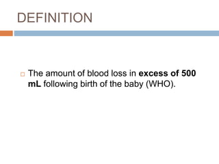
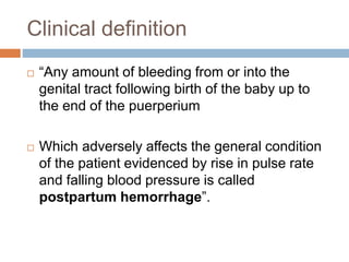


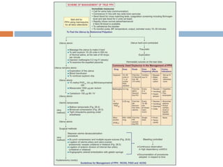
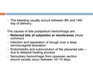
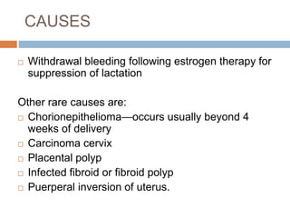


Postpartum hemorrhage (PPH) is defined as blood loss of over 500 ml after childbirth, with primary PPH occurring within 24 hours and secondary PPH beyond that. Major causes of primary PPH include uterine atony, retained tissue, and trauma, with management involving prevention strategies during prenatal and intranatal periods, as well as immediate interventions for active bleeding. Secondary PPH typically arises from retained placental tissue or infections, and management includes supportive care and potential surgical intervention.


