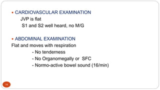
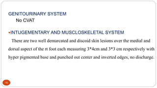
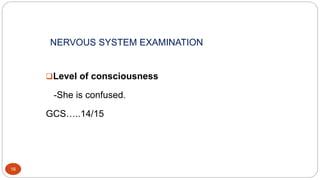
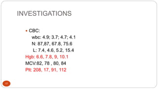
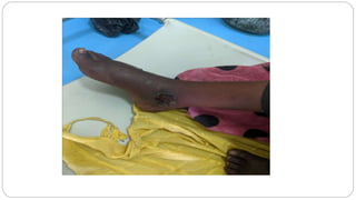
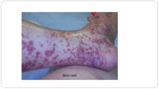
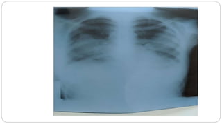
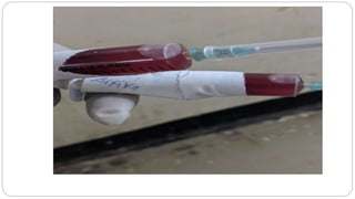
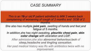
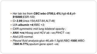
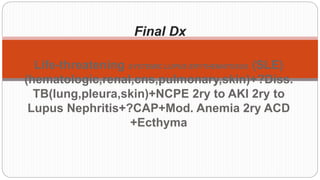
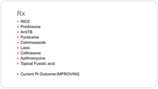
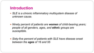
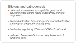
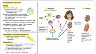
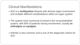
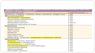
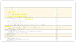
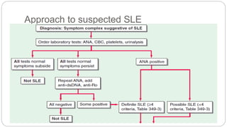
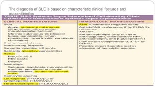
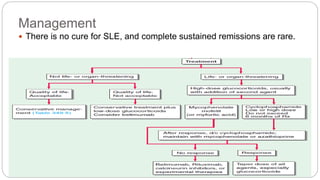
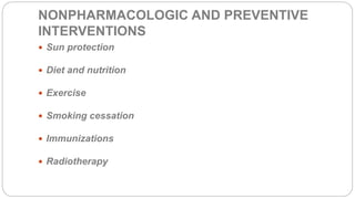
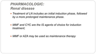
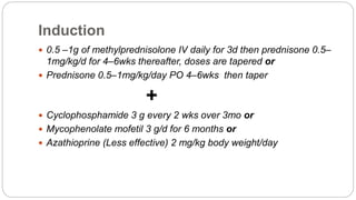
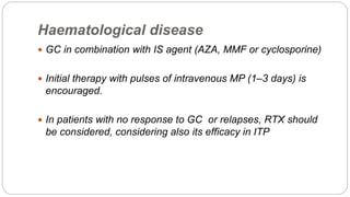
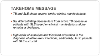
ADAMA HOSPITAL MEDICAL COLLEGE
1
GRAND ROUND
PRESENTED BY: Dr. Kebede M.(Med R2)
Dr. Melaku Y.(Med R1)
Dr. Mengistayehu T.(Intern)
MODERATOR: Dr. Shoba Ibrahim(MD internist, Assist.prof)
OUTLINE
2
 Summary of the History, Physical Examination and Investigation.
 Discussion of differential diagnosis and mgt.
 Final diagnosis.
 References.
HISTORY
3
IDENTIFICATION
4
Name: A.N .
Age: 18
Sex: Female
Address: Chole
Religion: Muslim
Ethnicity: Oromo
Status: Not married
Education level: 6thgrade
Occupation: Student
Department:Internal Medicine
Ward: B
CHIEF COMPLIANT
5
 Easy fatigability of two months duration.
HISTORY OF PRESENT ILLNESS
6
 She is an 18 years old female patient presented with gradual onset
of the above complaint for the past two months with no aggravating
and relieving factors noticed by the pt and the family.
Associated to it:
7
 She has dry cough of 5 months for which she visited near by hospital
one month back and was given unspecified injectable medication for
two wks, but no improvement
 She has LGIF, night sweat, chest pain of three wks, dysphagia and
SOB of one wk duration
 She has multiple joint pain, hand and feet swelling of 5 months
 She has tinnitus, vertigo, head ache, decreased appetite of three wks
CONT…
8
 She has also progressive type of skin lesion over the right leg
and foot which initially was fluid filled and later progressed to
pus filled for the past three months for which she visited the
near by health institution and was given un specified oral
medication ,but no improvement apart from slight improvement
from pain and decreased purulent discharge.
Otherwise:
9
 No orthopnea, PND, palpitation
 No abdominal distension, pain or bowel habit change
 No yellowish discoloration of eyes or use of herbal medication
 No hx of urine color change or decreased UOP
 No hx of TB tx or contact with chronic cougher
 No hx of ABM or LOC
CONT…D
10
 No hx of trauma to the leg
 No heat or cold intolerance or anterior neck swelling
 No self or family hx of DM, HTN or cardiac illness
PHYSICAL EXAMINATION
11
GENERAL APPEARANCE & VITAL SIGNS
12
 ASL  PR = 148b/min radial art,
regular, full volume
 BP=100/70 supine,
 RR= 38 b/min
 T0=38.10c Axillary,
 SPO2=87_(AA),91-
95(INO2)
13
 H.E.E.N.T
Pale conjunctiva and non icteric sclera
LGS: No LAP
 RESPIRATORY SYSTEM
Decreased air entry over the posterior lower third chest
bilaterally
Coarse crepitation over the lt lower lung field
14
 CARDIOVASCULAR EXAMINATION
JVP is flat
S1 and S2 well heard, no M/G
 ABDOMINAL EXAMINATION
Flat and moves with respiration
- No tenderness
- No Organomegally or SFC
- Normo-active bowel sound (16/min)
15
GENITOURINARY SYSTEM
No CVAT
INTUGEMENTARY AND MUSCLOSKELETAL SYSTEM
There are two well demarcated and discoid skin lesions over the medial and
dorsal aspect of the rt foot each measuring 3*4cm and 3*3 cm respectively with
hyper pigmented base and punched out center and inverted edges, no discharge.
NERVOUS SYSTEM EXAMINATION
16
Level of consciousness
-She is confused.
GCS…..14/15
INVESTIGATIONS
17
 CBC:
wbc: 4.9; 3.7; 4.7; 4.1
N: 87,87, 67.8, 75.6
L: 7.4, 4.6, 5.2, 15.4
Hgb: 6.6, 7.8, 9, 10.1
MCV:82, 78 , 80, 84
Plt: 208, 17, 91, 112
CONT…D
18
 U/A: Alb+2, bld+3
 RFT:
Cr: 3.99, 3.79, 3.42, 1.33
BUN: 154, 195, 190, 144
 LFT: 2x done and normal
 Ser/ electrolyte: normal
CONT….D
19
 ESR: 100
 RBS:126mg/dl.
 PICT: Nonreactive.
 VDRL: Negative.
 Viral markers: Negative
CONT…D
20
 Peripheral morphology : Microcytic hypochromic
 Gen X-pert: Negative
 ANA: +Ve
 CXR: Bilateral mid lung opacity w/c is symmetrical
 Abd U/s: normal
CONT…D
21
 Pleural fluid analysis:
App: Reddish
Gluc: 46mg/dl
Alb: 1.5gm/dl
RBC: 1500/ul
Wbc: 7500/ul
N:77%
L:33%
PH: 7
22
DIFERENCIAL DIAGNOSIS
1.Sytemic lupus erythematosus
2.Disseminated Tuberculosis( Lung, Pleura,Skin)
3.Rhupus
4.Mixed connective tissue disorders
5.Vasculitis
Discussion on DDX
In favor:
 Constitutional symptoms
 Anemia, increased ESR
 Abnormal Urinalysis
 Multiple organ involvement
 Ulcer
 Improvement with steroid
management
Against:
 Age
 No palpable purpura
 No Reynaud's phenomenon
 No rhinitis, sinusitis or asthma
 ANA Positive
 No nasal or oral involvement
5.Vasculitis
 Mixed connective tissue disease (MCTD) is defined as a
generalized connective tissue disorder
 characterized by the presence of high titer anti-U1
ribonucleoprotein (RNP) antibodies in combination with clinical
features
 commonly seen in systemic lupus erythematosus (SLE), systemic
sclerosis (SSc), and polymyositis (PM)
4.Mixed connective tissue disorders(MCTD)
In favor:
 All non specific constitutional symptoms
 Bilateral joint pain
 Dysphagia
 Heartburn
 ANA –Positive
 Increased LDH
 Remission after steroid
Against:
 No proximal muscle weakness
 No Reynaud's phenomenon
 No localized muscle pain
 Markedly increased ESR
 Age
 Renal and lung involvement
4.Mixed connective disorders
 Describes those patients having overlapping features of SLE and
Rheumatoid arthritis
 In these cases sever joint deformities, nodules and erosions may
occur
3.Rhupus
In favor:
 Joint swelling
 Constitutional symptoms
 Duration of symptoms(>6
weeks
 Increased ESR
 ANA-Positive
 Failed antibiotic treatment
 Improvement with steroid
Against:
 No morning stiffness
 Large joint involvement
 Decreased platelets
3.Rhupus
In favor:
 TB symptom Complex
 Epidemiology
 Lung, Pleura and skin
 Increased ESR
 Marked Improvement after anti-
TB treatment
Against:
 No TB contact history
 Sputum gene X-pert-negative
 Chest x-ray finding
 Pleural fluid analysis
 Relapsing and remitting course
 Kidney involvement
2.Disseminated Tuberculosis( Lung, Pleura,Skin)
In favor:
 Fulfills diagnostic
criteria(EULAR 2019)
 Marked Improvement after
treatment for SLE
Against:
 Pleural fluid analysis
 Skin lesions are not on photo-
Sensitive areas
1.Systemic Lupus Erythematosus
CASE SUMMARY
This is an 18-yr old F patient admitted to MW 3 weeks back
complaining of worsening of cough of 3 months and SOB of 2
months.
She also has multiple joint pain, swelling of hands and feet,and
fatigue of 5 months.
In addition,she has night sweating, pleuritic chest pain, skin
color change with ulceration and LGIF.
After admission,she also abnormal behaviour,lack of
sleep,headache and tingling sensation.
Her past medical history was Rx with antibiotics twice with no
improvement.
 Her lab Ixs from CBC:wbc-3700,L-6%;hgb-6.6;plt-
916000;ESR-100;
 Cr-3.99;Urea-154;AST-84,ALT-48;
 U/A-albumin +4;RBC +3;
 CXR-symmetric mid lung bilateral opacity ;
 ANA +ve;Hbsag and HCV ab –ve;PIHCT –ve;
 Abd,U/S-normal.
 Pleural fluid analysis-gluc-46,alb-1.5g/dl,RBC-1500,WBC-
7500:N-77%;sputum gene xpert –ve;
Final Dx
Life-threatening SYSTEMIC LUPUS ERYTHEMATOSUS (SLE)
(hematologic,renal,cns,pulmonary,skin)+?Diss.
TB(lung,pleura,skin)+NCPE 2ry to AKI 2ry to
Lupus Nephritis+?CAP+Mod. Anemia 2ry ACD
+Ecthyma
Rx
 INO2
 Prednisone
 AntiTB
 Pyridoxine
 Cotrimoxazole
 Lasix
 Ceftriaxone
 Azithromycine
 Topical Fusidic acid
 Current Pt Outcome:IMPROVING
DISCUSSION
 SLE:
 Introduction
 General treatment
 Specific organ treatment
 Special conditions
 Prognosis and Survival
Introduction
 SLE is a chronic inflammatory multisystem disease of
unknown cause.
 Ninety percent of patients are women of child-bearing years;
people of all genders, ages, and ethnic groups are
susceptible.
 Sixty-five percent of patients with SLE have disease onset
between the ages of 16 and 55
Etiology and pathogenesis
 Interactions between susceptibility genes and
environmental factors result in abnormal immune
responses:
 lowered activation thresholds and abnormal activation
pathways in adaptive immunity cells
 ineffective regulatory CD4+ and CD8+ T cells and
 reduced clearance of immune complexes and of
apoptotic cells
Clinical Manifestations
 SLE is a multisystem disease with diverse organ involvement
and multiple different manifestations within an organ system.
 The system most commonly involved is the musculoskeletal
system, with 95% of patients having involvement, usually as
arthralgias or myalgias.
 Arthritis is also common and is one of the diagnostic criteria for
SLE.
Approach to suspected SLE
The diagnosis of SLE is based on characteristic clinical features and
autoantibodies
According to SLICC 2012,SLE can be classified as
definite,probable or possible.
Management
 There is no cure for SLE, and complete sustained remissions are rare.
The goals of therapy for SLE are :
 to ensure long-term survival,
 achieve the lowest possible disease activity,
 prevent organ damage,
 minimize drug toxicity,
 improve quality of life, and
 educate patients about their role in disease management
NONPHARMACOLOGIC AND PREVENTIVE
INTERVENTIONS
 Sun protection
 Diet and nutrition
 Exercise
 Smoking cessation
 Immunizations
 Radiotherapy
PHARMACOLOGIC:
Renal disease
 Treatment of LN includes an initial induction phase, followed
by a more prolonged maintenance phase.
 MMF and CYC are the IS agents of choice for induction
treatment;
 MMF or AZA may be used as maintenance therapy
Induction
 0.5 –1g of methylprednisolone IV daily for 3d then prednisone 0.5–
1mg/kg/d for 4–6wks thereafter, doses are tapered or
 Prednisone 0.5–1mg/kg/day PO 4–6wks then taper
+
 Cyclophosphamide 3 g every 2 wks over 3mo or
 Mycophenolate mofetil 3 g/d for 6 months or
 Azathioprine (Less effective) 2 mg/kg body weight/day
Maintenance
 5.0 – 7.5 mg/day prednisone
+
 Mycophenolate mofetil 2 g/day or
 Azathioprine 2 mg/kg/day
Haematological disease
 GC in combination with IS agent (AZA, MMF or cyclosporine)
 Initial therapy with pulses of intravenous MP (1–3 days) is
encouraged.
 In patients with no response to GC or relapses, RTX should
be considered, considering also its efficacy in ITP
 Thrombopoietin agonists or splenectomy should be reserved
as last options.
 Autoimmune leucopaenia is common in SLE but rarely needs
treatment
 In refractory or relapsing disease, RTX may be considered.
 CNIs may be considered as second-line agents for induction
or maintenance therapy mainly in membranous LN,
podocytopathy, or in proliferative disease with refractory
nephrotic syndrome
Neuropsychiatric disease (NPSLE
 Treatment of NPSLE depends on whether the underlying
pathophysiological mechanism is inflammatory or
embolic/thrombotic/ischaemic
 GC and/or IS agents should be considered in the former, while
anticoagulant/antithrombotic treatment is favoured when aPL
antibodies are present
Skin:
 Protection from ultraviolet exposure with broad-spectrum sunscreens and
smoking cessation
 topical agents (GC and/or CNIs) and antimalarials, with or without
systemic GC
 MTX can be added.
 Others retinoids, dapsone and MMF or EC-mycophenolic acid
Lung
 mild, may respond to treatment with nonsteroidal anti-
inflammatory drugs (NSAIDs);
 when more severe, patients require a brief course of
glucocorticoid therapy.
 Life-threatening pulmonary manifestations probably require
early aggressive immunosuppressive therapy as well as
supportive care.
Infections
 Risk of infection in SLE is associated with both disease-related
and treatment-related factors
 Protection against infections should be proactive, focusing
both on primary prevention, as well as timely recognition and
treatment.
 Vaccination is preferable during stable disease
 Abstract
 infection one of the leading causes of morbidity and mortality
 The reasons for the high incidence of infection are immunosuppressive
therapy and immune disturbances of lupus itself.
 Bacterial infections are most frequent, followed by viral and fungal infections
 Vaccination is the most important tool in the prevention of infections.
 Prophylaxis of tuberculosis and pneumocystosis are also recommended to
prevent those deadly infections.
TB and SLE:(From Journal of Clinical and Diagnostic Research)
 SLE and TB are intricately related with an increase in the risk of TB in SLE.
 There were case series of five female patients of SLE with TB who presented
between January 2015 and December 2015 in a tertiary care teaching
hospital in North Eastern India.
 All the patients were young to middle aged females having SLE with or
without lupus nephritis who were on immunosuppressive therapy with
corticosteroids, mycophenolate mofetil or cyclophosphamide.
 Two of the cases had sputum positive pulmonary tuberculosis
while rest had Extra-Pulmonary TB (EPTB).
 The response to anti-tubercular therapy led to clinical
improvement in all the cases except one who had an adverse
outcome.
 The series further substantiates the increased risk of TB in
SLE thus, prompting further research towards better
management of these two disease entities in conjunction.
SLE are prone to develop intercurrent infections because of:
(i) compliment deficiency;
(ii) mannose binding lectin (MBL) deficiency;
(iii) chronic inflammation and tissue damage; and
(iv) use of immunosuppressive therapy
 TB is a common infection in SLE patients and the prevalence
of TB in patients with SLE ranges from 5% - 11.6% in studies
reported from India.
 The incidence of TB in SLE patients is considered to be 15-
fold higher;
 extra-pulmonary involvement and disseminated TB are more
common
 differentiating disease flare from active TB disease in patients with
SLE based remains a challenge.
 Evidence is available suggesting that monoclonal antibodies raised
against TB can cross react with DNA,
 features of autoimmunity are evident in mycobacterium induced
arthritis in experimental models,and
 detection of antibodies in patients with TB similar to that found in
SLE
 In a study from Taiwan among 2721 patients with SLE
antecedent TB was present in 44 (1.8%) of patients;
 TB patients were found to have an odds ratio of 2.09 for
subsequent development of SLE after controlling for other
potential risk factors.
 Some even advocated isoniazid prophylaxis in patients with
SLE receiving long term corticosteroid treatment.
Follow up
 At least quarterly visits recommended
 CBC,sCr,U/A
 Complement levels and anti-dsDNA as adjuncts to detect lupus
flares
 Opportunistic infection detection
 Contraception and Family planning
Challenges
 Incomplete Investigations
 Guidelines recommended drugs not available or otherwise not
affordable by the patient
TAKEHOME MESSAGE
 TB and SLE share several similar clinical manifestations
 So, differentiating disease flare from active TB disease in
patients with SLE based on clinical manifestations alone
remains a challenge.
 high index of suspicion and focussed evaluation in the
diagnosis of intercurrent infections, particularly, TB in patients
with SLE is crucial.
REFERENCES
 HARRISON 20th Edition
 EULAR 2019 Management of SLE
 ACR and EULAR 2019 Classification criteria
 UPTODATE 2018
 Journals
THANK YOU

grand round

  • 1.
    ADAMA HOSPITAL MEDICALCOLLEGE 1 GRAND ROUND PRESENTED BY: Dr. Kebede M.(Med R2) Dr. Melaku Y.(Med R1) Dr. Mengistayehu T.(Intern) MODERATOR: Dr. Shoba Ibrahim(MD internist, Assist.prof)
  • 2.
    OUTLINE 2  Summary ofthe History, Physical Examination and Investigation.  Discussion of differential diagnosis and mgt.  Final diagnosis.  References.
  • 3.
  • 4.
    IDENTIFICATION 4 Name: A.N . Age:18 Sex: Female Address: Chole Religion: Muslim Ethnicity: Oromo Status: Not married Education level: 6thgrade Occupation: Student Department:Internal Medicine Ward: B
  • 5.
    CHIEF COMPLIANT 5  Easyfatigability of two months duration.
  • 6.
    HISTORY OF PRESENTILLNESS 6  She is an 18 years old female patient presented with gradual onset of the above complaint for the past two months with no aggravating and relieving factors noticed by the pt and the family.
  • 7.
    Associated to it: 7 She has dry cough of 5 months for which she visited near by hospital one month back and was given unspecified injectable medication for two wks, but no improvement  She has LGIF, night sweat, chest pain of three wks, dysphagia and SOB of one wk duration  She has multiple joint pain, hand and feet swelling of 5 months  She has tinnitus, vertigo, head ache, decreased appetite of three wks
  • 8.
    CONT… 8  She hasalso progressive type of skin lesion over the right leg and foot which initially was fluid filled and later progressed to pus filled for the past three months for which she visited the near by health institution and was given un specified oral medication ,but no improvement apart from slight improvement from pain and decreased purulent discharge.
  • 9.
    Otherwise: 9  No orthopnea,PND, palpitation  No abdominal distension, pain or bowel habit change  No yellowish discoloration of eyes or use of herbal medication  No hx of urine color change or decreased UOP  No hx of TB tx or contact with chronic cougher  No hx of ABM or LOC
  • 10.
    CONT…D 10  No hxof trauma to the leg  No heat or cold intolerance or anterior neck swelling  No self or family hx of DM, HTN or cardiac illness
  • 11.
  • 12.
    GENERAL APPEARANCE &VITAL SIGNS 12  ASL  PR = 148b/min radial art, regular, full volume  BP=100/70 supine,  RR= 38 b/min  T0=38.10c Axillary,  SPO2=87_(AA),91- 95(INO2)
  • 13.
    13  H.E.E.N.T Pale conjunctivaand non icteric sclera LGS: No LAP  RESPIRATORY SYSTEM Decreased air entry over the posterior lower third chest bilaterally Coarse crepitation over the lt lower lung field
  • 14.
    14  CARDIOVASCULAR EXAMINATION JVPis flat S1 and S2 well heard, no M/G  ABDOMINAL EXAMINATION Flat and moves with respiration - No tenderness - No Organomegally or SFC - Normo-active bowel sound (16/min)
  • 15.
    15 GENITOURINARY SYSTEM No CVAT INTUGEMENTARYAND MUSCLOSKELETAL SYSTEM There are two well demarcated and discoid skin lesions over the medial and dorsal aspect of the rt foot each measuring 3*4cm and 3*3 cm respectively with hyper pigmented base and punched out center and inverted edges, no discharge.
  • 16.
    NERVOUS SYSTEM EXAMINATION 16 Levelof consciousness -She is confused. GCS…..14/15
  • 17.
    INVESTIGATIONS 17  CBC: wbc: 4.9;3.7; 4.7; 4.1 N: 87,87, 67.8, 75.6 L: 7.4, 4.6, 5.2, 15.4 Hgb: 6.6, 7.8, 9, 10.1 MCV:82, 78 , 80, 84 Plt: 208, 17, 91, 112
  • 18.
    CONT…D 18  U/A: Alb+2,bld+3  RFT: Cr: 3.99, 3.79, 3.42, 1.33 BUN: 154, 195, 190, 144  LFT: 2x done and normal  Ser/ electrolyte: normal
  • 19.
    CONT….D 19  ESR: 100 RBS:126mg/dl.  PICT: Nonreactive.  VDRL: Negative.  Viral markers: Negative
  • 20.
    CONT…D 20  Peripheral morphology: Microcytic hypochromic  Gen X-pert: Negative  ANA: +Ve  CXR: Bilateral mid lung opacity w/c is symmetrical  Abd U/s: normal
  • 21.
    CONT…D 21  Pleural fluidanalysis: App: Reddish Gluc: 46mg/dl Alb: 1.5gm/dl RBC: 1500/ul Wbc: 7500/ul N:77% L:33% PH: 7
  • 22.
  • 23.
    1.Sytemic lupus erythematosus 2.DisseminatedTuberculosis( Lung, Pleura,Skin) 3.Rhupus 4.Mixed connective tissue disorders 5.Vasculitis Discussion on DDX
  • 24.
    In favor:  Constitutionalsymptoms  Anemia, increased ESR  Abnormal Urinalysis  Multiple organ involvement  Ulcer  Improvement with steroid management Against:  Age  No palpable purpura  No Reynaud's phenomenon  No rhinitis, sinusitis or asthma  ANA Positive  No nasal or oral involvement 5.Vasculitis
  • 28.
     Mixed connectivetissue disease (MCTD) is defined as a generalized connective tissue disorder  characterized by the presence of high titer anti-U1 ribonucleoprotein (RNP) antibodies in combination with clinical features  commonly seen in systemic lupus erythematosus (SLE), systemic sclerosis (SSc), and polymyositis (PM) 4.Mixed connective tissue disorders(MCTD)
  • 30.
    In favor:  Allnon specific constitutional symptoms  Bilateral joint pain  Dysphagia  Heartburn  ANA –Positive  Increased LDH  Remission after steroid Against:  No proximal muscle weakness  No Reynaud's phenomenon  No localized muscle pain  Markedly increased ESR  Age  Renal and lung involvement 4.Mixed connective disorders
  • 31.
     Describes thosepatients having overlapping features of SLE and Rheumatoid arthritis  In these cases sever joint deformities, nodules and erosions may occur 3.Rhupus
  • 32.
    In favor:  Jointswelling  Constitutional symptoms  Duration of symptoms(>6 weeks  Increased ESR  ANA-Positive  Failed antibiotic treatment  Improvement with steroid Against:  No morning stiffness  Large joint involvement  Decreased platelets 3.Rhupus
  • 33.
    In favor:  TBsymptom Complex  Epidemiology  Lung, Pleura and skin  Increased ESR  Marked Improvement after anti- TB treatment Against:  No TB contact history  Sputum gene X-pert-negative  Chest x-ray finding  Pleural fluid analysis  Relapsing and remitting course  Kidney involvement 2.Disseminated Tuberculosis( Lung, Pleura,Skin)
  • 34.
    In favor:  Fulfillsdiagnostic criteria(EULAR 2019)  Marked Improvement after treatment for SLE Against:  Pleural fluid analysis  Skin lesions are not on photo- Sensitive areas 1.Systemic Lupus Erythematosus
  • 38.
    CASE SUMMARY This isan 18-yr old F patient admitted to MW 3 weeks back complaining of worsening of cough of 3 months and SOB of 2 months. She also has multiple joint pain, swelling of hands and feet,and fatigue of 5 months. In addition,she has night sweating, pleuritic chest pain, skin color change with ulceration and LGIF. After admission,she also abnormal behaviour,lack of sleep,headache and tingling sensation. Her past medical history was Rx with antibiotics twice with no improvement.
  • 39.
     Her labIxs from CBC:wbc-3700,L-6%;hgb-6.6;plt- 916000;ESR-100;  Cr-3.99;Urea-154;AST-84,ALT-48;  U/A-albumin +4;RBC +3;  CXR-symmetric mid lung bilateral opacity ;  ANA +ve;Hbsag and HCV ab –ve;PIHCT –ve;  Abd,U/S-normal.  Pleural fluid analysis-gluc-46,alb-1.5g/dl,RBC-1500,WBC- 7500:N-77%;sputum gene xpert –ve;
  • 40.
    Final Dx Life-threatening SYSTEMICLUPUS ERYTHEMATOSUS (SLE) (hematologic,renal,cns,pulmonary,skin)+?Diss. TB(lung,pleura,skin)+NCPE 2ry to AKI 2ry to Lupus Nephritis+?CAP+Mod. Anemia 2ry ACD +Ecthyma
  • 41.
    Rx  INO2  Prednisone AntiTB  Pyridoxine  Cotrimoxazole  Lasix  Ceftriaxone  Azithromycine  Topical Fusidic acid  Current Pt Outcome:IMPROVING
  • 42.
    DISCUSSION  SLE:  Introduction General treatment  Specific organ treatment  Special conditions  Prognosis and Survival
  • 43.
    Introduction  SLE isa chronic inflammatory multisystem disease of unknown cause.  Ninety percent of patients are women of child-bearing years; people of all genders, ages, and ethnic groups are susceptible.  Sixty-five percent of patients with SLE have disease onset between the ages of 16 and 55
  • 44.
    Etiology and pathogenesis Interactions between susceptibility genes and environmental factors result in abnormal immune responses:  lowered activation thresholds and abnormal activation pathways in adaptive immunity cells  ineffective regulatory CD4+ and CD8+ T cells and  reduced clearance of immune complexes and of apoptotic cells
  • 46.
    Clinical Manifestations  SLEis a multisystem disease with diverse organ involvement and multiple different manifestations within an organ system.  The system most commonly involved is the musculoskeletal system, with 95% of patients having involvement, usually as arthralgias or myalgias.  Arthritis is also common and is one of the diagnostic criteria for SLE.
  • 49.
  • 50.
    The diagnosis ofSLE is based on characteristic clinical features and autoantibodies
  • 51.
    According to SLICC2012,SLE can be classified as definite,probable or possible.
  • 52.
    Management  There isno cure for SLE, and complete sustained remissions are rare.
  • 53.
    The goals oftherapy for SLE are :  to ensure long-term survival,  achieve the lowest possible disease activity,  prevent organ damage,  minimize drug toxicity,  improve quality of life, and  educate patients about their role in disease management
  • 54.
    NONPHARMACOLOGIC AND PREVENTIVE INTERVENTIONS Sun protection  Diet and nutrition  Exercise  Smoking cessation  Immunizations  Radiotherapy
  • 55.
    PHARMACOLOGIC: Renal disease  Treatmentof LN includes an initial induction phase, followed by a more prolonged maintenance phase.  MMF and CYC are the IS agents of choice for induction treatment;  MMF or AZA may be used as maintenance therapy
  • 56.
    Induction  0.5 –1gof methylprednisolone IV daily for 3d then prednisone 0.5– 1mg/kg/d for 4–6wks thereafter, doses are tapered or  Prednisone 0.5–1mg/kg/day PO 4–6wks then taper +  Cyclophosphamide 3 g every 2 wks over 3mo or  Mycophenolate mofetil 3 g/d for 6 months or  Azathioprine (Less effective) 2 mg/kg body weight/day
  • 57.
    Maintenance  5.0 –7.5 mg/day prednisone +  Mycophenolate mofetil 2 g/day or  Azathioprine 2 mg/kg/day
  • 58.
    Haematological disease  GCin combination with IS agent (AZA, MMF or cyclosporine)  Initial therapy with pulses of intravenous MP (1–3 days) is encouraged.  In patients with no response to GC or relapses, RTX should be considered, considering also its efficacy in ITP
  • 59.
     Thrombopoietin agonistsor splenectomy should be reserved as last options.  Autoimmune leucopaenia is common in SLE but rarely needs treatment
  • 60.
     In refractoryor relapsing disease, RTX may be considered.  CNIs may be considered as second-line agents for induction or maintenance therapy mainly in membranous LN, podocytopathy, or in proliferative disease with refractory nephrotic syndrome
  • 61.
    Neuropsychiatric disease (NPSLE Treatment of NPSLE depends on whether the underlying pathophysiological mechanism is inflammatory or embolic/thrombotic/ischaemic  GC and/or IS agents should be considered in the former, while anticoagulant/antithrombotic treatment is favoured when aPL antibodies are present
  • 62.
    Skin:  Protection fromultraviolet exposure with broad-spectrum sunscreens and smoking cessation  topical agents (GC and/or CNIs) and antimalarials, with or without systemic GC  MTX can be added.  Others retinoids, dapsone and MMF or EC-mycophenolic acid
  • 63.
    Lung  mild, mayrespond to treatment with nonsteroidal anti- inflammatory drugs (NSAIDs);  when more severe, patients require a brief course of glucocorticoid therapy.  Life-threatening pulmonary manifestations probably require early aggressive immunosuppressive therapy as well as supportive care.
  • 64.
    Infections  Risk ofinfection in SLE is associated with both disease-related and treatment-related factors  Protection against infections should be proactive, focusing both on primary prevention, as well as timely recognition and treatment.  Vaccination is preferable during stable disease
  • 65.
     Abstract  infectionone of the leading causes of morbidity and mortality  The reasons for the high incidence of infection are immunosuppressive therapy and immune disturbances of lupus itself.  Bacterial infections are most frequent, followed by viral and fungal infections  Vaccination is the most important tool in the prevention of infections.  Prophylaxis of tuberculosis and pneumocystosis are also recommended to prevent those deadly infections.
  • 66.
    TB and SLE:(FromJournal of Clinical and Diagnostic Research)  SLE and TB are intricately related with an increase in the risk of TB in SLE.  There were case series of five female patients of SLE with TB who presented between January 2015 and December 2015 in a tertiary care teaching hospital in North Eastern India.  All the patients were young to middle aged females having SLE with or without lupus nephritis who were on immunosuppressive therapy with corticosteroids, mycophenolate mofetil or cyclophosphamide.
  • 67.
     Two ofthe cases had sputum positive pulmonary tuberculosis while rest had Extra-Pulmonary TB (EPTB).  The response to anti-tubercular therapy led to clinical improvement in all the cases except one who had an adverse outcome.  The series further substantiates the increased risk of TB in SLE thus, prompting further research towards better management of these two disease entities in conjunction.
  • 68.
    SLE are proneto develop intercurrent infections because of: (i) compliment deficiency; (ii) mannose binding lectin (MBL) deficiency; (iii) chronic inflammation and tissue damage; and (iv) use of immunosuppressive therapy
  • 69.
     TB isa common infection in SLE patients and the prevalence of TB in patients with SLE ranges from 5% - 11.6% in studies reported from India.  The incidence of TB in SLE patients is considered to be 15- fold higher;  extra-pulmonary involvement and disseminated TB are more common
  • 70.
     differentiating diseaseflare from active TB disease in patients with SLE based remains a challenge.  Evidence is available suggesting that monoclonal antibodies raised against TB can cross react with DNA,  features of autoimmunity are evident in mycobacterium induced arthritis in experimental models,and  detection of antibodies in patients with TB similar to that found in SLE
  • 71.
     In astudy from Taiwan among 2721 patients with SLE antecedent TB was present in 44 (1.8%) of patients;  TB patients were found to have an odds ratio of 2.09 for subsequent development of SLE after controlling for other potential risk factors.  Some even advocated isoniazid prophylaxis in patients with SLE receiving long term corticosteroid treatment.
  • 72.
    Follow up  Atleast quarterly visits recommended  CBC,sCr,U/A  Complement levels and anti-dsDNA as adjuncts to detect lupus flares  Opportunistic infection detection  Contraception and Family planning
  • 73.
    Challenges  Incomplete Investigations Guidelines recommended drugs not available or otherwise not affordable by the patient
  • 74.
    TAKEHOME MESSAGE  TBand SLE share several similar clinical manifestations  So, differentiating disease flare from active TB disease in patients with SLE based on clinical manifestations alone remains a challenge.  high index of suspicion and focussed evaluation in the diagnosis of intercurrent infections, particularly, TB in patients with SLE is crucial.
  • 75.
    REFERENCES  HARRISON 20thEdition  EULAR 2019 Management of SLE  ACR and EULAR 2019 Classification criteria  UPTODATE 2018  Journals
  • 76.

Editor's Notes