Download as PDF, PPTX


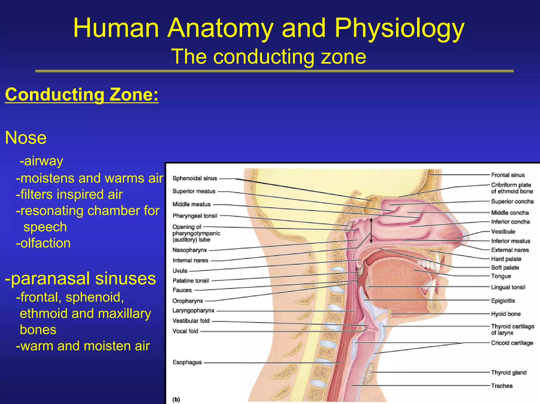


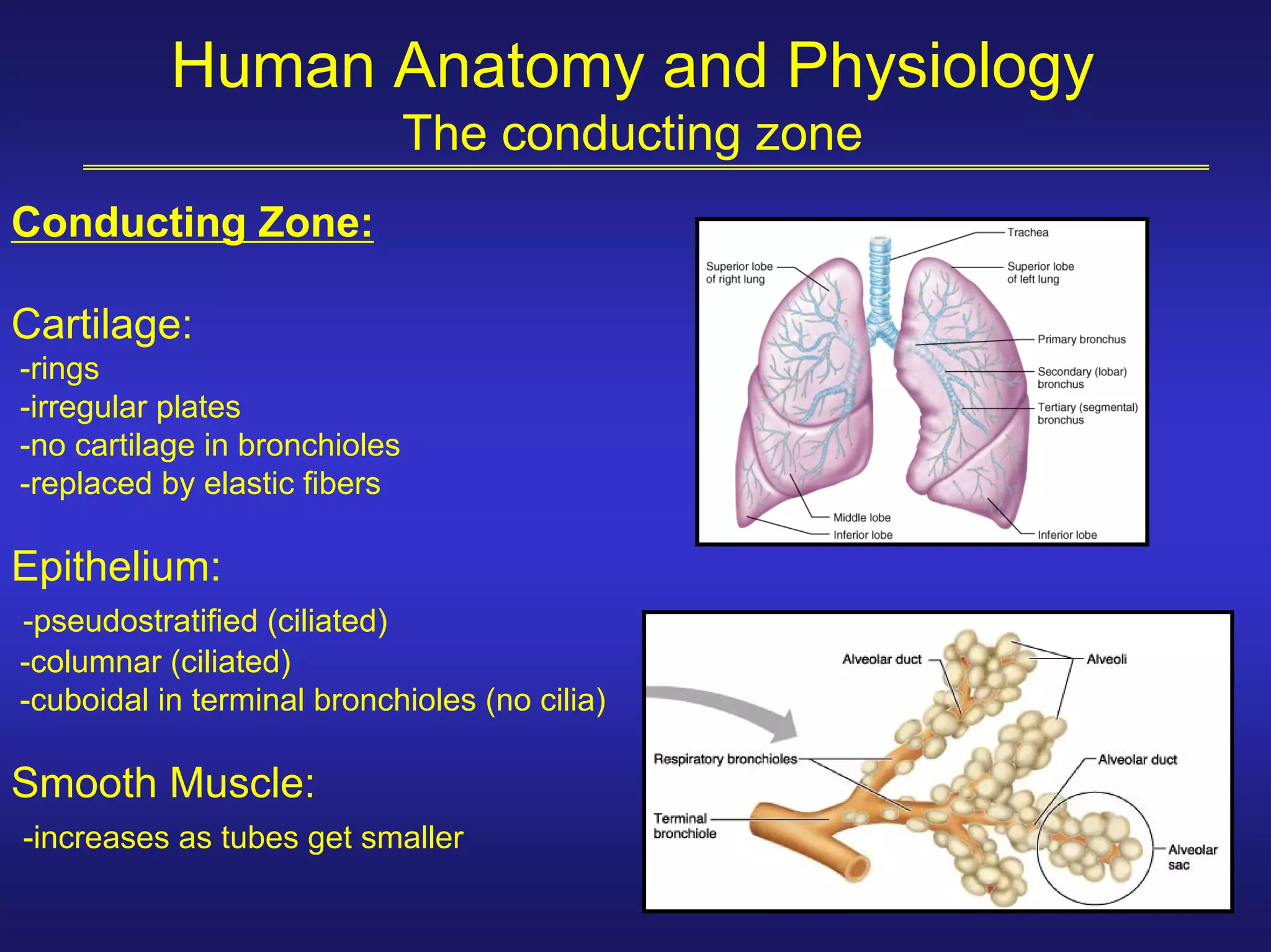

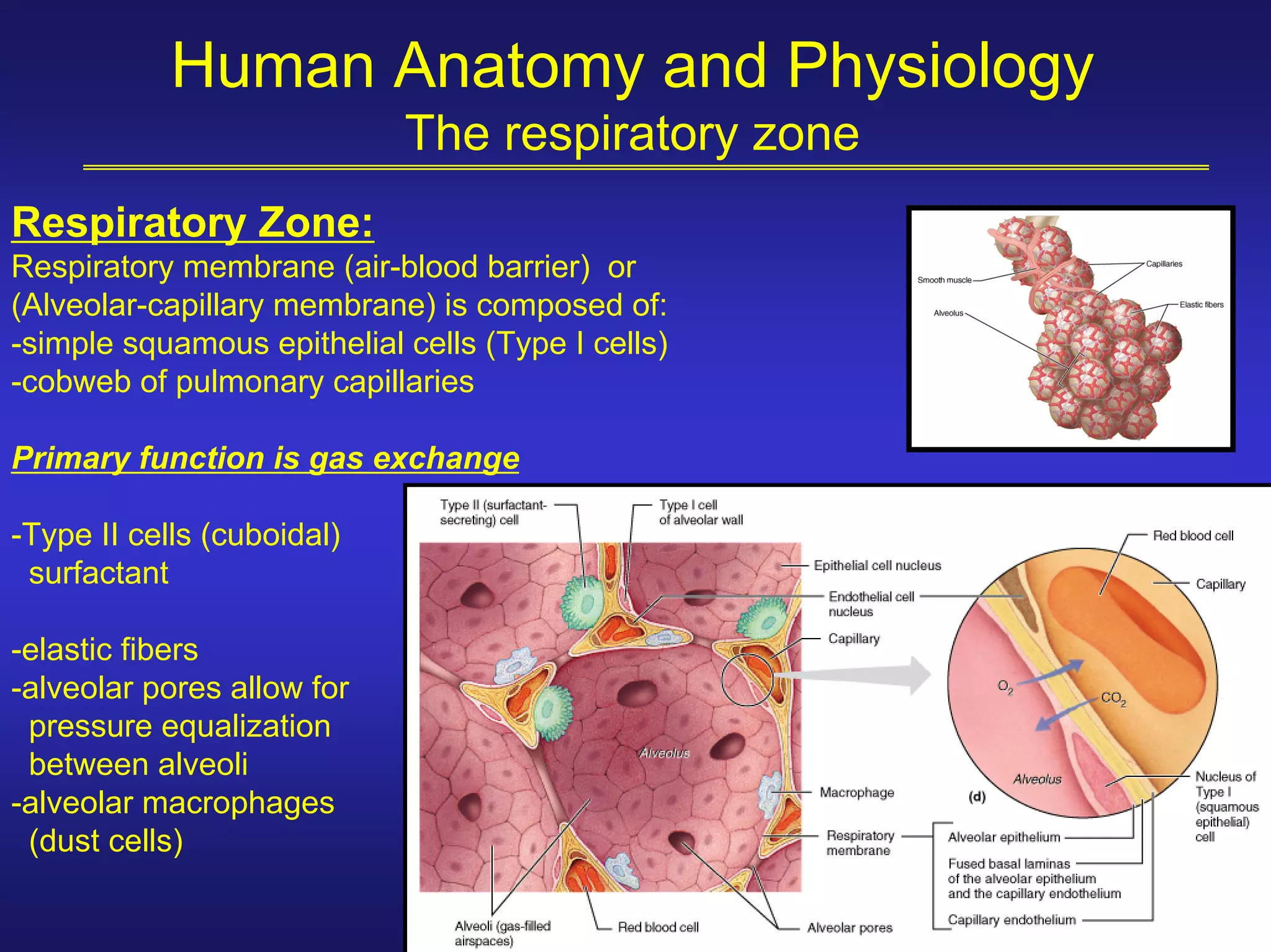


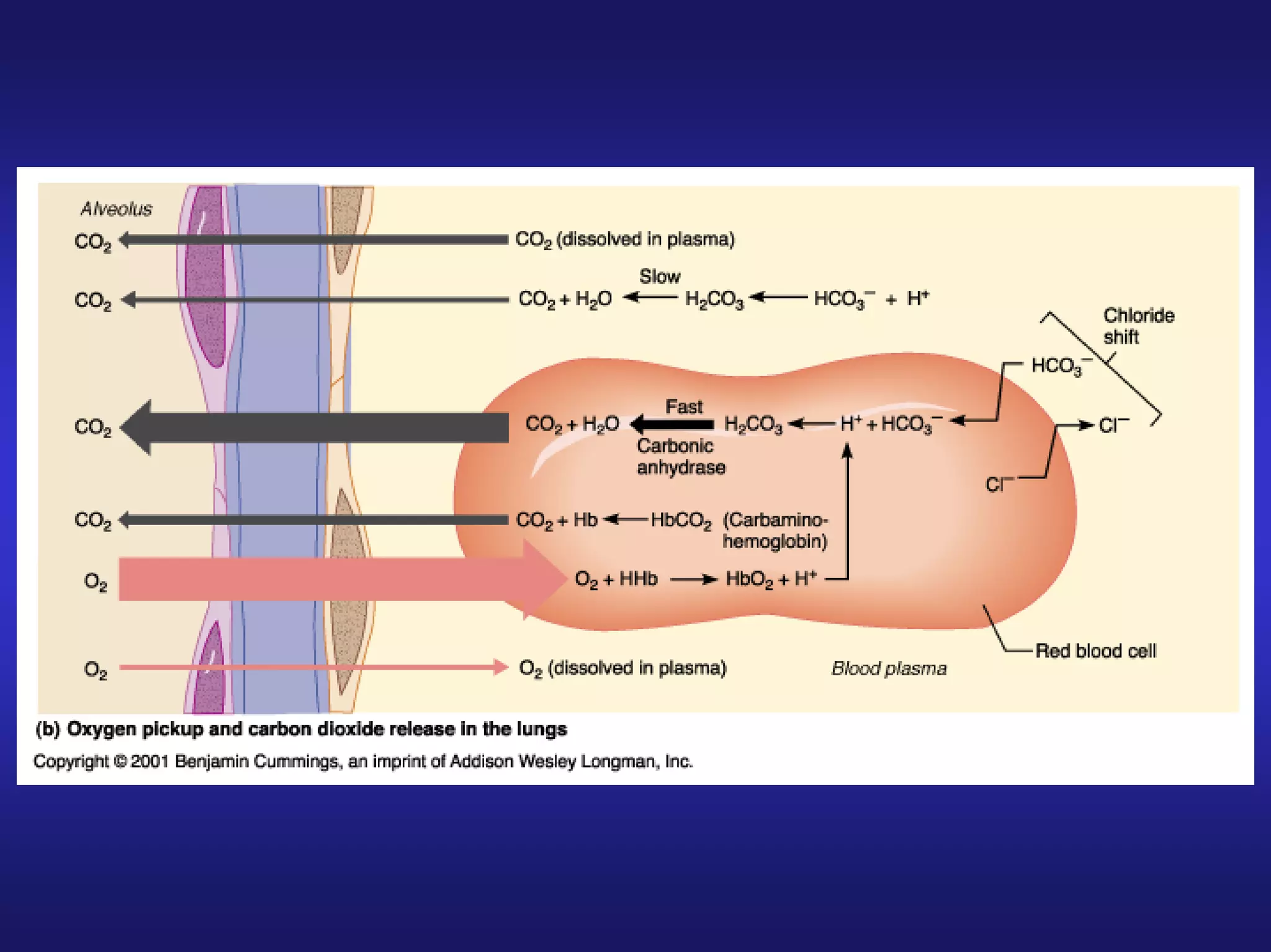


The respiratory system has three main functions: gas exchange, pulmonary ventilation, and transport of respiratory gases. It has two main zones - the conducting zone which includes the nose, pharynx, larynx, trachea, and bronchi, and the respiratory zone where gas exchange occurs in the alveoli. Gas exchange takes place across the alveolar-capillary membrane, which is only one cell thick. Oxygen moves from the alveoli into the blood in the pulmonary capillaries and carbon dioxide moves out of the blood into the alveoli.