This document describes an arthroscopic technique for decompressing a bony suprascapular foramen to relieve suprascapular nerve entrapment. The technique uses a lateral viewing portal and posterior working portal in the subacromial space. A superomedial portal is created to directly access and decompress the suprascapular notch using a Kerrison punch rongeur. This allows safe removal of bone compressing the suprascapular nerve in patients where non-arthroscopic techniques cannot fully address the problem. The procedure successfully resolved shoulder pain in one patient reported as a case study.
Internal fixation of fractures of the capitellum and trochlea - Retrospective...Apollo Hospitals
Fractures of capitellum and trochlea account for 0.5-1% of elbow fractures and 6% of distal humerus fractures. These usually occur due to axial loading of the distal humerus by forces transmitted across the joint producing a coronal shear fracture of the capitellum or the trochlea. Internal fixation is the best modality to restore articular congruity in these fractures.
elbow and wrist and hand fracture with managementkajalgoel8
describing anatomy of the wrist and hand ..
what is fracture
mechanism of injury of all the fracture
classification of fracture
clinical features
radiologicals exminations
management of the fracture
Curious about heli skiing? Looking for the winter adventure of a lifetime? Look no further than The Total Heliski World of Adventure. Get a glimpse into what this sport is all about and who can do it. Also see some highlights from the Total Heliski Show, held in London since 2010. Contact Total Heliski for the trip of a lifetime.
step by step presentation on ultrasound evaluation of shoulder and knee joints with illustrations of probe positioning.multiple examples of pathologies also added.
Birmingham mid-head resection arthroplasty of hip for avascular necrosis of f...Apollo Hospitals
To study the outcome of Birmingham mid-head resection (BMHR) arthroplasty of the hip in young and active patients with avascular necrosis of femoral head with gross defects.
Explore natural remedies for syphilis treatment in Singapore. Discover alternative therapies, herbal remedies, and lifestyle changes that may complement conventional treatments. Learn about holistic approaches to managing syphilis symptoms and supporting overall health.
Ethanol (CH3CH2OH), or beverage alcohol, is a two-carbon alcohol
that is rapidly distributed in the body and brain. Ethanol alters many
neurochemical systems and has rewarding and addictive properties. It
is the oldest recreational drug and likely contributes to more morbidity,
mortality, and public health costs than all illicit drugs combined. The
5th edition of the Diagnostic and Statistical Manual of Mental Disorders
(DSM-5) integrates alcohol abuse and alcohol dependence into a single
disorder called alcohol use disorder (AUD), with mild, moderate,
and severe subclassifications (American Psychiatric Association, 2013).
In the DSM-5, all types of substance abuse and dependence have been
combined into a single substance use disorder (SUD) on a continuum
from mild to severe. A diagnosis of AUD requires that at least two of
the 11 DSM-5 behaviors be present within a 12-month period (mild
AUD: 2–3 criteria; moderate AUD: 4–5 criteria; severe AUD: 6–11 criteria).
The four main behavioral effects of AUD are impaired control over
drinking, negative social consequences, risky use, and altered physiological
effects (tolerance, withdrawal). This chapter presents an overview
of the prevalence and harmful consequences of AUD in the U.S.,
the systemic nature of the disease, neurocircuitry and stages of AUD,
comorbidities, fetal alcohol spectrum disorders, genetic risk factors, and
pharmacotherapies for AUD.
These simplified slides by Dr. Sidra Arshad present an overview of the non-respiratory functions of the respiratory tract.
Learning objectives:
1. Enlist the non-respiratory functions of the respiratory tract
2. Briefly explain how these functions are carried out
3. Discuss the significance of dead space
4. Differentiate between minute ventilation and alveolar ventilation
5. Describe the cough and sneeze reflexes
Study Resources:
1. Chapter 39, Guyton and Hall Textbook of Medical Physiology, 14th edition
2. Chapter 34, Ganong’s Review of Medical Physiology, 26th edition
3. Chapter 17, Human Physiology by Lauralee Sherwood, 9th edition
4. Non-respiratory functions of the lungs https://academic.oup.com/bjaed/article/13/3/98/278874
The prostate is an exocrine gland of the male mammalian reproductive system
It is a walnut-sized gland that forms part of the male reproductive system and is located in front of the rectum and just below the urinary bladder
Function is to store and secrete a clear, slightly alkaline fluid that constitutes 10-30% of the volume of the seminal fluid that along with the spermatozoa, constitutes semen
A healthy human prostate measures (4cm-vertical, by 3cm-horizontal, 2cm ant-post ).
It surrounds the urethra just below the urinary bladder. It has anterior, median, posterior and two lateral lobes
It’s work is regulated by androgens which are responsible for male sex characteristics
Generalised disease of the prostate due to hormonal derangement which leads to non malignant enlargement of the gland (increase in the number of epithelial cells and stromal tissue)to cause compression of the urethra leading to symptoms (LUTS
Anti ulcer drugs and their Advance pharmacology ||
Anti-ulcer drugs are medications used to prevent and treat ulcers in the stomach and upper part of the small intestine (duodenal ulcers). These ulcers are often caused by an imbalance between stomach acid and the mucosal lining, which protects the stomach lining.
||Scope: Overview of various classes of anti-ulcer drugs, their mechanisms of action, indications, side effects, and clinical considerations.
Ozempic: Preoperative Management of Patients on GLP-1 Receptor Agonists Saeid Safari
Preoperative Management of Patients on GLP-1 Receptor Agonists like Ozempic and Semiglutide
ASA GUIDELINE
NYSORA Guideline
2 Case Reports of Gastric Ultrasound
Title: Sense of Smell
Presenter: Dr. Faiza, Assistant Professor of Physiology
Qualifications:
MBBS (Best Graduate, AIMC Lahore)
FCPS Physiology
ICMT, CHPE, DHPE (STMU)
MPH (GC University, Faisalabad)
MBA (Virtual University of Pakistan)
Learning Objectives:
Describe the primary categories of smells and the concept of odor blindness.
Explain the structure and location of the olfactory membrane and mucosa, including the types and roles of cells involved in olfaction.
Describe the pathway and mechanisms of olfactory signal transmission from the olfactory receptors to the brain.
Illustrate the biochemical cascade triggered by odorant binding to olfactory receptors, including the role of G-proteins and second messengers in generating an action potential.
Identify different types of olfactory disorders such as anosmia, hyposmia, hyperosmia, and dysosmia, including their potential causes.
Key Topics:
Olfactory Genes:
3% of the human genome accounts for olfactory genes.
400 genes for odorant receptors.
Olfactory Membrane:
Located in the superior part of the nasal cavity.
Medially: Folds downward along the superior septum.
Laterally: Folds over the superior turbinate and upper surface of the middle turbinate.
Total surface area: 5-10 square centimeters.
Olfactory Mucosa:
Olfactory Cells: Bipolar nerve cells derived from the CNS (100 million), with 4-25 olfactory cilia per cell.
Sustentacular Cells: Produce mucus and maintain ionic and molecular environment.
Basal Cells: Replace worn-out olfactory cells with an average lifespan of 1-2 months.
Bowman’s Gland: Secretes mucus.
Stimulation of Olfactory Cells:
Odorant dissolves in mucus and attaches to receptors on olfactory cilia.
Involves a cascade effect through G-proteins and second messengers, leading to depolarization and action potential generation in the olfactory nerve.
Quality of a Good Odorant:
Small (3-20 Carbon atoms), volatile, water-soluble, and lipid-soluble.
Facilitated by odorant-binding proteins in mucus.
Membrane Potential and Action Potential:
Resting membrane potential: -55mV.
Action potential frequency in the olfactory nerve increases with odorant strength.
Adaptation Towards the Sense of Smell:
Rapid adaptation within the first second, with further slow adaptation.
Psychological adaptation greater than receptor adaptation, involving feedback inhibition from the central nervous system.
Primary Sensations of Smell:
Camphoraceous, Musky, Floral, Pepperminty, Ethereal, Pungent, Putrid.
Odor Detection Threshold:
Examples: Hydrogen sulfide (0.0005 ppm), Methyl-mercaptan (0.002 ppm).
Some toxic substances are odorless at lethal concentrations.
Characteristics of Smell:
Odor blindness for single substances due to lack of appropriate receptor protein.
Behavioral and emotional influences of smell.
Transmission of Olfactory Signals:
From olfactory cells to glomeruli in the olfactory bulb, involving lateral inhibition.
Primitive, less old, and new olfactory systems with different path
Pulmonary Thromboembolism - etilogy, types, medical- Surgical and nursing man...VarunMahajani
Disruption of blood supply to lung alveoli due to blockage of one or more pulmonary blood vessels is called as Pulmonary thromboembolism. In this presentation we will discuss its causes, types and its management in depth.
Lung Cancer: Artificial Intelligence, Synergetics, Complex System Analysis, S...Oleg Kshivets
RESULTS: Overall life span (LS) was 2252.1±1742.5 days and cumulative 5-year survival (5YS) reached 73.2%, 10 years – 64.8%, 20 years – 42.5%. 513 LCP lived more than 5 years (LS=3124.6±1525.6 days), 148 LCP – more than 10 years (LS=5054.4±1504.1 days).199 LCP died because of LC (LS=562.7±374.5 days). 5YS of LCP after bi/lobectomies was significantly superior in comparison with LCP after pneumonectomies (78.1% vs.63.7%, P=0.00001 by log-rank test). AT significantly improved 5YS (66.3% vs. 34.8%) (P=0.00000 by log-rank test) only for LCP with N1-2. Cox modeling displayed that 5YS of LCP significantly depended on: phase transition (PT) early-invasive LC in terms of synergetics, PT N0—N12, cell ratio factors (ratio between cancer cells- CC and blood cells subpopulations), G1-3, histology, glucose, AT, blood cell circuit, prothrombin index, heparin tolerance, recalcification time (P=0.000-0.038). Neural networks, genetic algorithm selection and bootstrap simulation revealed relationships between 5YS and PT early-invasive LC (rank=1), PT N0—N12 (rank=2), thrombocytes/CC (3), erythrocytes/CC (4), eosinophils/CC (5), healthy cells/CC (6), lymphocytes/CC (7), segmented neutrophils/CC (8), stick neutrophils/CC (9), monocytes/CC (10); leucocytes/CC (11). Correct prediction of 5YS was 100% by neural networks computing (area under ROC curve=1.0; error=0.0).
CONCLUSIONS: 5YS of LCP after radical procedures significantly depended on: 1) PT early-invasive cancer; 2) PT N0--N12; 3) cell ratio factors; 4) blood cell circuit; 5) biochemical factors; 6) hemostasis system; 7) AT; 8) LC characteristics; 9) LC cell dynamics; 10) surgery type: lobectomy/pneumonectomy; 11) anthropometric data. Optimal diagnosis and treatment strategies for LC are: 1) screening and early detection of LC; 2) availability of experienced thoracic surgeons because of complexity of radical procedures; 3) aggressive en block surgery and adequate lymph node dissection for completeness; 4) precise prediction; 5) adjuvant chemoimmunoradiotherapy for LCP with unfavorable prognosis.
Prix Galien International 2024 Forum ProgramLevi Shapiro
June 20, 2024, Prix Galien International and Jerusalem Ethics Forum in ROME. Detailed agenda including panels:
- ADVANCES IN CARDIOLOGY: A NEW PARADIGM IS COMING
- WOMEN’S HEALTH: FERTILITY PRESERVATION
- WHAT’S NEW IN THE TREATMENT OF INFECTIOUS,
ONCOLOGICAL AND INFLAMMATORY SKIN DISEASES?
- ARTIFICIAL INTELLIGENCE AND ETHICS
- GENE THERAPY
- BEYOND BORDERS: GLOBAL INITIATIVES FOR DEMOCRATIZING LIFE SCIENCE TECHNOLOGIES AND PROMOTING ACCESS TO HEALTHCARE
- ETHICAL CHALLENGES IN LIFE SCIENCES
- Prix Galien International Awards Ceremony
HOT NEW PRODUCT! BIG SALES FAST SHIPPING NOW FROM CHINA!! EU KU DB BK substit...GL Anaacs
Contact us if you are interested:
Email / Skype : kefaya1771@gmail.com
Threema: PXHY5PDH
New BATCH Ku !!! MUCH IN DEMAND FAST SALE EVERY BATCH HAPPY GOOD EFFECT BIG BATCH !
Contact me on Threema or skype to start big business!!
Hot-sale products:
NEW HOT EUTYLONE WHITE CRYSTAL!!
5cl-adba precursor (semi finished )
5cl-adba raw materials
ADBB precursor (semi finished )
ADBB raw materials
APVP powder
5fadb/4f-adb
Jwh018 / Jwh210
Eutylone crystal
Protonitazene (hydrochloride) CAS: 119276-01-6
Flubrotizolam CAS: 57801-95-3
Metonitazene CAS: 14680-51-4
Payment terms: Western Union,MoneyGram,Bitcoin or USDT.
Deliver Time: Usually 7-15days
Shipping method: FedEx, TNT, DHL,UPS etc.Our deliveries are 100% safe, fast, reliable and discreet.
Samples will be sent for your evaluation!If you are interested in, please contact me, let's talk details.
We specializes in exporting high quality Research chemical, medical intermediate, Pharmaceutical chemicals and so on. Products are exported to USA, Canada, France, Korea, Japan,Russia, Southeast Asia and other countries.
MANAGEMENT OF ATRIOVENTRICULAR CONDUCTION BLOCK.pdfJim Jacob Roy
Cardiac conduction defects can occur due to various causes.
Atrioventricular conduction blocks ( AV blocks ) are classified into 3 types.
This document describes the acute management of AV block.
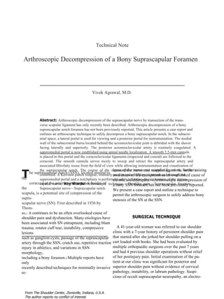
1. T Technical Note Arthroscopic Decompression of a Bony Suprascapular Foramen Vivek Agrawal, M.D. Abstract: Arthroscopic decompression of the suprascapular nerve by transection of the trans- verse scapular ligament has only recently been described. Arthroscopic decompression of a bony suprascapular notch foramen has not been previously reported. This article presents a case report and outlines an arthroscopic technique to safely decompress a bony suprascapular notch. In the subacro- mial space, a lateral portal is used for viewing and a posterior portal for instrumentation. The medial wall of the subacromial bursa located behind the acromioclavicular joint is debrided with the shaver facing laterally and superiorly. The posterior acromioclavicular artery is routinely coagulated. A superomedial portal is now established using spinal needle localization. A smooth 5.5-mm cannula is placed in this portal and the coracoclavicular ligaments (trapezoid and conoid) are followed to the coracoid. The smooth cannula serves nicely to sweep and retract the suprascapular artery and associated fibrofatty tissue from the field of view while allowing instrumentation and visualization of the suprascapular notch. The course of the suprascapular nerve and morphology of the notch is confirmed. A Kerrison punch rongeur, routinely used in spine surgery, is introduced through the superomedial portal and a notchplasty is performed safely, allowing decompression of the supras- capular nerve. Key Words: Arthroscopic technique— Shoulder pain —Suprascapular foramen— Suprascapular nerve—Suprascapular notch. lease of the transverse scapular ligament, further raising awareness of SN entrapment as an overlooked cause of chronic shoulder pain. 3-6 Arthroscopic decompression of a bony SSN foramen has not been previously reported. We present a case report and outline a technique to permit the arthroscopic surgeon to safely address bony stenosis of the SN at the SSN. SURGICAL TECHNIQUE A 41-year-old woman was referred to our shoulder clinic with a 7-year history of persistent shoulder pain that started after she jerked her shoulder pulling on a cart loaded with books. She had been evaluated by multiple orthopaedic surgeons over the past 7 years and had 4 previous shoulder operations without relief of her postinjury pain. Initial examination of the pa- tient at our clinic was significant for posterior and superior shoulder pain without evidence of cervical pathology, instability, or labrum pathology. Suspi- cious of occult suprascapular neuropathy, an electro- he suprascapular notch (SSN), located medial to the coracoid at the anterior and superior border of the scapula, is a potential site of compression of the supra- scapular nerve (SN). First described in 1936 by Thom- as, 1 it continues to be an often overlooked cause of shoulder pain and dysfunction. Many etiologies have been associated with SN entrapment, including blunt trauma, rotator cuff tear, instability, compressive lesions such as ganglion cysts, passage of the suprascapular artery through the SSN, crutch use, repetitive traction injury in athletics, and variations in SSN morphology, including a bony foramen. 2 Multiple reports have only recently described techniques for minimally invasive re- From The Shoulder Center, Zionsville, Indiana, U.S.A. The author reports no conflict of interest.
2. conoid) are identified. The conoid ligament is visu- alized and followed inferiorly to its attachment to the coracoid. Frequently, this can be done with a gentle sweeping motion along these ligaments. The coracoid insertion of the transverse scapular liga- ment is seen perpendicular to the coracoid insertion of the conoid ligament. Use of the shaver and ra- diofrequency device are restricted to the space lat- eral to the medial border of the coracoid to avoid injury to the suprascapular artery. Blunt dissection can often proceed inferiorly and medially from the coracoid while maintaining contact with bone, al- lowing the SSN, suprascapular artery, and SN to be visualized and safely retracted without directly dis- turbing these structures. The morphology of the SSN is now defined ( Fig 2 ). In most cases, the transverse scapular ligament is easily transected via the SM portal using arthroscopic scissors. For a bony or stenotic SSN foramen, as in the present case, a Kerrison punch rongeur is introduced through the SM portal to perform a foraminotomy. The superior and lateral walls of the SSN are de- compressed in a controlled manner. 11 Kerrison ron- geurs are routinely used for bone removal in spine surgery and are available in multiple sizes and configurations in most facilities. Upon completion of the SSN decompression, a probe is used to con- fi rm mobility of the SN by lifting it out of the SSN. Postoperatively, the patient reported resolution of her longstanding posterior and superior shoulder pain and was extremely pleased with her outcome. DISCUSSION Although the prevalence of suprascapular compres- sion neuropathy is thought to be rare, its role in F IGURE 1. Lateral decubitus position with portals for suprascap- ular notch approach outlined. (A, anterior portal; L, lateral portal; P, posterior portal; SM, superomedial portal.) diagnostic study of her upper extremity along with an magnetic resonance arthrogram were recommended. The electrodiagnostic study confirmed neuropathy of the SN at both the SSN and spinoglenoid notch (SGN), consistent with a “double crush” phenome- non. 7 To further confirm the diagnosis, the patient was referred for a confirmatory selective SN block. The SN block provided excellent but transient relief of her pain. After reviewing the risks, benefits, and options for treatment, including our arthroscopic approach to SN decompression, she wished to proceed with arthro- scopic treatment. The SN was decompressed at the SGN by transect- ing the spinoglenoid ligament as described by Plancher. 8 Our approach to visualize the SSN is sim- ilar to the approach described by Lafosse, 6 with sev- eral modifications. We use a semilateral decubitus position for shoulder arthroscopy ( Fig 1 ). The anat- omy and vascularity of the subacromial space has been elegantly described and is helpful in planning approaches to the SN and medial wall of the subacro- mial bursa. 9 After completing glenohumeral evalua- tion and treatment, the arthroscope is introduced into the subacromial space via the posterior portal. An anterolateral portal is established for outflow using an inside-outside technique, ensuring that both portals are within the subacromial bursa. A lateral portal with spinal needle localization is used to ensure an optimal angle of approach. An arthroscopic shaver is intro- duced through the lateral portal, and the posterior wall of the subacromial bursa is removed to improve visu- 326 V. AGRAWAL alization. The arthroscope is switched to the lateral portal for viewing and the shaver and radiofrequency device are introduced from the posterior portal. The coracoacromial (CA) ligament is visualized and fol- lowed medially. The acromioclavicular (AC) joint is localized but not exposed. The medial wall of the subacromial bursa is now removed to allow access to the SSN. The posterior AC artery is located immedi- ately posterior to the AC joint and is routinely coag- ulated. A modified superomedial (SM) portal is now established under direct visualization using spinal nee- dle localization. 10 A smooth 5.5-mm cannula is in- troduced, and the shaver and radiofrequency device are used for further dissection via the SM portal. The CA ligament is followed towards the coracoid. The coracoclavicular ligaments (trapezoid and
3. gachary et al., 2 Natsis et al. 15 examined 423 scapulas and noted an 8% incidence of a bony foramen (types IV and V). From May 2007 to May 2008, 2 of the 44 patients seen at our tertiary care shoulder clinic for arthro- scopic SN decompression at the SSN had a bony SSN foramen. To establish the diagnosis of suprascapular neuropathy, we request electrodiagnostic studies after a detailed clinical evaluation to confirm the diagnosis and predict an expected outcome for treatment. 16 In our experience, electrodiagnostic studies of the shoul- der girdle are highly variable in diagnostic quality. Therefore, a single experienced examiner familiar with the electrodiagnostic criteria for SN pathology performed most of the studies. 16 For patients with isolated SN lesions, we often use a selective SN block as a further diagnostic and confirmatory measure be- fore proceeding with arthroscopy. Several techniques for arthroscopic transverse scapu- lar ligament release along with early clinical results have been recently reported. 3-6,10 In the preliminary clinical series of 10 patients reported by Lafosse et al., 6 none had SN compression as a result of bony stenosis. However, he recommended using an arthroscopic burr to address bony stenosis. 6 This technique has several advantages: the SM por- tal is a safe distance from the SN and relatively familiar to most arthroscopic surgeons. 10 The SM portal allows a more direct approach to dissection of the deep surface of coracoclavicular ligaments and the SSN, and the smooth cannula in this portal serves nicely as a soft tissue retractor avoiding the need for another portal. Kerrison punch rongeurs are specifi- cally designed for decompression of spinal stenosis and uniquely suited to arthroscopic decompression of suprascapular nerve stenosis. For our initial case with a bony SSN foramen, we started with a burr as sug- gested by Lafosse; however, given the proximity of the neurovascular structures, we felt that the Kerrison punch offered a greater margin of safety and control. We completed the case with the Kerrison punch and used it exclusively for the second patient with a bony SSN. The technique is adaptable and appropriate for variations in SSN anatomy. Arthroscopic decompression of the suprascapular nerve with a bony SSN foramen has not been previ- ously reported. This technique provides the arthro- scopic surgeon another approach to safely decompress the suprascapular nerve at the SSN in cases of bony stenosis using commonly available instruments. F IGURE 2. (A) Right shoulder with bony suprascapular notch foramen. (B) Left shoulder with more commonly encountered superior transverse scapular ligament. ( A , suprascapular artery; N , suprascapular nerve; STSL , superior transverse scapular ligament; SSN , suprascapular notch.) shoulder pain and dysfunction is probably underap- preciated. The morphology, particularly a stenotic or bony notch of the SSN, may be associated with a predilection to suprascapular nerve injury. 2 The trans- verse scapular ligament, despite connecting 2 regions of the same bone, has been shown to have fibrocarti- lage entheses, indicating that it experiences both com- pressive and tensile loading. 12 Consistent with these fi ndings, bony bridges at the SSN are also seen more frequently with increasing age. 13 Of the 700 speci- mens examined by Edelson, 14 11.8% had a completely or partially ossified transverse scapular ligament. In a recent update to the classification proposed by Ren- 327 DECOMPRESSION OF A BONY SUPRASCAPULAR FORAMEN
4. REFERENCES 1. Thomas A. La paralysie du muscle sous-épineux. Presse Med 1936;64:1283-1284. 2. Rengachary SS, Burr D, Lucas S, Hassanein KM, Mohn MP, Matzke H. Suprascapular entrapment neuropathy: A clinical, anatomical, and comparative study. Part 2: Anatomical study. Neurosurgery 1979;5:447-451. 3. Barber FA. Percutaneous arthroscopic release of the supra- scapular nerve. Arthroscopy 2008;24:236e1-236e4. 4. Barwood SA, Burkhart SS, Lo IK. Arthroscopic suprascapular nerve release at the suprascapular notch in a cadaveric model: An anatomic approach. Arthroscopy 2007;23:221-225. 5. Bhatia DN, de Beer JF, van Rooyen KS, du Toit DF. Arthro- scopic suprascapular nerve decompression at the suprascapular notch. Arthroscopy 2006;22:1009-1013. 6. Lafosse L, Tomasi A, Corbett S, Baier G, Willems K, Gobezie R. Arthroscopic release of suprascapular nerve entrapment at the suprascapular notch: Technique and preliminary results. Arthroscopy 2007;23:34-42. 7. Upton AR, McComas AJ. The double crush in nerve entrap- ment syndromes. Lancet 1973;2:359-362. 8. Plancher KD, Luke TA, Peterson RK, Yacoubian SV. Poste- rior shoulder pain: A dynamic study of the spinoglenoid liga- ment and treatment with arthroscopic release of the scapular tunnel. Arthroscopy 2007;23:991-998. ing points for the arthroscopic surgeon. Arthroscopy 2007;23: 978-984. 10. Woolf SK, Guttmann D, Karch MM, Graham RD 2nd, Reid JB 3rd, Lubowitz JH. The superior-medial shoulder arthroscopy portal is safe. Arthroscopy 2007;23:247-250. 11. Klein GR, Ludwig SC, Vaccaro AR, Rushton SA, Lazar RD, Albert TJ. The efficacy of using an image-guided Kerrison punch in performing an anterior cervical foraminotomy. An anatomic analysis. Spine 1999;24:1358-1362. 12. Moriggl B, Jax P, Milz S, Büttner A, Benjamin M. Fibrocar- tilage at the entheses of the suprascapular (superior transverse scapular) ligament of man—A ligament spanning two regions of a single bone. J Anat 2001;199(Pt 5):539-545. 13. Hrdlicka A. The scapula: Visual observations. Am J Phys Anthropol 1942;29:73-94. 14. Edelson JG. Bony bridges and other variations of the suprascap- ular notch. J Bone Joint Surg Br 1995;77:505-506. 15. Natsis K, Totlis T, Tsikaras P, Appell HJ, Skandalakis P, Koebke J. Proposal for classification of the suprascapular notch: A study on 423 dried scapulas. Clin Anat 2007;20:135- 139. 16. Antoniou J, Tae SK, Williams GR, Bird S, Ramsey ML, Iannotti JP. Suprascapular neuropathy. Variability in the diag- nosis, treatment, and outcome. Clin Orthop Relat Res 2001; 386:131-138. 328 V. AGRAWAL 9. Yepes H, Al-Hibshi A, Tang M, Morris SF, Stanish WD. Vascular anatomy of the subacromial space: A map of bleed-