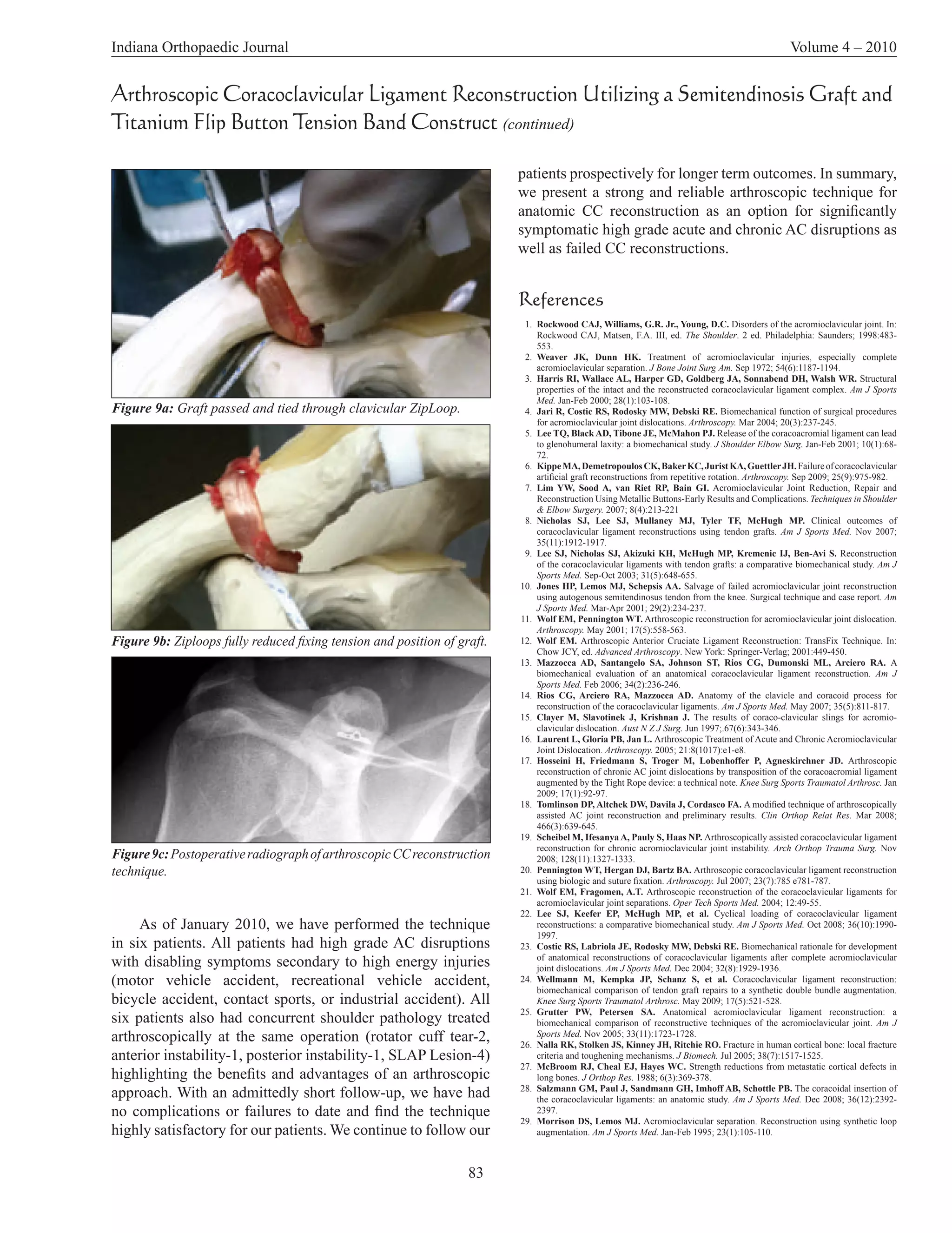
SlideShare a Scribd company logo
Indiana Orthopaedic Journal                                                                                        Volume 4 – 2010


  Arthroscopic Coracoclavicular Ligament Reconstruction Utilizing a
Semitendinosis Graft and Titanium Flip Button Tension Band Construct
                                                        Vivek Agrawal, MD
                                                The Shoulder Center, PC, Carmel, IN

Correspondence:
    Vivek Agrawal MD
    The Shoulder Center, PC
    12188-A North Meridian Street
    Suite 310
    Carmel, IN 46032-4406
    dragrawal@theshouldercenter.com
    (317) 802-9686

Introduction
     The surgical treatment of symptomatic complete or
high grade acromioclavicular (AC) joint disruptions remains
controversial with more than 60 published techniques.1
                                                                     Figure 1a: Patient with symptomatic Right Grade IV AC Separation.
Concerns regarding strength of initial fixation, cyclic failure,
and inconsistent outcomes with techniques similar to the
coracoacromial (CA) ligament transfer first described by
Weaver and Dunn have been reported.2-5 Failure due to
synthetic material abrasion at the clavicle or coracoid as well
as cyclic failure of the synthetic material is a concern with
techniques utilizing a synthetic material construct alone.6, 7
Excellent biomechanical strength and clinical outcomes have
been recently reported with tendon graft coracoclavicular
(CC) reconstruction.8-10 This paper describes an arthroscopic
technique that is equally suitable to acute or chronic high
grade AC joint disruptions as well as previously failed CC
reconstructions. A tension band construct is created utilizing
a single titanium flip button device placed at the inferior
cortex of the coracoid (#7 PE ZipLoop Extended ToggleLoc;
Biomet. Warsaw, IN) combined with a Semitendinosis graft.
Along with offering the benefits of an arthroscopic approach,        Figure 1b: Same patient positioned in the semi-lateral decubitus
the technique also offers the additional advantage of placing        position with portal sites and clavicle incision outlined.
no hardware at the superior aspect of the clavicle.
                                                                     the coracoid is then performed similar to the manner first
Surgical Technique                                                   described by Wolf.11
    After initially learning an arthroscopic AC reconstruction            The inferior and lateral aspect of the coracoid process is
technique in August 2000, as described by Wolf,11 we have            clearly visualized followed by a 1.5cm incision in Langer’s
gradually modified our technique both to address potential           lines at the superior aspect of the clavicle (3cm incision if
biomechanical and clinical limitations and to formulate the          also performing a distal clavicle resection). Subcutaneous
most reliable technique with inherent applicability for the          dissection is performed followed by subperiosteal elevation
broadest possible patient population.                                of soft tissues to the anterior and posterior margins of the
     We perform all shoulder arthroscopy procedures in the           clavicle. The periosteum anterior and inferior to the clavicle
modified lateral decubitus position utilizing 6-10 pounds of         is elevated to allow instrument passage to the coracoid.
counter-traction to suspend the arm. (Figure 1) For patients         The clavicular and coracoid tunnels are created utilizing an
that prefer an autograft, we recommend harvesting the                arthroscopic ACL guide and a 2.4mm drill point guide wire
ipsilateral Semitendinosis graft with the patient initially in the   as previously described.11 (Figure 2) A 4.5mm cannulated
supine position.12 Glenohumeral and subacromial arthroscopy          drill is utilized over the initial guide wire to create a 4.5mm
for diagnosis and treatment is initially performed as required       tunnel in the clavicle and coracoids. (Figure 3) The clavicular
for each unique patient circumstance. We prefer to address           tunnel is placed approximately 35mm proximal to the distal
all other concurrent pathology prior to proceeding with the          clavicle, placing it between the trapezoid and conoid ligament
CC reconstruction. Visualization of the inferior aspect of           insertions.13, 14


                                                              79
Indiana Orthopaedic Journal                                                                                    Volume 4 – 2010


Arthroscopic Coracoclavicular Ligament Reconstruction Utilizing a Semitendinosis Graft and
Titanium Flip Button Tension Band Construct (continued)

      The guide wire is removed leaving the cannulated drill in
place. After a pulling suture is passed through the cannulated
drill, superior to sub-coracoid, and retrieved via the anterior
cannula, the pulling suture is tied to the ToggleLoc passing
suture. The ToggleLoc device has two adjustable ZipLoops,
a passing suture loop, and a “zip suture” loop. (Figure 4)
With counter traction applied to the ZipLoops to ensure the
ToggleLoc does not flip prematurely, the ToggleLoc device is
pulled down through the clavicle and coracoid tunnels until
it is clearly visualized inferior to the coracoids. (Figure 5)
The ToggleLoc device is now allowed to flip and engage the
inferior coracoids. (Figure 6)


                                                                  Figure 4: #7 PE ZipLoop Extended ToggleLoc; Biomet. Warsaw, IN.
                                                                       The “zip suture” loop and one of the ZipLoops is
Figure 2: Sawbones model demonstrating placement of 2.4mm
                                                                  retrieved anterior to the clavicle leaving one ZipLoop within
drill point guide wire.
                                                                  the clavicular tunnel and one anterior to the clavicle. (Figure
                                                                  7) The Semitendinosis graft is passed through the anterior
                                                                  ZipLoop to create two equal limbs. The appropriate limb
                                                                  of the “zip suture” loop is pulled to shorten this anterior
                                                                  ZipLoop, pulling the Semitendinosis graft to the coracoid
                                                                  tunnel. The ToggleLoc can be rotated or adjusted under direct
                                                                  visualization, utilizing a probe via the anterior cannula, prior
                                                                  to fully reducing the anterior ZipLoop. The anterior ZipLoop
                                                                  is reduced fully, firmly approximating the midpoint of the
                                                                  Semitendinosis graft to the superior aspect of the coracoid
                                                                  and fixing the position of the ToggleLoc at the inferior
                                                                  coracoids. (Figure 8)
                                                                       One limb of the Semitendinosis graft is now passed
                                                                  posterior to the clavicle and medial to the clavicular
                                                                  tunnel to mimic the conoid ligament. With one limb of
                                                                  the Semitendinosis posterior to the clavicle and the other
Figure 3: Arthroscopic view of 4.5mm cannulated drill over the    anterior, the limbs are passed through the remaining ZipLoop
initial guide wire at inferior aspect of coracoid.                in the clavicle tunnel. A surgeon’s knot is utilized to tie the


                                                                   80
Indiana Orthopaedic Journal                                                                                     Volume 4 – 2010


Arthroscopic Coracoclavicular Ligament Reconstruction Utilizing a Semitendinosis Graft and
Titanium Flip Button Tension Band Construct (continued)


Figure 5a: ToggleLoc device is pulled down through the clavicle    Figure 5b: ToggleLoc device engaged at inferior aspect of
and coracoid tunnels.                                              coracoid.

                                                                         The passing suture is removed from the ToggleLoc
                                                                   device. Next, the two limbs of “zip suture” are separated and
                                                                   tied, sliding the knot to the superior aspect of the coracoid
                                                                   utilizing standard arthroscopic techniques. The excess ends
                                                                   of the graft are trimmed and the incision is closed in layers
                                                                   (fascia, subcutaneous, skin). The portals are closed with
                                                                   simple sutures. The patient wears a simple sling for one
                                                                   month postoperatively, performing Codman and supine range
                                                                   of motion exercises daily. Active motion and activities are
                                                                   gradually increased with most patients returning to all regular
                                                                   activities at 12-16 weeks postoperatively. We encourage
                                                                   patients to wait at least 6 months to return to contact sports
                                                                   (football, rugby, hockey, etc.) and Olympic style lifts (Dead
                                                                   lift, Snatch, Clean and Jerk). 15

                                                                   Discussion
                                                                        Of the multiple techniques previously described for
Figure 6: Arthroscopic view of ToggleLoc device engaged at         high grade AC disruptions, arthroscopic CC reconstruction
inferior aspect of coracoid.                                       has only been described recently.11, 16-21 Although many
                                                                   techniques focus on transfer of the CA ligament with or
                                                                   without augmentation, we prefer to avoid utilizing the CA
two limbs of the graft over the clavicle while the clavicle        ligament because the CA ligament may play an important
is held in a reduced position with the arm traction released.      role in shoulder function.5 Strength of initial fixation, cyclic
A running locking suture (MaxBraid, Biomet) is utilized to         failure, and inconsistent outcomes with techniques similar
suture the two limbs together. The “zip suture” is now pulled      to the coracoacromial CA ligament transfer first described
to shorten the clavicular ZipLoop until it is shortened as         by Weaver and Dunn are also of potential concern.3-6, 9,
much as possible. This ZipLoop compresses the graft to the         22
                                                                      Techniques utilizing only a synthetic material or rigid
superior aspect of the clavicle, while also creating a construct   fixation to maintain the relationship between the clavicle
to maintain tension in the graft. (Figure 9)                       and coracoid are inherently reliant on the biologic healing

                                                            81
Indiana Orthopaedic Journal                                                                                                 Volume 4 – 2010


Arthroscopic Coracoclavicular Ligament Reconstruction Utilizing a Semitendinosis Graft and
Titanium Flip Button Tension Band Construct (continued)


Figure 7: The “zip suture” loop and one of the ZipLoops is retrieved   Figure 8: The anterior ZipLoop is reduced fully, firmly approximating the
anterior to the clavicle leaving one ZipLoop within the clavicular     midpoint of the Semitendinosis graft to the superior aspect of the coracoid
tunnel and one anterior to the clavicle.                               and fixing the position of the ToggleLoc at the inferior coracoid.

potential of the patient’s disrupted CC ligaments and may              • Placing the graft at the superior cortex of the coracoid
not be appropriate for chronic AC disruptions or previously              more accurately reproduces the anatomic location of the
failed reconstructions. The biomechanical and functional                 native CC ligaments and avoids the possibility of anterior
limitations of these techniques have also been studied.4,                clavicle translation with passage of the graft around the
6, 7, 23
           Recent work focusing on reconstruction of the CC              clavicle.14, 28, 29
ligaments with biologic tissue (allograft or autograft) has            • The risk of neurovascular injury to structures medial to the
shown promising biomechanical and clinical results.8, 10, 13, 20,        coracoid is reduced as no dissection medial to the coracoid
22, 24, 25
           Our technique builds on and differs from previously           is required.
reported open and arthroscopic biologic CC reconstruction
techniques:                                                            • The placement of a single drill hole in the clavicle at 35mm
                                                                         medially allows the two limbs of the graft to reproduce the
• An arthroscopic technique potentially offers reduced                   anatomic course and function of the Trapezoid and Conoid
  morbidity, improved cosmesis, and the ability to address               ligaments.14
  concurrent shoulder pathology.11, 16
                                                                       • The #7 Adjustable Loop ToggleLoc Device has an average
• The lack of superior hardware placement avoids the potential           peak load of 1664.1N, 374.1lbs with 0mm cyclic loading
  for hardware loosening, irritation, or prominence.                     slippage under testing resulting in the highly desirable
                                                                         likelihood of failure of the Semitendinosis graft rather
• A single 4.5mm tunnel in the clavicle and coracoid instead             than at the points of fixation similar to the open technique
  of larger or multiple tunnels help reduce the risk of                  described by Lee et al (Biomet Sports Medicine, data on
  subsequent fracture.26, 27                                             file).9
• The continuous suture loops placed through the coracoid              • Minimal preparation of the graft is required reducing
  and clavicle provide uniform compression of the graft at               operative time.
  the coracoid and clavicle. Rather than relieve load from             • The technique utilizes readily available implants and
  the graft, they create a tension band construct to maintain            instruments, familiar to many arthroscopic surgeons,
  graft tension and position.                                            making it relatively simple to learn.

                                                                        82
Indiana Orthopaedic Journal                                                                                                                      Volume 4 – 2010


Arthroscopic Coracoclavicular Ligament Reconstruction Utilizing a Semitendinosis Graft and
Titanium Flip Button Tension Band Construct (continued)

                                                                          patients prospectively for longer term outcomes. In summary,
                                                                          we present a strong and reliable arthroscopic technique for
                                                                          anatomic CC reconstruction as an option for significantly
                                                                          symptomatic high grade acute and chronic AC disruptions as
                                                                          well as failed CC reconstructions.


                                                                          References
                                                                           1. Rockwood CAJ, Williams, G.R. Jr., Young, D.C. Disorders of the acromioclavicular joint. In:
                                                                              Rockwood CAJ, Matsen, F.A. III, ed. The Shoulder. 2 ed. Philadelphia: Saunders; 1998:483-
                                                                              553.
                                                                           2. Weaver JK, Dunn HK. Treatment of acromioclavicular injuries, especially complete
                                                                              acromioclavicular separation. J Bone Joint Surg Am. Sep 1972; 54(6):1187-1194.
                                                                           3. Harris RI, Wallace AL, Harper GD, Goldberg JA, Sonnabend DH, Walsh WR. Structural
                                                                              properties of the intact and the reconstructed coracoclavicular ligament complex. Am J Sports
                                                                              Med. Jan-Feb 2000; 28(1):103-108.
Figure 9a: Graft passed and tied through clavicular ZipLoop.               4. Jari R, Costic RS, Rodosky MW, Debski RE. Biomechanical function of surgical procedures
                                                                              for acromioclavicular joint dislocations. Arthroscopy. Mar 2004; 20(3):237-245.
                                                                           5. Lee TQ, Black AD, Tibone JE, McMahon PJ. Release of the coracoacromial ligament can lead
                                                                              to glenohumeral laxity: a biomechanical study. J Shoulder Elbow Surg. Jan-Feb 2001; 10(1):68-
                                                                              72.
                                                                           6. Kippe MA, Demetropoulos CK, Baker KC, Jurist KA, Guettler JH. Failure of coracoclavicular
                                                                              artificial graft reconstructions from repetitive rotation. Arthroscopy. Sep 2009; 25(9):975-982.
                                                                           7. Lim YW, Sood A, van Riet RP, Bain GI. Acromioclavicular Joint Reduction, Repair and
                                                                              Reconstruction Using Metallic Buttons-Early Results and Complications. Techniques in Shoulder
                                                                              & Elbow Surgery. 2007; 8(4):213-221
                                                                           8. Nicholas SJ, Lee SJ, Mullaney MJ, Tyler TF, McHugh MP. Clinical outcomes of
                                                                              coracoclavicular ligament reconstructions using tendon grafts. Am J Sports Med. Nov 2007;
                                                                              35(11):1912-1917.
                                                                           9. Lee SJ, Nicholas SJ, Akizuki KH, McHugh MP, Kremenic IJ, Ben-Avi S. Reconstruction
                                                                              of the coracoclavicular ligaments with tendon grafts: a comparative biomechanical study. Am J
                                                                              Sports Med. Sep-Oct 2003; 31(5):648-655.
                                                                          10. Jones HP, Lemos MJ, Schepsis AA. Salvage of failed acromioclavicular joint reconstruction
                                                                              using autogenous semitendinosus tendon from the knee. Surgical technique and case report. Am
                                                                              J Sports Med. Mar-Apr 2001; 29(2):234-237.
                                                                          11. Wolf EM, Pennington WT. Arthroscopic reconstruction for acromioclavicular joint dislocation.
                                                                              Arthroscopy. May 2001; 17(5):558-563.
Figure 9b: Ziploops fully reduced fixing tension and position of graft.   12. Wolf EM. Arthroscopic Anterior Cruciate Ligament Reconstruction: TransFix Technique. In:
                                                                              Chow JCY, ed. Advanced Arthroscopy. New York: Springer-Verlag; 2001:449-450.
                                                                          13. Mazzocca AD, Santangelo SA, Johnson ST, Rios CG, Dumonski ML, Arciero RA. A
                                                                              biomechanical evaluation of an anatomical coracoclavicular ligament reconstruction. Am J
                                                                              Sports Med. Feb 2006; 34(2):236-246.
                                                                          14. Rios CG, Arciero RA, Mazzocca AD. Anatomy of the clavicle and coracoid process for
                                                                              reconstruction of the coracoclavicular ligaments. Am J Sports Med. May 2007; 35(5):811-817.
                                                                          15. Clayer M, Slavotinek J, Krishnan J. The results of coraco-clavicular slings for acromio-
                                                                              clavicular dislocation. Aust N Z J Surg. Jun 1997;.67(6):343-346.
                                                                          16. Laurent L, Gloria PB, Jan L. Arthroscopic Treatment of Acute and Chronic Acromioclavicular
                                                                              Joint Dislocation. Arthroscopy. 2005; 21:8(1017):e1-e8.
                                                                          17. Hosseini H, Friedmann S, Troger M, Lobenhoffer P, Agneskirchner JD. Arthroscopic
                                                                              reconstruction of chronic AC joint dislocations by transposition of the coracoacromial ligament
                                                                              augmented by the Tight Rope device: a technical note. Knee Surg Sports Traumatol Arthrosc. Jan
                                                                              2009; 17(1):92-97.
                                                                          18. Tomlinson DP, Altchek DW, Davila J, Cordasco FA. A modified technique of arthroscopically
                                                                              assisted AC joint reconstruction and preliminary results. Clin Orthop Relat Res. Mar 2008;
                                                                              466(3):639-645.
                                                                          19. Scheibel M, Ifesanya A, Pauly S, Haas NP. Arthroscopically assisted coracoclavicular ligament
                                                                              reconstruction for chronic acromioclavicular joint instability. Arch Orthop Trauma Surg. Nov
Figure 9c: Postoperative radiograph of arthroscopic CC reconstruction         2008; 128(11):1327-1333.
technique.                                                                20. Pennington WT, Hergan DJ, Bartz BA. Arthroscopic coracoclavicular ligament reconstruction
                                                                              using biologic and suture fixation. Arthroscopy. Jul 2007; 23(7):785 e781-787.
                                                                          21. Wolf EM, Fragomen, A.T. Arthroscopic reconstruction of the coracoclavicular ligaments for
                                                                              acromioclavicular joint separations. Oper Tech Sports Med. 2004; 12:49-55.
                                                                          22. Lee SJ, Keefer EP, McHugh MP, et al. Cyclical loading of coracoclavicular ligament
     As of January 2010, we have performed the technique                      reconstructions: a comparative biomechanical study. Am J Sports Med. Oct 2008; 36(10):1990-
                                                                              1997.
in six patients. All patients had high grade AC disruptions               23. Costic RS, Labriola JE, Rodosky MW, Debski RE. Biomechanical rationale for development
with disabling symptoms secondary to high energy injuries                     of anatomical reconstructions of coracoclavicular ligaments after complete acromioclavicular
                                                                              joint dislocations. Am J Sports Med. Dec 2004; 32(8):1929-1936.
(motor vehicle accident, recreational vehicle accident,                   24. Wellmann M, Kempka JP, Schanz S, et al. Coracoclavicular ligament reconstruction:
                                                                              biomechanical comparison of tendon graft repairs to a synthetic double bundle augmentation.
bicycle accident, contact sports, or industrial accident). All                Knee Surg Sports Traumatol Arthrosc. May 2009; 17(5):521-528.
                                                                          25. Grutter PW, Petersen SA. Anatomical acromioclavicular ligament reconstruction: a
six patients also had concurrent shoulder pathology treated                   biomechanical comparison of reconstructive techniques of the acromioclavicular joint. Am J
arthroscopically at the same operation (rotator cuff tear-2,                  Sports Med. Nov 2005; 33(11):1723-1728.
                                                                          26. Nalla RK, Stolken JS, Kinney JH, Ritchie RO. Fracture in human cortical bone: local fracture
anterior instability-1, posterior instability-1, SLAP Lesion-4)               criteria and toughening mechanisms. J Biomech. Jul 2005; 38(7):1517-1525.
                                                                          27. McBroom RJ, Cheal EJ, Hayes WC. Strength reductions from metastatic cortical defects in
highlighting the benefits and advantages of an arthroscopic                   long bones. J Orthop Res. 1988; 6(3):369-378.
approach. With an admittedly short follow-up, we have had                 28. Salzmann GM, Paul J, Sandmann GH, Imhoff AB, Schottle PB. The coracoidal insertion of
                                                                              the coracoclavicular ligaments: an anatomic study. Am J Sports Med. Dec 2008; 36(12):2392-
no complications or failures to date and find the technique                   2397.
                                                                          29. Morrison DS, Lemos MJ. Acromioclavicular separation. Reconstruction using synthetic loop
highly satisfactory for our patients. We continue to follow our               augmentation. Am J Sports Med. Jan-Feb 1995; 23(1):105-110.


                                                                   83

More Related Content

What's hot

Meniscal injury
Meniscal injuryMeniscal injury
Meniscal injury
Sijan Bhattachan
 
Craniovetebraljunction
CraniovetebraljunctionCraniovetebraljunction
CraniovetebraljunctionKiran Kumar
 
Prashant gmc cvj
Prashant gmc cvjPrashant gmc cvj
Prashant gmc cvj
Prashant Sarda
 
Latissimus dorsi myocutaneous pedicled flap
Latissimus dorsi myocutaneous pedicled flapLatissimus dorsi myocutaneous pedicled flap
Latissimus dorsi myocutaneous pedicled flap
Jamil Kifayatullah
 
Retroperitoneal approach to the lumbar spine1
Retroperitoneal approach to the lumbar spine1Retroperitoneal approach to the lumbar spine1
Retroperitoneal approach to the lumbar spine1Nitin Paikrao
 
ULTRASONOGRAPHY OF DIAPHRAGM AND ITS CLINICAL IMPLICATION
ULTRASONOGRAPHY OF DIAPHRAGM AND ITS CLINICAL IMPLICATIONULTRASONOGRAPHY OF DIAPHRAGM AND ITS CLINICAL IMPLICATION
ULTRASONOGRAPHY OF DIAPHRAGM AND ITS CLINICAL IMPLICATION
Abhilasha Singh
 
Presentation1.pptx. shoulder joint.
Presentation1.pptx. shoulder joint.Presentation1.pptx. shoulder joint.
Presentation1.pptx. shoulder joint.
Abdellah Nazeer
 
Anatomy and clinical importance of knee joint
Anatomy and clinical importance of knee jointAnatomy and clinical importance of knee joint
Anatomy and clinical importance of knee joint
Jiwan Pandey
 
Practical guide to neck dissection
Practical guide to neck dissectionPractical guide to neck dissection
Practical guide to neck dissectionSpringer
 
Approaches to spine
Approaches to spineApproaches to spine
Approaches to spine
sunnysmartraj
 
Mucoid ACL Degeneration
Mucoid ACL Degeneration Mucoid ACL Degeneration
Mucoid ACL Degeneration
Vladimir Bobic
 
Mri – spine
Mri – spineMri – spine
Mri – spine
vinod naneria
 
Anatomy and imaging of knee joint
Anatomy and imaging of knee jointAnatomy and imaging of knee joint
Anatomy and imaging of knee joint
Kajal Jha
 
Meniscal pathologies and cartilage injuries
Meniscal pathologies and cartilage injuries Meniscal pathologies and cartilage injuries
Meniscal pathologies and cartilage injuries
sivavarigonda
 
LIGAMENTOUS ANATOMY OF KNEE
LIGAMENTOUS ANATOMY OF KNEELIGAMENTOUS ANATOMY OF KNEE
LIGAMENTOUS ANATOMY OF KNEE
TamilSelvan470
 
Anatomy of orbit paresh varsat
Anatomy of orbit paresh varsatAnatomy of orbit paresh varsat
Anatomy of orbit paresh varsat
DrParesh Varsat
 
Anatomy and congenital anomalies of orbit
Anatomy and congenital anomalies of orbitAnatomy and congenital anomalies of orbit
Anatomy and congenital anomalies of orbitGanesh Gaikwad
 
Cv junction
Cv junctionCv junction

What's hot (20)

Meniscal injury
Meniscal injuryMeniscal injury
Meniscal injury
 
Craniovetebraljunction
CraniovetebraljunctionCraniovetebraljunction
Craniovetebraljunction
 
Prashant gmc cvj
Prashant gmc cvjPrashant gmc cvj
Prashant gmc cvj
 
Latissimus dorsi myocutaneous pedicled flap
Latissimus dorsi myocutaneous pedicled flapLatissimus dorsi myocutaneous pedicled flap
Latissimus dorsi myocutaneous pedicled flap
 
Retroperitoneal approach to the lumbar spine1
Retroperitoneal approach to the lumbar spine1Retroperitoneal approach to the lumbar spine1
Retroperitoneal approach to the lumbar spine1
 
ULTRASONOGRAPHY OF DIAPHRAGM AND ITS CLINICAL IMPLICATION
ULTRASONOGRAPHY OF DIAPHRAGM AND ITS CLINICAL IMPLICATIONULTRASONOGRAPHY OF DIAPHRAGM AND ITS CLINICAL IMPLICATION
ULTRASONOGRAPHY OF DIAPHRAGM AND ITS CLINICAL IMPLICATION
 
Presentation1.pptx. shoulder joint.
Presentation1.pptx. shoulder joint.Presentation1.pptx. shoulder joint.
Presentation1.pptx. shoulder joint.
 
01. knee anatomy
01. knee anatomy01. knee anatomy
01. knee anatomy
 
Anatomy and clinical importance of knee joint
Anatomy and clinical importance of knee jointAnatomy and clinical importance of knee joint
Anatomy and clinical importance of knee joint
 
Practical guide to neck dissection
Practical guide to neck dissectionPractical guide to neck dissection
Practical guide to neck dissection
 
Approaches to spine
Approaches to spineApproaches to spine
Approaches to spine
 
Mucoid ACL Degeneration
Mucoid ACL Degeneration Mucoid ACL Degeneration
Mucoid ACL Degeneration
 
Orbital# intro
Orbital# introOrbital# intro
Orbital# intro
 
Mri – spine
Mri – spineMri – spine
Mri – spine
 
Anatomy and imaging of knee joint
Anatomy and imaging of knee jointAnatomy and imaging of knee joint
Anatomy and imaging of knee joint
 
Meniscal pathologies and cartilage injuries
Meniscal pathologies and cartilage injuries Meniscal pathologies and cartilage injuries
Meniscal pathologies and cartilage injuries
 
LIGAMENTOUS ANATOMY OF KNEE
LIGAMENTOUS ANATOMY OF KNEELIGAMENTOUS ANATOMY OF KNEE
LIGAMENTOUS ANATOMY OF KNEE
 
Anatomy of orbit paresh varsat
Anatomy of orbit paresh varsatAnatomy of orbit paresh varsat
Anatomy of orbit paresh varsat
 
Anatomy and congenital anomalies of orbit
Anatomy and congenital anomalies of orbitAnatomy and congenital anomalies of orbit
Anatomy and congenital anomalies of orbit
 
Cv junction
Cv junctionCv junction
Cv junction
 

Viewers also liked

Ac shoulder-dislocation
Ac shoulder-dislocationAc shoulder-dislocation
Ac shoulder-dislocation
SoulderPain
 
Pinched nerve-shoulder
Pinched nerve-shoulderPinched nerve-shoulder
Pinched nerve-shoulderSoulderPain
 
The Total Heliski World Of Adventure
The Total Heliski World Of AdventureThe Total Heliski World Of Adventure
The Total Heliski World Of Adventure
Total Heliski - The Heliski Media and Travel Company
 
Elsa i Ivet
Elsa i IvetElsa i Ivet
Elsa i Ivetelsaivet
 
Rotator cuff-repair
Rotator cuff-repairRotator cuff-repair
Rotator cuff-repairSoulderPain
 
Pinched nerve-shoulder
Pinched nerve-shoulderPinched nerve-shoulder
Pinched nerve-shoulderSoulderPain
 
Rotator cuff-repair
Rotator cuff-repairRotator cuff-repair
Rotator cuff-repairSoulderPain
 

Viewers also liked (7)

Ac shoulder-dislocation
Ac shoulder-dislocationAc shoulder-dislocation
Ac shoulder-dislocation
 
Pinched nerve-shoulder
Pinched nerve-shoulderPinched nerve-shoulder
Pinched nerve-shoulder
 
The Total Heliski World Of Adventure
The Total Heliski World Of AdventureThe Total Heliski World Of Adventure
The Total Heliski World Of Adventure
 
Elsa i Ivet
Elsa i IvetElsa i Ivet
Elsa i Ivet
 
Rotator cuff-repair
Rotator cuff-repairRotator cuff-repair
Rotator cuff-repair
 
Pinched nerve-shoulder
Pinched nerve-shoulderPinched nerve-shoulder
Pinched nerve-shoulder
 
Rotator cuff-repair
Rotator cuff-repairRotator cuff-repair
Rotator cuff-repair
 

Similar to Ac shoulder-dislocation

Ac shoulder-dislocation
Ac shoulder-dislocationAc shoulder-dislocation
Ac shoulder-dislocationSoulderPain
 
Iridodialysis repair
Iridodialysis repair Iridodialysis repair
Iridodialysis repair
Harshitha KM
 
Castagna 2007- arthroscopic rotator cuff repair using a triple-loaded suture ...
Castagna 2007- arthroscopic rotator cuff repair using a triple-loaded suture ...Castagna 2007- arthroscopic rotator cuff repair using a triple-loaded suture ...
Castagna 2007- arthroscopic rotator cuff repair using a triple-loaded suture ...
luantran92
 
Lt0520 d peek bio-pushlock knotless anchor for bankart repair
Lt0520 d   peek bio-pushlock knotless anchor for bankart repairLt0520 d   peek bio-pushlock knotless anchor for bankart repair
Lt0520 d peek bio-pushlock knotless anchor for bankart repairdrnaula
 
Mattress Double Anchor Footprint Repair | Shouder Surgery | Vail Colorado
Mattress Double Anchor Footprint Repair | Shouder Surgery | Vail ColoradoMattress Double Anchor Footprint Repair | Shouder Surgery | Vail Colorado
Mattress Double Anchor Footprint Repair | Shouder Surgery | Vail Colorado
Peter Millett MD
 
DOUBLE-BUNDLE "ALL-INSIDE" POSTERIOR CRUCIATE LIGAMENT RECONSTRUCTION
DOUBLE-BUNDLE "ALL-INSIDE" POSTERIOR CRUCIATE LIGAMENT RECONSTRUCTIONDOUBLE-BUNDLE "ALL-INSIDE" POSTERIOR CRUCIATE LIGAMENT RECONSTRUCTION
DOUBLE-BUNDLE "ALL-INSIDE" POSTERIOR CRUCIATE LIGAMENT RECONSTRUCTION
María Belén Torres
 
Ramp lesion
Ramp lesionRamp lesion
Ramp lesion
Mohamed Iraqi
 
Distalhu
DistalhuDistalhu
Distalhu
sultanibri
 
Ectropion
EctropionEctropion
Ectropion
SSSIHMS-PG
 
ACL reconstruction quadrapled hamstring graft
ACL reconstruction quadrapled hamstring graftACL reconstruction quadrapled hamstring graft
ACL reconstruction quadrapled hamstring graft
Sunirmal Mukherjee
 
Lt0460 b achilles suturebridge
Lt0460 b   achilles suturebridgeLt0460 b   achilles suturebridge
Lt0460 b achilles suturebridgedrnaula
 
Robotic Surgery in Ophthalmology, Angelo Tsirbas MD
Robotic Surgery in Ophthalmology, Angelo Tsirbas MDRobotic Surgery in Ophthalmology, Angelo Tsirbas MD
Robotic Surgery in Ophthalmology, Angelo Tsirbas MD
Esteem Cosmetic Studio
 
All inside posterior cruciate ligament killer turm
All inside posterior cruciate ligament killer turmAll inside posterior cruciate ligament killer turm
All inside posterior cruciate ligament killer turm
Jorge W Torres Loaiza
 
howtodotensionbandwire-160809071243.pdf
howtodotensionbandwire-160809071243.pdfhowtodotensionbandwire-160809071243.pdf
howtodotensionbandwire-160809071243.pdf
docshahir
 
How to do tension band wire
How to do tension band wireHow to do tension band wire
How to do tension band wire
Khadijah Nordin
 
ACL INJURIES
ACL INJURIESACL INJURIES
ACL INJURIES
Sidheshwar Thosar
 
Lt0500 e bio-tenodesis screw system
Lt0500 e   bio-tenodesis screw systemLt0500 e   bio-tenodesis screw system
Lt0500 e bio-tenodesis screw systemdrnaula
 
(Knee plc surg tech) la prade rf
(Knee plc surg tech) la prade rf(Knee plc surg tech) la prade rf
(Knee plc surg tech) la prade rfRicardo Vieta
 
C-Arm Free Oblique Lumbar Interbody Fusion (OLIF) and Lateral Percutaneous Pe...
C-Arm Free Oblique Lumbar Interbody Fusion (OLIF) and Lateral Percutaneous Pe...C-Arm Free Oblique Lumbar Interbody Fusion (OLIF) and Lateral Percutaneous Pe...
C-Arm Free Oblique Lumbar Interbody Fusion (OLIF) and Lateral Percutaneous Pe...
Crimson-Arthritis
 
Socket variants in upper extremity prosthesis.pptx1
Socket variants in upper extremity prosthesis.pptx1Socket variants in upper extremity prosthesis.pptx1
Socket variants in upper extremity prosthesis.pptx1
POLY GHOSH
 

Similar to Ac shoulder-dislocation (20)

Ac shoulder-dislocation
Ac shoulder-dislocationAc shoulder-dislocation
Ac shoulder-dislocation
 
Iridodialysis repair
Iridodialysis repair Iridodialysis repair
Iridodialysis repair
 
Castagna 2007- arthroscopic rotator cuff repair using a triple-loaded suture ...
Castagna 2007- arthroscopic rotator cuff repair using a triple-loaded suture ...Castagna 2007- arthroscopic rotator cuff repair using a triple-loaded suture ...
Castagna 2007- arthroscopic rotator cuff repair using a triple-loaded suture ...
 
Lt0520 d peek bio-pushlock knotless anchor for bankart repair
Lt0520 d   peek bio-pushlock knotless anchor for bankart repairLt0520 d   peek bio-pushlock knotless anchor for bankart repair
Lt0520 d peek bio-pushlock knotless anchor for bankart repair
 
Mattress Double Anchor Footprint Repair | Shouder Surgery | Vail Colorado
Mattress Double Anchor Footprint Repair | Shouder Surgery | Vail ColoradoMattress Double Anchor Footprint Repair | Shouder Surgery | Vail Colorado
Mattress Double Anchor Footprint Repair | Shouder Surgery | Vail Colorado
 
DOUBLE-BUNDLE "ALL-INSIDE" POSTERIOR CRUCIATE LIGAMENT RECONSTRUCTION
DOUBLE-BUNDLE "ALL-INSIDE" POSTERIOR CRUCIATE LIGAMENT RECONSTRUCTIONDOUBLE-BUNDLE "ALL-INSIDE" POSTERIOR CRUCIATE LIGAMENT RECONSTRUCTION
DOUBLE-BUNDLE "ALL-INSIDE" POSTERIOR CRUCIATE LIGAMENT RECONSTRUCTION
 
Ramp lesion
Ramp lesionRamp lesion
Ramp lesion
 
Distalhu
DistalhuDistalhu
Distalhu
 
Ectropion
EctropionEctropion
Ectropion
 
ACL reconstruction quadrapled hamstring graft
ACL reconstruction quadrapled hamstring graftACL reconstruction quadrapled hamstring graft
ACL reconstruction quadrapled hamstring graft
 
Lt0460 b achilles suturebridge
Lt0460 b   achilles suturebridgeLt0460 b   achilles suturebridge
Lt0460 b achilles suturebridge
 
Robotic Surgery in Ophthalmology, Angelo Tsirbas MD
Robotic Surgery in Ophthalmology, Angelo Tsirbas MDRobotic Surgery in Ophthalmology, Angelo Tsirbas MD
Robotic Surgery in Ophthalmology, Angelo Tsirbas MD
 
All inside posterior cruciate ligament killer turm
All inside posterior cruciate ligament killer turmAll inside posterior cruciate ligament killer turm
All inside posterior cruciate ligament killer turm
 
howtodotensionbandwire-160809071243.pdf
howtodotensionbandwire-160809071243.pdfhowtodotensionbandwire-160809071243.pdf
howtodotensionbandwire-160809071243.pdf
 
How to do tension band wire
How to do tension band wireHow to do tension band wire
How to do tension band wire
 
ACL INJURIES
ACL INJURIESACL INJURIES
ACL INJURIES
 
Lt0500 e bio-tenodesis screw system
Lt0500 e   bio-tenodesis screw systemLt0500 e   bio-tenodesis screw system
Lt0500 e bio-tenodesis screw system
 
(Knee plc surg tech) la prade rf
(Knee plc surg tech) la prade rf(Knee plc surg tech) la prade rf
(Knee plc surg tech) la prade rf
 
C-Arm Free Oblique Lumbar Interbody Fusion (OLIF) and Lateral Percutaneous Pe...
C-Arm Free Oblique Lumbar Interbody Fusion (OLIF) and Lateral Percutaneous Pe...C-Arm Free Oblique Lumbar Interbody Fusion (OLIF) and Lateral Percutaneous Pe...
C-Arm Free Oblique Lumbar Interbody Fusion (OLIF) and Lateral Percutaneous Pe...
 
Socket variants in upper extremity prosthesis.pptx1
Socket variants in upper extremity prosthesis.pptx1Socket variants in upper extremity prosthesis.pptx1
Socket variants in upper extremity prosthesis.pptx1
 

More from SoulderPain

Rotator cuff-study
Rotator cuff-studyRotator cuff-study
Rotator cuff-studySoulderPain
 
Rotator cuff-repair-study
Rotator cuff-repair-studyRotator cuff-repair-study
Rotator cuff-repair-studySoulderPain
 
Rotator cuff-study
Rotator cuff-studyRotator cuff-study
Rotator cuff-study
SoulderPain
 
Rotator cuff-repair
Rotator cuff-repairRotator cuff-repair
Rotator cuff-repair
SoulderPain
 
Shoulder replacement-surgery
Shoulder replacement-surgeryShoulder replacement-surgery
Shoulder replacement-surgery
SoulderPain
 
Rotator cuff-study
Rotator cuff-studyRotator cuff-study
Rotator cuff-study
SoulderPain
 
Rotator cuff-repair-study
Rotator cuff-repair-studyRotator cuff-repair-study
Rotator cuff-repair-study
SoulderPain
 
Pinched nerve-shoulder
Pinched nerve-shoulderPinched nerve-shoulder
Pinched nerve-shoulder
SoulderPain
 

More from SoulderPain (8)

Rotator cuff-study
Rotator cuff-studyRotator cuff-study
Rotator cuff-study
 
Rotator cuff-repair-study
Rotator cuff-repair-studyRotator cuff-repair-study
Rotator cuff-repair-study
 
Rotator cuff-study
Rotator cuff-studyRotator cuff-study
Rotator cuff-study
 
Rotator cuff-repair
Rotator cuff-repairRotator cuff-repair
Rotator cuff-repair
 
Shoulder replacement-surgery
Shoulder replacement-surgeryShoulder replacement-surgery
Shoulder replacement-surgery
 
Rotator cuff-study
Rotator cuff-studyRotator cuff-study
Rotator cuff-study
 
Rotator cuff-repair-study
Rotator cuff-repair-studyRotator cuff-repair-study
Rotator cuff-repair-study
 
Pinched nerve-shoulder
Pinched nerve-shoulderPinched nerve-shoulder
Pinched nerve-shoulder
 

Ac shoulder-dislocation

  • 1. Indiana Orthopaedic Journal Volume 4 – 2010 Arthroscopic Coracoclavicular Ligament Reconstruction Utilizing a Semitendinosis Graft and Titanium Flip Button Tension Band Construct Vivek Agrawal, MD The Shoulder Center, PC, Carmel, IN Correspondence: Vivek Agrawal MD The Shoulder Center, PC 12188-A North Meridian Street Suite 310 Carmel, IN 46032-4406 dragrawal@theshouldercenter.com (317) 802-9686 Introduction The surgical treatment of symptomatic complete or high grade acromioclavicular (AC) joint disruptions remains controversial with more than 60 published techniques.1 Figure 1a: Patient with symptomatic Right Grade IV AC Separation. Concerns regarding strength of initial fixation, cyclic failure, and inconsistent outcomes with techniques similar to the coracoacromial (CA) ligament transfer first described by Weaver and Dunn have been reported.2-5 Failure due to synthetic material abrasion at the clavicle or coracoid as well as cyclic failure of the synthetic material is a concern with techniques utilizing a synthetic material construct alone.6, 7 Excellent biomechanical strength and clinical outcomes have been recently reported with tendon graft coracoclavicular (CC) reconstruction.8-10 This paper describes an arthroscopic technique that is equally suitable to acute or chronic high grade AC joint disruptions as well as previously failed CC reconstructions. A tension band construct is created utilizing a single titanium flip button device placed at the inferior cortex of the coracoid (#7 PE ZipLoop Extended ToggleLoc; Biomet. Warsaw, IN) combined with a Semitendinosis graft. Along with offering the benefits of an arthroscopic approach, Figure 1b: Same patient positioned in the semi-lateral decubitus the technique also offers the additional advantage of placing position with portal sites and clavicle incision outlined. no hardware at the superior aspect of the clavicle. the coracoid is then performed similar to the manner first Surgical Technique described by Wolf.11 After initially learning an arthroscopic AC reconstruction The inferior and lateral aspect of the coracoid process is technique in August 2000, as described by Wolf,11 we have clearly visualized followed by a 1.5cm incision in Langer’s gradually modified our technique both to address potential lines at the superior aspect of the clavicle (3cm incision if biomechanical and clinical limitations and to formulate the also performing a distal clavicle resection). Subcutaneous most reliable technique with inherent applicability for the dissection is performed followed by subperiosteal elevation broadest possible patient population. of soft tissues to the anterior and posterior margins of the We perform all shoulder arthroscopy procedures in the clavicle. The periosteum anterior and inferior to the clavicle modified lateral decubitus position utilizing 6-10 pounds of is elevated to allow instrument passage to the coracoid. counter-traction to suspend the arm. (Figure 1) For patients The clavicular and coracoid tunnels are created utilizing an that prefer an autograft, we recommend harvesting the arthroscopic ACL guide and a 2.4mm drill point guide wire ipsilateral Semitendinosis graft with the patient initially in the as previously described.11 (Figure 2) A 4.5mm cannulated supine position.12 Glenohumeral and subacromial arthroscopy drill is utilized over the initial guide wire to create a 4.5mm for diagnosis and treatment is initially performed as required tunnel in the clavicle and coracoids. (Figure 3) The clavicular for each unique patient circumstance. We prefer to address tunnel is placed approximately 35mm proximal to the distal all other concurrent pathology prior to proceeding with the clavicle, placing it between the trapezoid and conoid ligament CC reconstruction. Visualization of the inferior aspect of insertions.13, 14 79
  • 2. Indiana Orthopaedic Journal Volume 4 – 2010 Arthroscopic Coracoclavicular Ligament Reconstruction Utilizing a Semitendinosis Graft and Titanium Flip Button Tension Band Construct (continued) The guide wire is removed leaving the cannulated drill in place. After a pulling suture is passed through the cannulated drill, superior to sub-coracoid, and retrieved via the anterior cannula, the pulling suture is tied to the ToggleLoc passing suture. The ToggleLoc device has two adjustable ZipLoops, a passing suture loop, and a “zip suture” loop. (Figure 4) With counter traction applied to the ZipLoops to ensure the ToggleLoc does not flip prematurely, the ToggleLoc device is pulled down through the clavicle and coracoid tunnels until it is clearly visualized inferior to the coracoids. (Figure 5) The ToggleLoc device is now allowed to flip and engage the inferior coracoids. (Figure 6) Figure 4: #7 PE ZipLoop Extended ToggleLoc; Biomet. Warsaw, IN. The “zip suture” loop and one of the ZipLoops is Figure 2: Sawbones model demonstrating placement of 2.4mm retrieved anterior to the clavicle leaving one ZipLoop within drill point guide wire. the clavicular tunnel and one anterior to the clavicle. (Figure 7) The Semitendinosis graft is passed through the anterior ZipLoop to create two equal limbs. The appropriate limb of the “zip suture” loop is pulled to shorten this anterior ZipLoop, pulling the Semitendinosis graft to the coracoid tunnel. The ToggleLoc can be rotated or adjusted under direct visualization, utilizing a probe via the anterior cannula, prior to fully reducing the anterior ZipLoop. The anterior ZipLoop is reduced fully, firmly approximating the midpoint of the Semitendinosis graft to the superior aspect of the coracoid and fixing the position of the ToggleLoc at the inferior coracoids. (Figure 8) One limb of the Semitendinosis graft is now passed posterior to the clavicle and medial to the clavicular tunnel to mimic the conoid ligament. With one limb of the Semitendinosis posterior to the clavicle and the other Figure 3: Arthroscopic view of 4.5mm cannulated drill over the anterior, the limbs are passed through the remaining ZipLoop initial guide wire at inferior aspect of coracoid. in the clavicle tunnel. A surgeon’s knot is utilized to tie the 80
  • 3. Indiana Orthopaedic Journal Volume 4 – 2010 Arthroscopic Coracoclavicular Ligament Reconstruction Utilizing a Semitendinosis Graft and Titanium Flip Button Tension Band Construct (continued) Figure 5a: ToggleLoc device is pulled down through the clavicle Figure 5b: ToggleLoc device engaged at inferior aspect of and coracoid tunnels. coracoid. The passing suture is removed from the ToggleLoc device. Next, the two limbs of “zip suture” are separated and tied, sliding the knot to the superior aspect of the coracoid utilizing standard arthroscopic techniques. The excess ends of the graft are trimmed and the incision is closed in layers (fascia, subcutaneous, skin). The portals are closed with simple sutures. The patient wears a simple sling for one month postoperatively, performing Codman and supine range of motion exercises daily. Active motion and activities are gradually increased with most patients returning to all regular activities at 12-16 weeks postoperatively. We encourage patients to wait at least 6 months to return to contact sports (football, rugby, hockey, etc.) and Olympic style lifts (Dead lift, Snatch, Clean and Jerk). 15 Discussion Of the multiple techniques previously described for Figure 6: Arthroscopic view of ToggleLoc device engaged at high grade AC disruptions, arthroscopic CC reconstruction inferior aspect of coracoid. has only been described recently.11, 16-21 Although many techniques focus on transfer of the CA ligament with or without augmentation, we prefer to avoid utilizing the CA two limbs of the graft over the clavicle while the clavicle ligament because the CA ligament may play an important is held in a reduced position with the arm traction released. role in shoulder function.5 Strength of initial fixation, cyclic A running locking suture (MaxBraid, Biomet) is utilized to failure, and inconsistent outcomes with techniques similar suture the two limbs together. The “zip suture” is now pulled to the coracoacromial CA ligament transfer first described to shorten the clavicular ZipLoop until it is shortened as by Weaver and Dunn are also of potential concern.3-6, 9, much as possible. This ZipLoop compresses the graft to the 22 Techniques utilizing only a synthetic material or rigid superior aspect of the clavicle, while also creating a construct fixation to maintain the relationship between the clavicle to maintain tension in the graft. (Figure 9) and coracoid are inherently reliant on the biologic healing 81
  • 4. Indiana Orthopaedic Journal Volume 4 – 2010 Arthroscopic Coracoclavicular Ligament Reconstruction Utilizing a Semitendinosis Graft and Titanium Flip Button Tension Band Construct (continued) Figure 7: The “zip suture” loop and one of the ZipLoops is retrieved Figure 8: The anterior ZipLoop is reduced fully, firmly approximating the anterior to the clavicle leaving one ZipLoop within the clavicular midpoint of the Semitendinosis graft to the superior aspect of the coracoid tunnel and one anterior to the clavicle. and fixing the position of the ToggleLoc at the inferior coracoid. potential of the patient’s disrupted CC ligaments and may • Placing the graft at the superior cortex of the coracoid not be appropriate for chronic AC disruptions or previously more accurately reproduces the anatomic location of the failed reconstructions. The biomechanical and functional native CC ligaments and avoids the possibility of anterior limitations of these techniques have also been studied.4, clavicle translation with passage of the graft around the 6, 7, 23 Recent work focusing on reconstruction of the CC clavicle.14, 28, 29 ligaments with biologic tissue (allograft or autograft) has • The risk of neurovascular injury to structures medial to the shown promising biomechanical and clinical results.8, 10, 13, 20, coracoid is reduced as no dissection medial to the coracoid 22, 24, 25 Our technique builds on and differs from previously is required. reported open and arthroscopic biologic CC reconstruction techniques: • The placement of a single drill hole in the clavicle at 35mm medially allows the two limbs of the graft to reproduce the • An arthroscopic technique potentially offers reduced anatomic course and function of the Trapezoid and Conoid morbidity, improved cosmesis, and the ability to address ligaments.14 concurrent shoulder pathology.11, 16 • The #7 Adjustable Loop ToggleLoc Device has an average • The lack of superior hardware placement avoids the potential peak load of 1664.1N, 374.1lbs with 0mm cyclic loading for hardware loosening, irritation, or prominence. slippage under testing resulting in the highly desirable likelihood of failure of the Semitendinosis graft rather • A single 4.5mm tunnel in the clavicle and coracoid instead than at the points of fixation similar to the open technique of larger or multiple tunnels help reduce the risk of described by Lee et al (Biomet Sports Medicine, data on subsequent fracture.26, 27 file).9 • The continuous suture loops placed through the coracoid • Minimal preparation of the graft is required reducing and clavicle provide uniform compression of the graft at operative time. the coracoid and clavicle. Rather than relieve load from • The technique utilizes readily available implants and the graft, they create a tension band construct to maintain instruments, familiar to many arthroscopic surgeons, graft tension and position. making it relatively simple to learn. 82
  • 5. Indiana Orthopaedic Journal Volume 4 – 2010 Arthroscopic Coracoclavicular Ligament Reconstruction Utilizing a Semitendinosis Graft and Titanium Flip Button Tension Band Construct (continued) patients prospectively for longer term outcomes. In summary, we present a strong and reliable arthroscopic technique for anatomic CC reconstruction as an option for significantly symptomatic high grade acute and chronic AC disruptions as well as failed CC reconstructions. References 1. Rockwood CAJ, Williams, G.R. Jr., Young, D.C. Disorders of the acromioclavicular joint. In: Rockwood CAJ, Matsen, F.A. III, ed. The Shoulder. 2 ed. Philadelphia: Saunders; 1998:483- 553. 2. Weaver JK, Dunn HK. Treatment of acromioclavicular injuries, especially complete acromioclavicular separation. J Bone Joint Surg Am. Sep 1972; 54(6):1187-1194. 3. Harris RI, Wallace AL, Harper GD, Goldberg JA, Sonnabend DH, Walsh WR. Structural properties of the intact and the reconstructed coracoclavicular ligament complex. Am J Sports Med. Jan-Feb 2000; 28(1):103-108. Figure 9a: Graft passed and tied through clavicular ZipLoop. 4. Jari R, Costic RS, Rodosky MW, Debski RE. Biomechanical function of surgical procedures for acromioclavicular joint dislocations. Arthroscopy. Mar 2004; 20(3):237-245. 5. Lee TQ, Black AD, Tibone JE, McMahon PJ. Release of the coracoacromial ligament can lead to glenohumeral laxity: a biomechanical study. J Shoulder Elbow Surg. Jan-Feb 2001; 10(1):68- 72. 6. Kippe MA, Demetropoulos CK, Baker KC, Jurist KA, Guettler JH. Failure of coracoclavicular artificial graft reconstructions from repetitive rotation. Arthroscopy. Sep 2009; 25(9):975-982. 7. Lim YW, Sood A, van Riet RP, Bain GI. Acromioclavicular Joint Reduction, Repair and Reconstruction Using Metallic Buttons-Early Results and Complications. Techniques in Shoulder & Elbow Surgery. 2007; 8(4):213-221 8. Nicholas SJ, Lee SJ, Mullaney MJ, Tyler TF, McHugh MP. Clinical outcomes of coracoclavicular ligament reconstructions using tendon grafts. Am J Sports Med. Nov 2007; 35(11):1912-1917. 9. Lee SJ, Nicholas SJ, Akizuki KH, McHugh MP, Kremenic IJ, Ben-Avi S. Reconstruction of the coracoclavicular ligaments with tendon grafts: a comparative biomechanical study. Am J Sports Med. Sep-Oct 2003; 31(5):648-655. 10. Jones HP, Lemos MJ, Schepsis AA. Salvage of failed acromioclavicular joint reconstruction using autogenous semitendinosus tendon from the knee. Surgical technique and case report. Am J Sports Med. Mar-Apr 2001; 29(2):234-237. 11. Wolf EM, Pennington WT. Arthroscopic reconstruction for acromioclavicular joint dislocation. Arthroscopy. May 2001; 17(5):558-563. Figure 9b: Ziploops fully reduced fixing tension and position of graft. 12. Wolf EM. Arthroscopic Anterior Cruciate Ligament Reconstruction: TransFix Technique. In: Chow JCY, ed. Advanced Arthroscopy. New York: Springer-Verlag; 2001:449-450. 13. Mazzocca AD, Santangelo SA, Johnson ST, Rios CG, Dumonski ML, Arciero RA. A biomechanical evaluation of an anatomical coracoclavicular ligament reconstruction. Am J Sports Med. Feb 2006; 34(2):236-246. 14. Rios CG, Arciero RA, Mazzocca AD. Anatomy of the clavicle and coracoid process for reconstruction of the coracoclavicular ligaments. Am J Sports Med. May 2007; 35(5):811-817. 15. Clayer M, Slavotinek J, Krishnan J. The results of coraco-clavicular slings for acromio- clavicular dislocation. Aust N Z J Surg. Jun 1997;.67(6):343-346. 16. Laurent L, Gloria PB, Jan L. Arthroscopic Treatment of Acute and Chronic Acromioclavicular Joint Dislocation. Arthroscopy. 2005; 21:8(1017):e1-e8. 17. Hosseini H, Friedmann S, Troger M, Lobenhoffer P, Agneskirchner JD. Arthroscopic reconstruction of chronic AC joint dislocations by transposition of the coracoacromial ligament augmented by the Tight Rope device: a technical note. Knee Surg Sports Traumatol Arthrosc. Jan 2009; 17(1):92-97. 18. Tomlinson DP, Altchek DW, Davila J, Cordasco FA. A modified technique of arthroscopically assisted AC joint reconstruction and preliminary results. Clin Orthop Relat Res. Mar 2008; 466(3):639-645. 19. Scheibel M, Ifesanya A, Pauly S, Haas NP. Arthroscopically assisted coracoclavicular ligament reconstruction for chronic acromioclavicular joint instability. Arch Orthop Trauma Surg. Nov Figure 9c: Postoperative radiograph of arthroscopic CC reconstruction 2008; 128(11):1327-1333. technique. 20. Pennington WT, Hergan DJ, Bartz BA. Arthroscopic coracoclavicular ligament reconstruction using biologic and suture fixation. Arthroscopy. Jul 2007; 23(7):785 e781-787. 21. Wolf EM, Fragomen, A.T. Arthroscopic reconstruction of the coracoclavicular ligaments for acromioclavicular joint separations. Oper Tech Sports Med. 2004; 12:49-55. 22. Lee SJ, Keefer EP, McHugh MP, et al. Cyclical loading of coracoclavicular ligament As of January 2010, we have performed the technique reconstructions: a comparative biomechanical study. Am J Sports Med. Oct 2008; 36(10):1990- 1997. in six patients. All patients had high grade AC disruptions 23. Costic RS, Labriola JE, Rodosky MW, Debski RE. Biomechanical rationale for development with disabling symptoms secondary to high energy injuries of anatomical reconstructions of coracoclavicular ligaments after complete acromioclavicular joint dislocations. Am J Sports Med. Dec 2004; 32(8):1929-1936. (motor vehicle accident, recreational vehicle accident, 24. Wellmann M, Kempka JP, Schanz S, et al. Coracoclavicular ligament reconstruction: biomechanical comparison of tendon graft repairs to a synthetic double bundle augmentation. bicycle accident, contact sports, or industrial accident). All Knee Surg Sports Traumatol Arthrosc. May 2009; 17(5):521-528. 25. Grutter PW, Petersen SA. Anatomical acromioclavicular ligament reconstruction: a six patients also had concurrent shoulder pathology treated biomechanical comparison of reconstructive techniques of the acromioclavicular joint. Am J arthroscopically at the same operation (rotator cuff tear-2, Sports Med. Nov 2005; 33(11):1723-1728. 26. Nalla RK, Stolken JS, Kinney JH, Ritchie RO. Fracture in human cortical bone: local fracture anterior instability-1, posterior instability-1, SLAP Lesion-4) criteria and toughening mechanisms. J Biomech. Jul 2005; 38(7):1517-1525. 27. McBroom RJ, Cheal EJ, Hayes WC. Strength reductions from metastatic cortical defects in highlighting the benefits and advantages of an arthroscopic long bones. J Orthop Res. 1988; 6(3):369-378. approach. With an admittedly short follow-up, we have had 28. Salzmann GM, Paul J, Sandmann GH, Imhoff AB, Schottle PB. The coracoidal insertion of the coracoclavicular ligaments: an anatomic study. Am J Sports Med. Dec 2008; 36(12):2392- no complications or failures to date and find the technique 2397. 29. Morrison DS, Lemos MJ. Acromioclavicular separation. Reconstruction using synthetic loop highly satisfactory for our patients. We continue to follow our augmentation. Am J Sports Med. Jan-Feb 1995; 23(1):105-110. 83