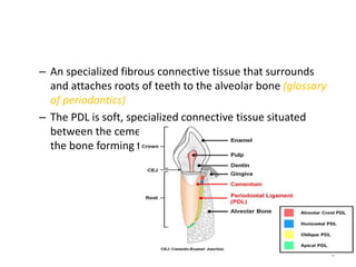
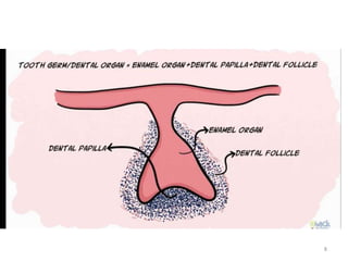
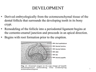
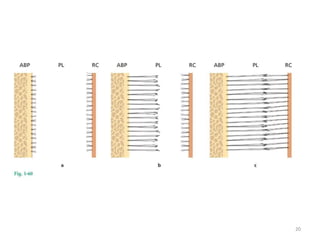
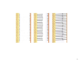
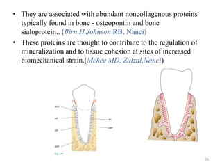
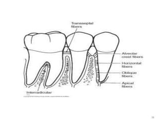
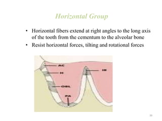
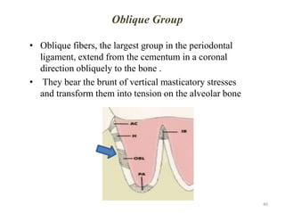
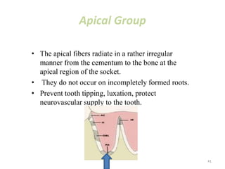
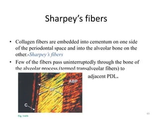
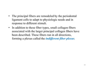
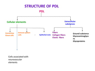
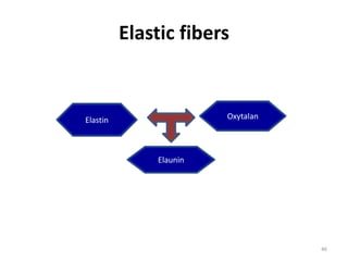
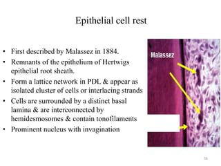
PERIODONTAL
LIGAMENT
1
Abdul Jabir
First year PG
Dept of periodontics
CONTENTS
• ntroduction
• Development
• Width and shape
• Periodontal fibers
• Composition
• Blood supply and nerve supply
• Functions
• Age changes in PDL
• In disease-periodontitis
2
INTRODUCTION
• The periodontal ligament is composed of a complex vascular
and highly cellular connective tissue that surrounds the tooth
root and connects it to the inner wall of the alveolar bone.
• It is continuous with the connective tissue of the gingiva and
communicates with the marrow spaces through vascular
channels in the bone.
3
– An specialized fibrous connective tissue that surrounds
and attaches roots of teeth to the alveolar bone (glossary
of periodontics)
– The PDL is soft, specialized connective tissue situated
between the cementum covering the root of the tooth and
the bone forming the socket wall.
4
Other names......
• Desmodont
• Gomphosis
• Pericementum
• Periodontal membrane
• Alvelo-dental ligament
• Dental periosteum
5
6
7
8
DEVELOPMENT
• Derived embryologically from the ectomesenchymal tissue of the
dental follicle that surrounds the developing tooth in its bony
crypt.
• Remodeling of the follicle into a periodontal ligament begins at
the cemento-enamel junction and proceeds in an apical direction.
• Begins with root formation prior to the eruption.
9
• Proliferation of inner and external enamel epithelium- cervical loop
of the tooth bud.
• This sheath of epithelium grows apically, in form of hertwigs
epithelial root sheath.
• The sheath forms a circumferential structure encompassing dental
papilla separating it externally from dental follicle cells.
• The dental follicle cells, located between the alveolar bone and the
epithelial root sheath, are composed of two subpopulations with
distinct morphological characteristics and locations;
 mesenchymal cells of the dental follicle proper close to bone
 the perifollicular mesenchyme close to HERS
10
11
• Mesenchymal cells- stellate shaped ,small and randomly
oriented.
• Perifollicular mesenchyme - More widely seperated.
• As root formation continues, cells in the perifollicular area,
gain polarity and cellular volume-Increase in synthetic
activity.
• They actively synthesis and deposit collagen fibrils and
glycoprotein in the developing PDL.
12
 The developing periodontal ligament, as well as
the mature periodontal ligament, contains undifferentiated
stem cells that retain the potential to differentiate into
osteoblasts, cementoblasts and fibroblasts.(Mc Culloch 1984)
 Stem cells occupy perivascular sites in the periodontal
ligament and in adjacent endosteal spaces.(Gould TRL 1983)
13
14
15
Width and shape
• hour glass shape
• It ranges in width from 0.15 to 0.38mm, reaching the
thinnest portion around the middle third of the root (Nanci
and Bosshardt, 2006).
• Periodontal ligament thickness decrease with increasing
age.
• It is thinner on the mesial root surface than on the distal
surface.
• Its varies with the functional demands made on the tooth.
16
• PDL space diminished around teeth that are
not in function and in unerupted teeth, but
that have been increased in teeth
hyperfunction.
• Narrowest in region of axis of rotation
17
• Radiographic appearance- 0.4 to 0.5 mm space,a
radiolucent area between the radioopaque lamina dura of
alveolar bone proper and radiopaque cementum.
• Narrower in permanent teeth than in deciduous teeth.
18
Development of principal
fibers
• Closely related to root formation
• Fiber bundles originate at the surface of the
newly formed root dentin in close relation to
the elongated and highly polarized
fibroblasts.
• During tooth eruption, as PDL matures, the
fringe fibers, merge across the width of the
ligament to form principal fiber bundles.
19
20
21
• Majority of the fibers course in a coronal direction from
cementum to bone, forming oblique fiber group.
• With continued development of the root major principal fibers
are established as continuous structures embedded as sharpey’s
fibers in bone and cementum.
• By the time of first occlusal contact of the tooth with its
antagonist, the fibers around the coronal third of the root, the
horizontal group are almost completely developed.
• As eruption continues, and definite occlusion is established, there
is progressive apical maturation of oblique fibers .
• With the formation of the apical fiber group .the definitive PDL
architecture is established.
22
• The mature periodontal ligament can be subdivided into
three regions (Garant 2003)
• a) A bone-related region, rich in cells and blood vessels,
• b) A cementum-related region characterized by dense well-
ordered collagen bundles, and
• c) A middle zone containing fewer cells and thinner collagen
fibrils .(Sloan P 1995)
23
Periodontal Fibers
• The most important elements of the periodontal ligament -
principal fibers,
• Collagenous and arranged in bundles and follow a wavy
course when viewed in longitudinal section.
• The terminal portions of the principal fibers that are inserted
into cementum and bone are termed Sharpey's fibers
• The principal fiber bundles consist of individual fibers that
form a continuous anastomosing network between tooth and
bone.
• Once embedded in the wall of the alveolus or in the tooth,
Sharpey's fibers calcify to a significant degree.
24
•
25
• They are associated with abundant noncollagenous proteins
typically found in bone - osteopontin and bone
sialoprotein.. (Birn H,Johnson RB, Nanci)
• These proteins are thought to contribute to the regulation of
mineralization and to tissue cohesion at sites of increased
biomechanical strain.(Mckee MD, Zalzal,Nanci)
26
27
collagen
• Collagen - protein composed of different
amino acids, the most important of which
are glycine, proline, hydroxylysine, and
hydroxyproline.
• Collagen is responsible for maintenance of
the framework and tone of tissue, and it
exhibits a wide range of diversity.
28
• Collagen biosynthesis occurs inside the fibroblasts to form
tropocollagen molecules.
• These aggregate into microfibrils that are packed together to form
fibrils.
• Collagen fibrils have a transverse striation with a characteristic
periodicity of 64 µm; this striation is caused by the overlapping
arrangement of the tropocollagen molecules.
• In collagen type I and III, these fibrils associate to form fibers. In
collagen type I fibers associate to form bundles.
29
30
• Main type of collagen in PDL is type l and lll
• 70% of PDL is type l collagen
• Collagen is synthesized by fibroblasts, chondroblasts,
osteoblasts, odontoblasts, and other cells
31
• The several types of collagen are all distinguishable by their
chemical composition, distribution, function, and
morphology
• The principal fibers are composed mainly of collagen type
I, whereas reticular fibers are composed of collagen type III.
• Collagen type IV is found in the basal lamina.
• Type VI collagen also has been immunolocalized in
periodontal ligament and gingiva
• The expression of type XII collagen during tooth
development is timed with alignment and organization of
periodontal fibers and is limited in tooth development to
cells within periodontal ligament.
32
• The molecular configuration of collagen
fibers provides them with a tensile
strength greater than that of steel.
• Collagen imparts a unique combination of
flexibility and strength to the tissues.
33
PRINCIPAL FIBERS
• The principal fibers of the periodontal ligament are arranged in six
groups that develop sequentially in the developing root:
• .(holmstrup 1996
34
Transseptal
alveolar crest
horizontal
oblique
apical
interradicular fibers
35
Transseptal Group
• Transseptal fibers extend interproximally over the alveolar bone
crest and are embedded in the cementum of adjacent teeth .
• They are reconstructed even after destruction of the alveolar
bone resulting from periodontal disease.
• These fibers may be considered as belonging to the gingiva
because they do not have osseous attachment
36
Alveolar Crest Group
• Alveolar crest fibers extend obliquely from the cementum just beneath
the junctional epithelium to the alveolar crest.
• Fibers also run from the cementum over the alveolar crest and to the
fibrous layer of the periosteum covering the alveolar bone
• The alveolar crest fibers prevent the extrusion of the tooth and resist
lateral tooth movements.
• The incision of these fibers during periodontal surgery does not
increase tooth mobility unless significant attachment loss has
occurred.
37
38
Horizontal Group
• Horizontal fibers extend at right angles to the long axis
of the tooth from the cementum to the alveolar bone
• Resist horizontal forces, tilting and rotational forces
39
Oblique Group
• Oblique fibers, the largest group in the periodontal
ligament, extend from the cementum in a coronal
direction obliquely to the bone .
• They bear the brunt of vertical masticatory stresses
and transform them into tension on the alveolar bone
40
Apical Group
• The apical fibers radiate in a rather irregular
manner from the cementum to the bone at the
apical region of the socket.
• They do not occur on incompletely formed roots.
• Prevent tooth tipping, luxation, protect
neurovascular supply to the tooth.
41
Interradicular Group
• The inter radicular fibers fan out from the cementum to
the tooth in the furcation areas of multirooted teeth.
• Prevent tooth tipping, luxation and torquing
42
Sharpey’s fibers
• Collagen fibers are embedded into cementum on one side
of the periodontal space and into the alveolar bone on the
other.-Sharpey’s fibers
• Few of the fibers pass uninterruptedly through the bone of
the alveolar process,(termed transalveolar fibers) to
continue as principal fibers of the adjacent PDL.
43
• The principal fibers are remodeled by the periodontal
ligament cells to adapt to physiologic needs and in
response to different stimuli.
• In addition to these fiber types, small collagen fibers
associated with the larger principal collagen fibers have
been described. These fibers run in all directions,
forming a plexus called the indifferent fiber plexus.
44
STRUCTURE OF PDL
PDL
Cellular elements Extracellular
substances
Connective
tissue cells - Epithelial rests
Immune system
cells
Fibers
Collagen fibers
Elastic fibers
Ground substance
Glycosaminoglyca
ns
Glycoproteins
Cells associated with
neurovascular
elements
Elastic fibers
46
Elastin Oxytalan
Elaunin
• Elastin- It is a protein in the connective tissue that is
elastic and allows many tissues in the body to resume its
shape after stretching and contracting.
• Oxytalin- Bundles of microfibrils with extensive
distribution in PDL. They are numerous and dense in the
cervical region of the ligament, where they run parallel
to root surface in a vertical direction and bend to attach
to the cementum.
• Elaunin- Component of elastic fibers formed by
deposition of elastin between oxytalan fibers. Found with
in the fibers of gigival ligament.
47
Cellular Elements
Connective
tissue cells
epithelial cell
rests
immune
system cells
cells associated
with
neurovascular
elements
48
Fibroblasts
Cementoblasts
Osteoblasts
Fibroblasts
• The Principal cells of the PDL.
• The most common cells in the periodontal ligament
• appear as ovoid or elongated cells oriented along the principal
fibers, exhibiting pseudopodia-like processes.(Roberts WE)
• Large cells with an extensive cytoplasm containing an abundance
of organelles associated with protein synthesis.
• Well developed cytoskeleton
49
• Regularly distributed throughout the ligament and are
oriented with their long axis parallel to the direction of
collagen fibrils.
• They produce collagen and elastin-structural connective
tissue proteins.
• They also secrete an active collagenase and a family of
enzymes collectively know as matrix metalloproteinases
(MMP’s)
• Fibroblasts are responsible for the formation and remodelling
of the PDL fibers, and maintaining the width of PDL.
• They maintain the width by preventing encroachment of bone
and cementum into the periodontal space
50
• Importantly, in inflammatory situations, such as those
associated with periodontal diseases, an increased
expression of matrix metalloproteinases occurs that
aggressively destroys collagen.
• They possess the capacity to phagocytose “old” collagen
fibers and degrade them by enzyme hydrolysis.
• Collagen turnover appears to be regulated by fibroblasts in
a process of intracellular degradation of collagen not
involving the action of collagenase.
• Because of the exceptionally high rate of turnover of
collagen in the ligament, any interference with fibroblast
function by disease rapidly produces a loss of the
supporting tissue of a tooth
51
Gingival fibroblast and
periodontal ligament fibroblast
• As compared with GF, PDLF contain high levels of alkaline
phosphatase.(Ohshima et al., 1988; Somerman et al., 1988)
• GF derived from general mesenchyme and PDLF derived from
ecto-mesenchymal in origin.
• Under the SEM, GF and PDLF were similar. (Rose et al., 1'987;
Ohshim et al., 1988; Somerman et al., 1988).
• Walsi. (1984) reported that porcine periodonital ligament
fibroblasts synthesize osteonectin, a molecuIe described as a
major bone matrix glycoprotein, having the ability to bind to
both hydroxyapatite and collagen. Unlike GF, PDLF may
possess the ability for mineralization (O1hshima ci ai., 1988)
and for producing mineral-like nodules in-vitro.
52
• Several studies have shown that when gingival connective
tissue is allowed, after surgery, to come into contact with the
root surface, root resorption will occur. On the contrary, when
periodontal ligament cells are selectively allowed to come into
contact with the root surface, regeneration of a functional
periodontium can be observed histologically
• By the use of membranes of various kinds, such techniques
of guided tissue regeneration can limit the apical migration of
gingival epithelial cells and favor the establishment of a new
attachment by periodontal fibroblasts.
• Guide Tissue Regeneration (GTR) employs a barrier
membrane around the periodontal defect to prevent epithelial
downgrowth and fibroblast transgrowth into the wound space,
thereby maintaining a space for true periodontal tissue
regeneration.
53
osteoblasts
• Bone forming cells lining the tooth
socket
• Appears cuboidal & exhibits a
basophilic cytoplasm with extensive
rough endoplasmic reticulum
• Prominent nucleus
• Cell contacts one another by means
of desmosomes & tight junctions
• Remains in active functional state
upto 20 days
54
Cementoblasts
• Cementum forming cells
• Not elongated as fibroblasts, squat cuboidal
cells
• Rich in cytoplasm and
have large vesicular nuclei
55
Epithelial cell rest
• First described by Malassez in 1884.
• Remnants of the epithelium of Hertwigs
epithelial root sheath.
• Form a lattice network in PDL & appear as
isolated cluster of cells or interlacing strands
• Cells are surrounded by a distinct basal
lamina & are interconnected by
hemidesmosomes & contain tonofilaments
• Prominent nucleus with invagination
56
• They are attached to one another by tonofilaments or
desmosomes
• Most numerous in the apical area & cervical area
• Diminish with age by degenerating /disappearing or by
undergoing calcifications to form cementicles
• Exact function unknown but involved in periodontal repair
or regeneration
57
58
Defence cells
• These include mast cells, lymphocytes, neutrophils and
macrophages.
• Responsible for phagocytosing particulate matter and
invading organisms
• Mast cells responsible for production of histamine,
heparin
• Macrophages secretes growth factors that regulate the
proliferation of adjacent fibroblasts.
59
Ground substance
Glycosaminoglycans
Hyaluronic
acid, heparan
sulphate
Proteoglycan
Glycoproteins
Fibronectin
laminin
60
Ground substance also has a high water content – 70%
• Proteoglycans are compounds containing anionic
polysaccharides covalently attached to a protein core through
a specific trisaccharide galactose-galactose-xylose sequence.
• Proteoglycan namely decorin, biglycan involved in the
organization and regulation of collagen fibers and are
abundantly present in the periodontal ligament.
• Decorin is present bound to collagen and is important to
regulate fibrillogenesis.
• Decorin is bound to transforming growth factor-beta and
prevents excessive fibrosis.This property prevents undue
cross-linking of the collagen fibres.
61
• Hyaluronic acid is an anionic, nonsulphated
glycosaminoglycan. It is of high molecular weight and
forms in the plasma membrane.
• Glycoprotein have adhesive properties
• Fibronectin is uniformly distributed in the PDL. They bind
to collagen and proteoglycan. It promotes adhesion of
fibroblast to extracellular matrix and plays a role in
alignment of collagen fibres.
• Laminin has been found in the basement membrane of
epithelial cell rests of malassez.
• Tenascin – adhesive glycoprotein that binds to fibronectin
and to proteoglycan.
62
Blood supply
• Main source- Superior and inferior alveolar arteries
• Blood supply reaches periodontal ligament from 3 sources
 Apical vessels
 Penetrating vessels from alveolar bone
 Anastomosing vessels from gingiva
• More abundant in PDL of posterior teeth and in mandible.
• Single rooted tooth-More abundant in the gingival third and apical
third.
• Blood vessels are closer to bone than cementum
• Blood supply slightly greater on mesial and distal surfaces than on
lingual and facial surfaces.
63
• The PDL has some specialised features in the vasculature,
Large number of fenestrations
Cervical plexus of capillary loops
• The fenestrated capillary beds have increased capacity for
diffusion and filtration.
• Cervical plexus is found in the gingival crevice and anstamoses
with the gingival and PDL vessels.
64
Nerve supply
• Two types of nerve fibers-
• Sensory fibers –associated with nociception and
mechanoception.
• Autonomic fibers –associated with PDL vessels
65
Sensory autonomic
cementicles
• Globular masses of cementum arranged in concentric lamella
that lie free in the periodontal ligament or adhere to the root
surface
• Calcified bodies
• Seen in older individuals ,and remain free in the connective
tissue.
• They may fuse to form large calcified masses or maybe joined
with the cementum.
66
Functions of Periodontal Ligament
Physical
Formative
and
remodeling
Nutritional Sensory.
67
Physical Functions
Provision of a soft tissue “casing” to protect the vessels
and nerves from injury by mechanical forces.
Transmission of occlusal forces to the bone.
Attachment of the teeth to the bone.
Maintenance of the gingival tissues in their proper
relationship to the teeth.
Resistance to the impact of occlusal forces (shock
absorption).
68
Resistance to Impact of Occlusal
Forces (Shock Absorption)
Tensional theory
 The principal fibers of the periodontal ligament are
the major factor in supporting the tooth and
transmitting forces to the bone.
 When a force is applied to the crown, the principal
fibers first unfold and straighten and then transmit
forces to the alveolar bone, causing an elastic
deformation of the bony socket.
 Finally, when the alveolar bone has reached its
limit, the load is transmitted to the basal bone.
69
70
The tensional theory : …..The principal fibers of the
PDL bear the major responsibility in supporting
the tooth and transmitting forces to the bone.
When a force is applied to the crown
principal fibers first unfold and straighten
transmit forces to the alveolar bone
causes elastic deformation of the bony socket.
Finally, when the alveolar bone has reached its limit,
the load is transmitted to the basal bone.
Viscoelastic theory
 The displacement of the tooth is largely controlled by
fluid movements, with fibers having only a secondary role.
 When forces are transmitted to the tooth, the extracellular
fluid passes from the periodontal ligament into the marrow
spaces of bone through foramina in the cribriform plate.
 After depletion of tissue fluids, the fiber bundles absorb
the slack and tighten.
 This leads to a blood vessel stenosis
 Arterial back pressure causes ballooning of the vessels
and passage of the blood ultra filtrates into the tissues,
thereby replenishing the tissue fluids
71
Transmission of Occlusal
Forces to Bone
• The arrangement of principal fibers similar to a hammock
or a suspension bridge
• When an axial force is applied to a tooth, tendency of
displacement of root into alveolus occurs
• Oblique fibres alter their wavy, untensed pattern and
assume their full length & sustain major part of axial force
• When horizontal or tipping force is applied, 2 phases of
tooth movement occur
• ---within confines of PDL
• ---displacement of facial & lingual bony plates
72
• In areas of tension, principal fibers are taut, whereas in areas
of pressure, they get compressed, the tooth is displaced and
corresponding distortion of bone exists in the direction of root
movement.
In single rooted teeth, the axis of rotation is located in the area
between apical third and middle third of root. In multirooted
teeth axis of rotation is located in the bone between roots
73
Formative and Remodeling Function
• Cells of periodontal ligament participate in the formation &
resorption of cementum & bone
• Help in the accommodation of periodontium to occlusal
forces
• Help in repair of injuries
PDL is constantly undergoing remodeling.
Old cells and fibres are break down and replaced by new ones.
There is also rapid collagen turnover
74
Nutritional functions
• Supplies nutrients to the cementum, bone, and gingiva by
way of the blood vessels and also provides lymphatic
drainage.
• The blood vessels are also involved in the removal of
catabolites.
• Occlusion of blood vessels leads to necrosis of cells in the
affected part of the ligament.
75
Sensory Functions
• The periodontal ligament is abundantly supplied with
sensory nerve fibers capable of transmitting tactile,
pressure, and pain sensations by the trigeminal pathways.
• Provides most efficient proprioceptive mechanism,
allowing detection of the delicate forces to the teeth.
• Nerve bundles pass into the periodontal ligament from the
periapical area and through channels from the alveolar bone
that follow the course of the blood vessels
76
• The bundles divide into single myelinated fibers, which
ultimately lose their myelin sheaths and end in one of four
types of neural termination:
• (1) free endings, which have a treelike configuration and
carry pain sensation;
• (2) Ruffini-like mechanoreceptors, located primarily in the
apical area; coiled
• (3)Meissner's corpuscles, also mechanoreceptors, found
mainly in the midroot region; and
• (4) spindlelike pressure and vibration endings, which are
surrounded by a fibrous capsule and located mainly in the
apex.(Maeda 1990)
77
Age changes in the pdl
• in cell number and cell activity.
• Decreased number of fibroblasts
• Scalloping of alveolar bone and cementum.
• PDL fibers attached to the peaks of these scallops.
• decreased organic matrix production and epithelial cell
rests
• increased amounts of elastic fiber
78
In disease-periodontitis
• An inflammatory disease of supporting tissues of the teeth
caused by specific microorganisms or groups of specific
microorganisms resulting in progressive destruction of PDL
and alveolar bone with pocket formation, recession or both.
79
Matrix changes
• Tissue destruction begins within perivascular extra
cellular matrix where most of the collagen within the
foci of inflammation is degraded
• Gingivitis- matrix destruction by matrix
metalloproteinases (MMP)--- PMNL
• Periodontitis- MMP’s produced by PMNL,
macrophages, keratinocytes, fibroblasts
80
• Type I & type III collagens are lost at foci of inflammation
• Increase in type V collagen
• Destruction of non collagenous proteins
81
Periodontal response to occlusal forces
• The effect of occlusal forces on periodontium is influenced by
Magnitude
Direction
Duration
Frequency of forces
82
When magnitude increases
• Widening of PDL space
• Increase in the no: & width of periodontal fibres
• Increase in the density of alveolar bone
Change in the direction of force
reorientation of stresses & strains within the periodontium
83
• The principal fibers of the PDL are arranged so that they best
accommodate occlusal forces along the long axis of the tooth.
• Lateral and torque forces will injure the periodontium.
• Constant pressure on the bone is more injurious than
intermittent forces.
• The more frequent the application of the intermittent force,
the more injurious is the force to the periodontium.
84
Trauma from occlusion
• When occlusal forces exceed the adaptive
capacity of the tissues, tissue injury results
• The resultant tissue injury is trauma from
occlusion
85
Acute trauma
• Results from an abrupt occlusal impact,
• It causes
Tooth pain
Sensitivity to percussion
Increased tooth mobility
86
Chronic trauma
From gradual changes in occlusion by
Tooth wear & drifting movement
Extrusion of teeth
Bruxism & clenching
87
Primary trauma
• When trauma from occlusion is the result of alteration in
occlusal force
• No alteration in connective tissue attachment & no pocket
formation
88
Secondary trauma
• When trauma from occlusion results when adaptive capacity of
tissues to resist occlusal forces is impaired by Bone loss
resulting from marginal inflammation.
• Decrease PDL attachment area & this alters the leverage on
the remaining tissues
89
conclusion
. The PDL is a physically small, but
functionally important tissue in tooth
support, proprioception and regulation
of alveolar bone volume.
The PDL is an absolute requirement for
rapid remodeling of alveolar bone when
forces are applied to teeth.
90
references
• Carranza’s Clinical Periodontology – Michael G.
Newman et al – thirteenth Edition
• Clinical periodontology and implant dentistry- 5th edition jan lindhe,
niklaus p lang, thokild karring
• Ten Cate’s Oral Histology – Development, Structure
and Function – Antonio nanci –eighth Edition
91
92

Periodontal ligament

  • 1.
  • 2.
    CONTENTS • ntroduction • Development •Width and shape • Periodontal fibers • Composition • Blood supply and nerve supply • Functions • Age changes in PDL • In disease-periodontitis 2
  • 3.
    INTRODUCTION • The periodontalligament is composed of a complex vascular and highly cellular connective tissue that surrounds the tooth root and connects it to the inner wall of the alveolar bone. • It is continuous with the connective tissue of the gingiva and communicates with the marrow spaces through vascular channels in the bone. 3
  • 4.
    – An specializedfibrous connective tissue that surrounds and attaches roots of teeth to the alveolar bone (glossary of periodontics) – The PDL is soft, specialized connective tissue situated between the cementum covering the root of the tooth and the bone forming the socket wall. 4
  • 5.
    Other names...... • Desmodont •Gomphosis • Pericementum • Periodontal membrane • Alvelo-dental ligament • Dental periosteum 5
  • 6.
  • 7.
  • 8.
  • 9.
    DEVELOPMENT • Derived embryologicallyfrom the ectomesenchymal tissue of the dental follicle that surrounds the developing tooth in its bony crypt. • Remodeling of the follicle into a periodontal ligament begins at the cemento-enamel junction and proceeds in an apical direction. • Begins with root formation prior to the eruption. 9
  • 10.
    • Proliferation ofinner and external enamel epithelium- cervical loop of the tooth bud. • This sheath of epithelium grows apically, in form of hertwigs epithelial root sheath. • The sheath forms a circumferential structure encompassing dental papilla separating it externally from dental follicle cells. • The dental follicle cells, located between the alveolar bone and the epithelial root sheath, are composed of two subpopulations with distinct morphological characteristics and locations;  mesenchymal cells of the dental follicle proper close to bone  the perifollicular mesenchyme close to HERS 10
  • 11.
  • 12.
    • Mesenchymal cells-stellate shaped ,small and randomly oriented. • Perifollicular mesenchyme - More widely seperated. • As root formation continues, cells in the perifollicular area, gain polarity and cellular volume-Increase in synthetic activity. • They actively synthesis and deposit collagen fibrils and glycoprotein in the developing PDL. 12
  • 13.
     The developingperiodontal ligament, as well as the mature periodontal ligament, contains undifferentiated stem cells that retain the potential to differentiate into osteoblasts, cementoblasts and fibroblasts.(Mc Culloch 1984)  Stem cells occupy perivascular sites in the periodontal ligament and in adjacent endosteal spaces.(Gould TRL 1983) 13
  • 14.
  • 15.
  • 16.
    Width and shape •hour glass shape • It ranges in width from 0.15 to 0.38mm, reaching the thinnest portion around the middle third of the root (Nanci and Bosshardt, 2006). • Periodontal ligament thickness decrease with increasing age. • It is thinner on the mesial root surface than on the distal surface. • Its varies with the functional demands made on the tooth. 16
  • 17.
    • PDL spacediminished around teeth that are not in function and in unerupted teeth, but that have been increased in teeth hyperfunction. • Narrowest in region of axis of rotation 17
  • 18.
    • Radiographic appearance-0.4 to 0.5 mm space,a radiolucent area between the radioopaque lamina dura of alveolar bone proper and radiopaque cementum. • Narrower in permanent teeth than in deciduous teeth. 18
  • 19.
    Development of principal fibers •Closely related to root formation • Fiber bundles originate at the surface of the newly formed root dentin in close relation to the elongated and highly polarized fibroblasts. • During tooth eruption, as PDL matures, the fringe fibers, merge across the width of the ligament to form principal fiber bundles. 19
  • 20.
  • 21.
  • 22.
    • Majority ofthe fibers course in a coronal direction from cementum to bone, forming oblique fiber group. • With continued development of the root major principal fibers are established as continuous structures embedded as sharpey’s fibers in bone and cementum. • By the time of first occlusal contact of the tooth with its antagonist, the fibers around the coronal third of the root, the horizontal group are almost completely developed. • As eruption continues, and definite occlusion is established, there is progressive apical maturation of oblique fibers . • With the formation of the apical fiber group .the definitive PDL architecture is established. 22
  • 23.
    • The matureperiodontal ligament can be subdivided into three regions (Garant 2003) • a) A bone-related region, rich in cells and blood vessels, • b) A cementum-related region characterized by dense well- ordered collagen bundles, and • c) A middle zone containing fewer cells and thinner collagen fibrils .(Sloan P 1995) 23
  • 24.
    Periodontal Fibers • Themost important elements of the periodontal ligament - principal fibers, • Collagenous and arranged in bundles and follow a wavy course when viewed in longitudinal section. • The terminal portions of the principal fibers that are inserted into cementum and bone are termed Sharpey's fibers • The principal fiber bundles consist of individual fibers that form a continuous anastomosing network between tooth and bone. • Once embedded in the wall of the alveolus or in the tooth, Sharpey's fibers calcify to a significant degree. 24
  • 25.
  • 26.
    • They areassociated with abundant noncollagenous proteins typically found in bone - osteopontin and bone sialoprotein.. (Birn H,Johnson RB, Nanci) • These proteins are thought to contribute to the regulation of mineralization and to tissue cohesion at sites of increased biomechanical strain.(Mckee MD, Zalzal,Nanci) 26
  • 27.
  • 28.
    collagen • Collagen -protein composed of different amino acids, the most important of which are glycine, proline, hydroxylysine, and hydroxyproline. • Collagen is responsible for maintenance of the framework and tone of tissue, and it exhibits a wide range of diversity. 28
  • 29.
    • Collagen biosynthesisoccurs inside the fibroblasts to form tropocollagen molecules. • These aggregate into microfibrils that are packed together to form fibrils. • Collagen fibrils have a transverse striation with a characteristic periodicity of 64 µm; this striation is caused by the overlapping arrangement of the tropocollagen molecules. • In collagen type I and III, these fibrils associate to form fibers. In collagen type I fibers associate to form bundles. 29
  • 30.
  • 31.
    • Main typeof collagen in PDL is type l and lll • 70% of PDL is type l collagen • Collagen is synthesized by fibroblasts, chondroblasts, osteoblasts, odontoblasts, and other cells 31
  • 32.
    • The severaltypes of collagen are all distinguishable by their chemical composition, distribution, function, and morphology • The principal fibers are composed mainly of collagen type I, whereas reticular fibers are composed of collagen type III. • Collagen type IV is found in the basal lamina. • Type VI collagen also has been immunolocalized in periodontal ligament and gingiva • The expression of type XII collagen during tooth development is timed with alignment and organization of periodontal fibers and is limited in tooth development to cells within periodontal ligament. 32
  • 33.
    • The molecularconfiguration of collagen fibers provides them with a tensile strength greater than that of steel. • Collagen imparts a unique combination of flexibility and strength to the tissues. 33
  • 34.
    PRINCIPAL FIBERS • Theprincipal fibers of the periodontal ligament are arranged in six groups that develop sequentially in the developing root: • .(holmstrup 1996 34 Transseptal alveolar crest horizontal oblique apical interradicular fibers
  • 35.
  • 36.
    Transseptal Group • Transseptalfibers extend interproximally over the alveolar bone crest and are embedded in the cementum of adjacent teeth . • They are reconstructed even after destruction of the alveolar bone resulting from periodontal disease. • These fibers may be considered as belonging to the gingiva because they do not have osseous attachment 36
  • 37.
    Alveolar Crest Group •Alveolar crest fibers extend obliquely from the cementum just beneath the junctional epithelium to the alveolar crest. • Fibers also run from the cementum over the alveolar crest and to the fibrous layer of the periosteum covering the alveolar bone • The alveolar crest fibers prevent the extrusion of the tooth and resist lateral tooth movements. • The incision of these fibers during periodontal surgery does not increase tooth mobility unless significant attachment loss has occurred. 37
  • 38.
  • 39.
    Horizontal Group • Horizontalfibers extend at right angles to the long axis of the tooth from the cementum to the alveolar bone • Resist horizontal forces, tilting and rotational forces 39
  • 40.
    Oblique Group • Obliquefibers, the largest group in the periodontal ligament, extend from the cementum in a coronal direction obliquely to the bone . • They bear the brunt of vertical masticatory stresses and transform them into tension on the alveolar bone 40
  • 41.
    Apical Group • Theapical fibers radiate in a rather irregular manner from the cementum to the bone at the apical region of the socket. • They do not occur on incompletely formed roots. • Prevent tooth tipping, luxation, protect neurovascular supply to the tooth. 41
  • 42.
    Interradicular Group • Theinter radicular fibers fan out from the cementum to the tooth in the furcation areas of multirooted teeth. • Prevent tooth tipping, luxation and torquing 42
  • 43.
    Sharpey’s fibers • Collagenfibers are embedded into cementum on one side of the periodontal space and into the alveolar bone on the other.-Sharpey’s fibers • Few of the fibers pass uninterruptedly through the bone of the alveolar process,(termed transalveolar fibers) to continue as principal fibers of the adjacent PDL. 43
  • 44.
    • The principalfibers are remodeled by the periodontal ligament cells to adapt to physiologic needs and in response to different stimuli. • In addition to these fiber types, small collagen fibers associated with the larger principal collagen fibers have been described. These fibers run in all directions, forming a plexus called the indifferent fiber plexus. 44
  • 45.
    STRUCTURE OF PDL PDL Cellularelements Extracellular substances Connective tissue cells - Epithelial rests Immune system cells Fibers Collagen fibers Elastic fibers Ground substance Glycosaminoglyca ns Glycoproteins Cells associated with neurovascular elements
  • 46.
  • 47.
    • Elastin- Itis a protein in the connective tissue that is elastic and allows many tissues in the body to resume its shape after stretching and contracting. • Oxytalin- Bundles of microfibrils with extensive distribution in PDL. They are numerous and dense in the cervical region of the ligament, where they run parallel to root surface in a vertical direction and bend to attach to the cementum. • Elaunin- Component of elastic fibers formed by deposition of elastin between oxytalan fibers. Found with in the fibers of gigival ligament. 47
  • 48.
    Cellular Elements Connective tissue cells epithelialcell rests immune system cells cells associated with neurovascular elements 48 Fibroblasts Cementoblasts Osteoblasts
  • 49.
    Fibroblasts • The Principalcells of the PDL. • The most common cells in the periodontal ligament • appear as ovoid or elongated cells oriented along the principal fibers, exhibiting pseudopodia-like processes.(Roberts WE) • Large cells with an extensive cytoplasm containing an abundance of organelles associated with protein synthesis. • Well developed cytoskeleton 49
  • 50.
    • Regularly distributedthroughout the ligament and are oriented with their long axis parallel to the direction of collagen fibrils. • They produce collagen and elastin-structural connective tissue proteins. • They also secrete an active collagenase and a family of enzymes collectively know as matrix metalloproteinases (MMP’s) • Fibroblasts are responsible for the formation and remodelling of the PDL fibers, and maintaining the width of PDL. • They maintain the width by preventing encroachment of bone and cementum into the periodontal space 50
  • 51.
    • Importantly, ininflammatory situations, such as those associated with periodontal diseases, an increased expression of matrix metalloproteinases occurs that aggressively destroys collagen. • They possess the capacity to phagocytose “old” collagen fibers and degrade them by enzyme hydrolysis. • Collagen turnover appears to be regulated by fibroblasts in a process of intracellular degradation of collagen not involving the action of collagenase. • Because of the exceptionally high rate of turnover of collagen in the ligament, any interference with fibroblast function by disease rapidly produces a loss of the supporting tissue of a tooth 51
  • 52.
    Gingival fibroblast and periodontalligament fibroblast • As compared with GF, PDLF contain high levels of alkaline phosphatase.(Ohshima et al., 1988; Somerman et al., 1988) • GF derived from general mesenchyme and PDLF derived from ecto-mesenchymal in origin. • Under the SEM, GF and PDLF were similar. (Rose et al., 1'987; Ohshim et al., 1988; Somerman et al., 1988). • Walsi. (1984) reported that porcine periodonital ligament fibroblasts synthesize osteonectin, a molecuIe described as a major bone matrix glycoprotein, having the ability to bind to both hydroxyapatite and collagen. Unlike GF, PDLF may possess the ability for mineralization (O1hshima ci ai., 1988) and for producing mineral-like nodules in-vitro. 52
  • 53.
    • Several studieshave shown that when gingival connective tissue is allowed, after surgery, to come into contact with the root surface, root resorption will occur. On the contrary, when periodontal ligament cells are selectively allowed to come into contact with the root surface, regeneration of a functional periodontium can be observed histologically • By the use of membranes of various kinds, such techniques of guided tissue regeneration can limit the apical migration of gingival epithelial cells and favor the establishment of a new attachment by periodontal fibroblasts. • Guide Tissue Regeneration (GTR) employs a barrier membrane around the periodontal defect to prevent epithelial downgrowth and fibroblast transgrowth into the wound space, thereby maintaining a space for true periodontal tissue regeneration. 53
  • 54.
    osteoblasts • Bone formingcells lining the tooth socket • Appears cuboidal & exhibits a basophilic cytoplasm with extensive rough endoplasmic reticulum • Prominent nucleus • Cell contacts one another by means of desmosomes & tight junctions • Remains in active functional state upto 20 days 54
  • 55.
    Cementoblasts • Cementum formingcells • Not elongated as fibroblasts, squat cuboidal cells • Rich in cytoplasm and have large vesicular nuclei 55
  • 56.
    Epithelial cell rest •First described by Malassez in 1884. • Remnants of the epithelium of Hertwigs epithelial root sheath. • Form a lattice network in PDL & appear as isolated cluster of cells or interlacing strands • Cells are surrounded by a distinct basal lamina & are interconnected by hemidesmosomes & contain tonofilaments • Prominent nucleus with invagination 56
  • 57.
    • They areattached to one another by tonofilaments or desmosomes • Most numerous in the apical area & cervical area • Diminish with age by degenerating /disappearing or by undergoing calcifications to form cementicles • Exact function unknown but involved in periodontal repair or regeneration 57
  • 58.
  • 59.
    Defence cells • Theseinclude mast cells, lymphocytes, neutrophils and macrophages. • Responsible for phagocytosing particulate matter and invading organisms • Mast cells responsible for production of histamine, heparin • Macrophages secretes growth factors that regulate the proliferation of adjacent fibroblasts. 59
  • 60.
  • 61.
    • Proteoglycans arecompounds containing anionic polysaccharides covalently attached to a protein core through a specific trisaccharide galactose-galactose-xylose sequence. • Proteoglycan namely decorin, biglycan involved in the organization and regulation of collagen fibers and are abundantly present in the periodontal ligament. • Decorin is present bound to collagen and is important to regulate fibrillogenesis. • Decorin is bound to transforming growth factor-beta and prevents excessive fibrosis.This property prevents undue cross-linking of the collagen fibres. 61
  • 62.
    • Hyaluronic acidis an anionic, nonsulphated glycosaminoglycan. It is of high molecular weight and forms in the plasma membrane. • Glycoprotein have adhesive properties • Fibronectin is uniformly distributed in the PDL. They bind to collagen and proteoglycan. It promotes adhesion of fibroblast to extracellular matrix and plays a role in alignment of collagen fibres. • Laminin has been found in the basement membrane of epithelial cell rests of malassez. • Tenascin – adhesive glycoprotein that binds to fibronectin and to proteoglycan. 62
  • 63.
    Blood supply • Mainsource- Superior and inferior alveolar arteries • Blood supply reaches periodontal ligament from 3 sources  Apical vessels  Penetrating vessels from alveolar bone  Anastomosing vessels from gingiva • More abundant in PDL of posterior teeth and in mandible. • Single rooted tooth-More abundant in the gingival third and apical third. • Blood vessels are closer to bone than cementum • Blood supply slightly greater on mesial and distal surfaces than on lingual and facial surfaces. 63
  • 64.
    • The PDLhas some specialised features in the vasculature, Large number of fenestrations Cervical plexus of capillary loops • The fenestrated capillary beds have increased capacity for diffusion and filtration. • Cervical plexus is found in the gingival crevice and anstamoses with the gingival and PDL vessels. 64
  • 65.
    Nerve supply • Twotypes of nerve fibers- • Sensory fibers –associated with nociception and mechanoception. • Autonomic fibers –associated with PDL vessels 65 Sensory autonomic
  • 66.
    cementicles • Globular massesof cementum arranged in concentric lamella that lie free in the periodontal ligament or adhere to the root surface • Calcified bodies • Seen in older individuals ,and remain free in the connective tissue. • They may fuse to form large calcified masses or maybe joined with the cementum. 66
  • 67.
    Functions of PeriodontalLigament Physical Formative and remodeling Nutritional Sensory. 67
  • 68.
    Physical Functions Provision ofa soft tissue “casing” to protect the vessels and nerves from injury by mechanical forces. Transmission of occlusal forces to the bone. Attachment of the teeth to the bone. Maintenance of the gingival tissues in their proper relationship to the teeth. Resistance to the impact of occlusal forces (shock absorption). 68
  • 69.
    Resistance to Impactof Occlusal Forces (Shock Absorption) Tensional theory  The principal fibers of the periodontal ligament are the major factor in supporting the tooth and transmitting forces to the bone.  When a force is applied to the crown, the principal fibers first unfold and straighten and then transmit forces to the alveolar bone, causing an elastic deformation of the bony socket.  Finally, when the alveolar bone has reached its limit, the load is transmitted to the basal bone. 69
  • 70.
    70 The tensional theory: …..The principal fibers of the PDL bear the major responsibility in supporting the tooth and transmitting forces to the bone. When a force is applied to the crown principal fibers first unfold and straighten transmit forces to the alveolar bone causes elastic deformation of the bony socket. Finally, when the alveolar bone has reached its limit, the load is transmitted to the basal bone.
  • 71.
    Viscoelastic theory  Thedisplacement of the tooth is largely controlled by fluid movements, with fibers having only a secondary role.  When forces are transmitted to the tooth, the extracellular fluid passes from the periodontal ligament into the marrow spaces of bone through foramina in the cribriform plate.  After depletion of tissue fluids, the fiber bundles absorb the slack and tighten.  This leads to a blood vessel stenosis  Arterial back pressure causes ballooning of the vessels and passage of the blood ultra filtrates into the tissues, thereby replenishing the tissue fluids 71
  • 72.
    Transmission of Occlusal Forcesto Bone • The arrangement of principal fibers similar to a hammock or a suspension bridge • When an axial force is applied to a tooth, tendency of displacement of root into alveolus occurs • Oblique fibres alter their wavy, untensed pattern and assume their full length & sustain major part of axial force • When horizontal or tipping force is applied, 2 phases of tooth movement occur • ---within confines of PDL • ---displacement of facial & lingual bony plates 72
  • 73.
    • In areasof tension, principal fibers are taut, whereas in areas of pressure, they get compressed, the tooth is displaced and corresponding distortion of bone exists in the direction of root movement. In single rooted teeth, the axis of rotation is located in the area between apical third and middle third of root. In multirooted teeth axis of rotation is located in the bone between roots 73
  • 74.
    Formative and RemodelingFunction • Cells of periodontal ligament participate in the formation & resorption of cementum & bone • Help in the accommodation of periodontium to occlusal forces • Help in repair of injuries PDL is constantly undergoing remodeling. Old cells and fibres are break down and replaced by new ones. There is also rapid collagen turnover 74
  • 75.
    Nutritional functions • Suppliesnutrients to the cementum, bone, and gingiva by way of the blood vessels and also provides lymphatic drainage. • The blood vessels are also involved in the removal of catabolites. • Occlusion of blood vessels leads to necrosis of cells in the affected part of the ligament. 75
  • 76.
    Sensory Functions • Theperiodontal ligament is abundantly supplied with sensory nerve fibers capable of transmitting tactile, pressure, and pain sensations by the trigeminal pathways. • Provides most efficient proprioceptive mechanism, allowing detection of the delicate forces to the teeth. • Nerve bundles pass into the periodontal ligament from the periapical area and through channels from the alveolar bone that follow the course of the blood vessels 76
  • 77.
    • The bundlesdivide into single myelinated fibers, which ultimately lose their myelin sheaths and end in one of four types of neural termination: • (1) free endings, which have a treelike configuration and carry pain sensation; • (2) Ruffini-like mechanoreceptors, located primarily in the apical area; coiled • (3)Meissner's corpuscles, also mechanoreceptors, found mainly in the midroot region; and • (4) spindlelike pressure and vibration endings, which are surrounded by a fibrous capsule and located mainly in the apex.(Maeda 1990) 77
  • 78.
    Age changes inthe pdl • in cell number and cell activity. • Decreased number of fibroblasts • Scalloping of alveolar bone and cementum. • PDL fibers attached to the peaks of these scallops. • decreased organic matrix production and epithelial cell rests • increased amounts of elastic fiber 78
  • 79.
    In disease-periodontitis • Aninflammatory disease of supporting tissues of the teeth caused by specific microorganisms or groups of specific microorganisms resulting in progressive destruction of PDL and alveolar bone with pocket formation, recession or both. 79
  • 80.
    Matrix changes • Tissuedestruction begins within perivascular extra cellular matrix where most of the collagen within the foci of inflammation is degraded • Gingivitis- matrix destruction by matrix metalloproteinases (MMP)--- PMNL • Periodontitis- MMP’s produced by PMNL, macrophages, keratinocytes, fibroblasts 80
  • 81.
    • Type I& type III collagens are lost at foci of inflammation • Increase in type V collagen • Destruction of non collagenous proteins 81
  • 82.
    Periodontal response toocclusal forces • The effect of occlusal forces on periodontium is influenced by Magnitude Direction Duration Frequency of forces 82
  • 83.
    When magnitude increases •Widening of PDL space • Increase in the no: & width of periodontal fibres • Increase in the density of alveolar bone Change in the direction of force reorientation of stresses & strains within the periodontium 83
  • 84.
    • The principalfibers of the PDL are arranged so that they best accommodate occlusal forces along the long axis of the tooth. • Lateral and torque forces will injure the periodontium. • Constant pressure on the bone is more injurious than intermittent forces. • The more frequent the application of the intermittent force, the more injurious is the force to the periodontium. 84
  • 85.
    Trauma from occlusion •When occlusal forces exceed the adaptive capacity of the tissues, tissue injury results • The resultant tissue injury is trauma from occlusion 85
  • 86.
    Acute trauma • Resultsfrom an abrupt occlusal impact, • It causes Tooth pain Sensitivity to percussion Increased tooth mobility 86
  • 87.
    Chronic trauma From gradualchanges in occlusion by Tooth wear & drifting movement Extrusion of teeth Bruxism & clenching 87
  • 88.
    Primary trauma • Whentrauma from occlusion is the result of alteration in occlusal force • No alteration in connective tissue attachment & no pocket formation 88
  • 89.
    Secondary trauma • Whentrauma from occlusion results when adaptive capacity of tissues to resist occlusal forces is impaired by Bone loss resulting from marginal inflammation. • Decrease PDL attachment area & this alters the leverage on the remaining tissues 89
  • 90.
    conclusion . The PDLis a physically small, but functionally important tissue in tooth support, proprioception and regulation of alveolar bone volume. The PDL is an absolute requirement for rapid remodeling of alveolar bone when forces are applied to teeth. 90
  • 91.
    references • Carranza’s ClinicalPeriodontology – Michael G. Newman et al – thirteenth Edition • Clinical periodontology and implant dentistry- 5th edition jan lindhe, niklaus p lang, thokild karring • Ten Cate’s Oral Histology – Development, Structure and Function – Antonio nanci –eighth Edition 91
  • 92.

Editor's Notes

  • #13 Dental follicle proper contains all the precursors for cementum, bone and periodontal ligament formation Migrating fibroblasts from the perifollicular mesenchyme proliferate during root development to contribute to the pool of periodontal ligament fibroblasts Perifollicular mesenchyme and perivascular cells may give rise to osteoblasts of the alveolar bone
  • #15 a)Tooth bud in bone crypt,collagen fibers produced by fibroblast in loose ct,fiber bundles oriented in coronal direction form dentogingival,transseptal,dentoperiostel b)Tooth start to erupt,principal fibres formed c)More apically positioned bundles of orirnted fibres d)Well oriented in occlusion
  • #16 Initially, fibers become embedded in the cementum as sharpey’s fibres & are in coronal direction within the developing periodontal region giving them an orientation almost parallel to the root surface. Fiber formation & deposition occur sequentially from newly forming CEJ to the apex of the tooth root. First fibres to be deposited - dentogingival & transeptal fibres & those deposited apical to CEJ form gingivodental fibre group.
  • #22 Intermediate plexus=middle zone of pdl formed by splicing and unsplicing of fibers to accomadate minor tooth mvmt, Rapid remodeling Adjustment for small tooth mvmt Name by sicher Recent studies proved it artifact
  • #29 Amount of collagen in a tissue is determined by its hyoxyproline content.
  • #30 Small amounts of type 5,type 6 and traces of type 4 and type 7 collagen is also seen.
  • #43 Prevent tooth tipping,luxation,torquing
  • #45 Described scackleford Absent teeth comes to occlusion
  • #48 Elastin=composed of microfibrillar glycoprotein,elastin,rodlike shape and are aggregated into globular protein waves. Oxytalan-support blood vessels. Regulatevascular flow. Elaunin-apical area,associated with increased mechanical stress in these areas.
  • #55 Found on surface al bone
  • #56 Similar to fibroblast but less rer,more mitochondria
  • #58 Widely distributed but close to cementum
  • #62 Help in cell adhesion,cell repair
  • #86 Cf=mobility,pain on chewing,fremitus,toth migration