The document discusses the blood supply, nerve supply, and lymphatic drainage of the periodontium. It states that the periodontium receives its blood supply from branches of the internal maxillary artery and its lymphatic drainage involves drainage to local lymph nodes. It also describes the rich nerve supply to the periodontium derived from the trigeminal nerve and its branches. Changes in microcirculation and lymphangiogenesis occur in the periodontium during periodontal disease.
THIS PRESENTATION INCLUDES:
INTRODUCTION
MAIN BLOOD SUPPLY BRANCHES TO PERIODONTIUM
BLOOD SUPPLY TO MAXILLARY TEETH AND PERIODONTIUM
BLOOD SUPPLY TO MANDIBULAR TEETH AND PERIODONTIUM
VENOUS DRAINAGE OF MAXILLARY AND MANDIBULAR TEETH AND PERIODONTIUM
BLOOD SUPPLY TO EACH COMPONENT OF PERIODONTIUM
CLINICAL SIGNIFICANCE OF BLOOD SUPPLYING THE PERIODONTIUM
CLINICAL CORELATIONS WITH GINGIVITIS AND PERIODONTITIS
CONCLUSION
REFERENCES
Various Plaque Hypothesis are proposed to prove how plaque becomes pathogenic and cause periodontitis. Helpful in understanding pathogenesis of periodontitis especially how Gingivitis change to Periodontitis. All the details have been added and made in easy language to understand.
Useful for BDS and MDS students
Periodontitis is a chronic infectious inflammatory disease caused by microbes; however the presence of microbes is not enough for the cause of its complex nature of disease. Inflammation is the prime cause of periodontal disease. It commences with the aggregation of pathogenic microbes that induce the host to stimulate a cascade of inflammatory response reactions which in-turn leads to the destruction of the host tissues itself. There is a complex interplay of innate and adaptive immune responses which fights against the pathogens by direct interaction or by release of certain molecules including cytokines.
Cytokines are cell signalling molecules that aid cell to cell communication in immune responses and stimulate the movement of cells towards sites of inflammation, infection and trauma. Cytokine biology reveals that there are some subsets of cytokines which are pro-inflammatory cytokines which stimulate the inflammatory responses and cause tissue destruction.
A periodontist is expected to have a sound basis of the cytokine profile to understand the pathogenesis of periodontitis and also to discover the new treatment modality of anti-cytokine therapy.
seminar on gingiva
contents:
Introduction
Definition
Development of gingiva
Macroscopic anatomy
Microscopic anatomy
Blood supply
Lymphatic drainage
Nerve supply
Correlation of clinical and microscopic features
Repair/healing of gingiva
Age changes
Gingival diseases
Clinical considerations
Conclusion
References
Definition of periodontal pocket, classification, Histopathology of periodontal pocket, microflora involved, pathogenesis, periodontal pocket as a healing lesion, microtopography of root surface, treatment of periodontal pocket
Inflammation and Immunity in periodontitis pptPerio Files
Local destruction of periodontium occurs mostly by activation of immune and inflammatory response, initiated by plaque. First innate immune response is activated followed by specific immune response.
Useful for BDS and MDS students
Dr. Ahmed M. Adawy
Professor Emeritus, Dep. Oral & Maxillofacial Surg.
Former Dean, Faculty of Dental Medicine
Al-Azhar University
Salivary glands are exocrine glands that produce saliva through a series of ducts. The glands may be affected by a wide range of disorders. They can be involved with acute and chronic inflammatory processes, give rise to benign and malignant tumors, manifest congenital abnormalities or represent involvement of a systemic disorder. Further, partial or complete obstruction of the ductal element can occurs. Physical examination and diagnostic aids are presented. Current surgical managements of these disorders are discussed.
This slide is about secondary lymphoid organs. Majorly focusing on lymphnode, spleen and splenic circulation, tonsils, mucosal associated lymphoid tissue, appendix and their medical applications.
Dr. Ahmed M. Adawy, Professor Emeritus, Dep. Oral & Maxillofacial Surgery. Former Dean, Faculty of Dental Medicine
Al-Azhar University. There are four pairs of air sinuses making the boundaries of the nasal cavity. Maxillary sinus is the largest air cell. Anatomy and physiology of the maxillary sinus are given. Maxillary sinusitis is an inflammation of the sinus. Odontogenic causes represent nearly 30% of the etiology. Clinical and radiographic examinations are discussed together with treatment plan.
Oro-antral fistula is a rare complication of surgery at the posterior maxillary region. Several techniques for closure are presented. Additionally, information about sinus lift procedure is given.
Management Of Malignant Salivary Gland Tumors Take note of the peculiarities
Management Of Malignant Salivary Gland Tumors Take note of the peculiarities
micro teaching on communication m.sc nursing.pdfAnurag Sharma
Microteaching is a unique model of practice teaching. It is a viable instrument for the. desired change in the teaching behavior or the behavior potential which, in specified types of real. classroom situations, tends to facilitate the achievement of specified types of objectives.
New Directions in Targeted Therapeutic Approaches for Older Adults With Mantl...i3 Health
i3 Health is pleased to make the speaker slides from this activity available for use as a non-accredited self-study or teaching resource.
This slide deck presented by Dr. Kami Maddocks, Professor-Clinical in the Division of Hematology and
Associate Division Director for Ambulatory Operations
The Ohio State University Comprehensive Cancer Center, will provide insight into new directions in targeted therapeutic approaches for older adults with mantle cell lymphoma.
STATEMENT OF NEED
Mantle cell lymphoma (MCL) is a rare, aggressive B-cell non-Hodgkin lymphoma (NHL) accounting for 5% to 7% of all lymphomas. Its prognosis ranges from indolent disease that does not require treatment for years to very aggressive disease, which is associated with poor survival (Silkenstedt et al, 2021). Typically, MCL is diagnosed at advanced stage and in older patients who cannot tolerate intensive therapy (NCCN, 2022). Although recent advances have slightly increased remission rates, recurrence and relapse remain very common, leading to a median overall survival between 3 and 6 years (LLS, 2021). Though there are several effective options, progress is still needed towards establishing an accepted frontline approach for MCL (Castellino et al, 2022). Treatment selection and management of MCL are complicated by the heterogeneity of prognosis, advanced age and comorbidities of patients, and lack of an established standard approach for treatment, making it vital that clinicians be familiar with the latest research and advances in this area. In this activity chaired by Michael Wang, MD, Professor in the Department of Lymphoma & Myeloma at MD Anderson Cancer Center, expert faculty will discuss prognostic factors informing treatment, the promising results of recent trials in new therapeutic approaches, and the implications of treatment resistance in therapeutic selection for MCL.
Target Audience
Hematology/oncology fellows, attending faculty, and other health care professionals involved in the treatment of patients with mantle cell lymphoma (MCL).
Learning Objectives
1.) Identify clinical and biological prognostic factors that can guide treatment decision making for older adults with MCL
2.) Evaluate emerging data on targeted therapeutic approaches for treatment-naive and relapsed/refractory MCL and their applicability to older adults
3.) Assess mechanisms of resistance to targeted therapies for MCL and their implications for treatment selection
Couples presenting to the infertility clinic- Do they really have infertility...Sujoy Dasgupta
Dr Sujoy Dasgupta presented the study on "Couples presenting to the infertility clinic- Do they really have infertility? – The unexplored stories of non-consummation" in the 13th Congress of the Asia Pacific Initiative on Reproduction (ASPIRE 2024) at Manila on 24 May, 2024.
Pulmonary Thromboembolism - etilogy, types, medical- Surgical and nursing man...VarunMahajani
Disruption of blood supply to lung alveoli due to blockage of one or more pulmonary blood vessels is called as Pulmonary thromboembolism. In this presentation we will discuss its causes, types and its management in depth.
The prostate is an exocrine gland of the male mammalian reproductive system
It is a walnut-sized gland that forms part of the male reproductive system and is located in front of the rectum and just below the urinary bladder
Function is to store and secrete a clear, slightly alkaline fluid that constitutes 10-30% of the volume of the seminal fluid that along with the spermatozoa, constitutes semen
A healthy human prostate measures (4cm-vertical, by 3cm-horizontal, 2cm ant-post ).
It surrounds the urethra just below the urinary bladder. It has anterior, median, posterior and two lateral lobes
It’s work is regulated by androgens which are responsible for male sex characteristics
Generalised disease of the prostate due to hormonal derangement which leads to non malignant enlargement of the gland (increase in the number of epithelial cells and stromal tissue)to cause compression of the urethra leading to symptoms (LUTS
Prix Galien International 2024 Forum ProgramLevi Shapiro
June 20, 2024, Prix Galien International and Jerusalem Ethics Forum in ROME. Detailed agenda including panels:
- ADVANCES IN CARDIOLOGY: A NEW PARADIGM IS COMING
- WOMEN’S HEALTH: FERTILITY PRESERVATION
- WHAT’S NEW IN THE TREATMENT OF INFECTIOUS,
ONCOLOGICAL AND INFLAMMATORY SKIN DISEASES?
- ARTIFICIAL INTELLIGENCE AND ETHICS
- GENE THERAPY
- BEYOND BORDERS: GLOBAL INITIATIVES FOR DEMOCRATIZING LIFE SCIENCE TECHNOLOGIES AND PROMOTING ACCESS TO HEALTHCARE
- ETHICAL CHALLENGES IN LIFE SCIENCES
- Prix Galien International Awards Ceremony
Acute scrotum is a general term referring to an emergency condition affecting the contents or the wall of the scrotum.
There are a number of conditions that present acutely, predominantly with pain and/or swelling
A careful and detailed history and examination, and in some cases, investigations allow differentiation between these diagnoses. A prompt diagnosis is essential as the patient may require urgent surgical intervention
Testicular torsion refers to twisting of the spermatic cord, causing ischaemia of the testicle.
Testicular torsion results from inadequate fixation of the testis to the tunica vaginalis producing ischemia from reduced arterial inflow and venous outflow obstruction.
The prevalence of testicular torsion in adult patients hospitalized with acute scrotal pain is approximately 25 to 50 percent
263778731218 Abortion Clinic /Pills In Harare ,sisternakatoto
263778731218 Abortion Clinic /Pills In Harare ,ABORTION WOMEN’S CLINIC +27730423979 IN women clinic we believe that every woman should be able to make choices in her pregnancy. Our job is to provide compassionate care, safety,affordable and confidential services. That’s why we have won the trust from all generations of women all over the world. we use non surgical method(Abortion pills) to terminate…Dr.LISA +27730423979women Clinic is committed to providing the highest quality of obstetrical and gynecological care to women of all ages. Our dedicated staff aim to treat each patient and her health concerns with compassion and respect.Our dedicated group ABORTION WOMEN’S CLINIC +27730423979 IN women clinic we believe that every woman should be able to make choices in her pregnancy. Our job is to provide compassionate care, safety,affordable and confidential services. That’s why we have won the trust from all generations of women all over the world. we use non surgical method(Abortion pills) to terminate…Dr.LISA +27730423979women Clinic is committed to providing the highest quality of obstetrical and gynecological care to women of all ages. Our dedicated staff aim to treat each patient and her health concerns with compassion and respect.Our dedicated group of receptionists, nurses, and physicians have worked together as a teamof receptionists, nurses, and physicians have worked together as a team wwww.lisywomensclinic.co.za/
Title: Sense of Smell
Presenter: Dr. Faiza, Assistant Professor of Physiology
Qualifications:
MBBS (Best Graduate, AIMC Lahore)
FCPS Physiology
ICMT, CHPE, DHPE (STMU)
MPH (GC University, Faisalabad)
MBA (Virtual University of Pakistan)
Learning Objectives:
Describe the primary categories of smells and the concept of odor blindness.
Explain the structure and location of the olfactory membrane and mucosa, including the types and roles of cells involved in olfaction.
Describe the pathway and mechanisms of olfactory signal transmission from the olfactory receptors to the brain.
Illustrate the biochemical cascade triggered by odorant binding to olfactory receptors, including the role of G-proteins and second messengers in generating an action potential.
Identify different types of olfactory disorders such as anosmia, hyposmia, hyperosmia, and dysosmia, including their potential causes.
Key Topics:
Olfactory Genes:
3% of the human genome accounts for olfactory genes.
400 genes for odorant receptors.
Olfactory Membrane:
Located in the superior part of the nasal cavity.
Medially: Folds downward along the superior septum.
Laterally: Folds over the superior turbinate and upper surface of the middle turbinate.
Total surface area: 5-10 square centimeters.
Olfactory Mucosa:
Olfactory Cells: Bipolar nerve cells derived from the CNS (100 million), with 4-25 olfactory cilia per cell.
Sustentacular Cells: Produce mucus and maintain ionic and molecular environment.
Basal Cells: Replace worn-out olfactory cells with an average lifespan of 1-2 months.
Bowman’s Gland: Secretes mucus.
Stimulation of Olfactory Cells:
Odorant dissolves in mucus and attaches to receptors on olfactory cilia.
Involves a cascade effect through G-proteins and second messengers, leading to depolarization and action potential generation in the olfactory nerve.
Quality of a Good Odorant:
Small (3-20 Carbon atoms), volatile, water-soluble, and lipid-soluble.
Facilitated by odorant-binding proteins in mucus.
Membrane Potential and Action Potential:
Resting membrane potential: -55mV.
Action potential frequency in the olfactory nerve increases with odorant strength.
Adaptation Towards the Sense of Smell:
Rapid adaptation within the first second, with further slow adaptation.
Psychological adaptation greater than receptor adaptation, involving feedback inhibition from the central nervous system.
Primary Sensations of Smell:
Camphoraceous, Musky, Floral, Pepperminty, Ethereal, Pungent, Putrid.
Odor Detection Threshold:
Examples: Hydrogen sulfide (0.0005 ppm), Methyl-mercaptan (0.002 ppm).
Some toxic substances are odorless at lethal concentrations.
Characteristics of Smell:
Odor blindness for single substances due to lack of appropriate receptor protein.
Behavioral and emotional influences of smell.
Transmission of Olfactory Signals:
From olfactory cells to glomeruli in the olfactory bulb, involving lateral inhibition.
Primitive, less old, and new olfactory systems with different path
These lecture slides, by Dr Sidra Arshad, offer a quick overview of physiological basis of a normal electrocardiogram.
Learning objectives:
1. Define an electrocardiogram (ECG) and electrocardiography
2. Describe how dipoles generated by the heart produce the waveforms of the ECG
3. Describe the components of a normal electrocardiogram of a typical bipolar leads (limb II)
4. Differentiate between intervals and segments
5. Enlist some common indications for obtaining an ECG
Study Resources:
1. Chapter 11, Guyton and Hall Textbook of Medical Physiology, 14th edition
2. Chapter 9, Human Physiology - From Cells to Systems, Lauralee Sherwood, 9th edition
3. Chapter 29, Ganong’s Review of Medical Physiology, 26th edition
4. Electrocardiogram, StatPearls - https://www.ncbi.nlm.nih.gov/books/NBK549803/
5. ECG in Medical Practice by ABM Abdullah, 4th edition
6. ECG Basics, http://www.nataliescasebook.com/tag/e-c-g-basics
New Drug Discovery and Development .....NEHA GUPTA
The "New Drug Discovery and Development" process involves the identification, design, testing, and manufacturing of novel pharmaceutical compounds with the aim of introducing new and improved treatments for various medical conditions. This comprehensive endeavor encompasses various stages, including target identification, preclinical studies, clinical trials, regulatory approval, and post-market surveillance. It involves multidisciplinary collaboration among scientists, researchers, clinicians, regulatory experts, and pharmaceutical companies to bring innovative therapies to market and address unmet medical needs.
Pharynx and Clinical Correlations BY Dr.Rabia Inam Gandapore.pptx
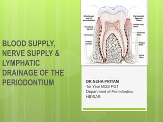
Blood supply,nerve supply and lymphatic drainage of the periodontium final
1. BLOOD SUPPLY,
NERVE SUPPLY &
LYMPHATIC
DRAINAGE OF THE
PERIODONTIUM DR.NEHA PRITAM
1sr Year MDS PGT
Department of Periodontics
HIDSAR
2. THE PERIODONTIUM
The periodontium is
defined as those
tissues supporting
and investing the
tooth and consists of
cementum,
periodontal ligament
(PDL), bone lining
the alveolus
(socket), and that
part of the gingiva
facing the tooth.
Ten Cate's Oral Histology,Development,Structure and Function
3. The normal periodontium provides
the support necessary to maintain
teeth in function.
Four principle components
1. Gingiva
2. Periodontal ligament
3. Cementum
4. Alveolar bone
5. INTRODUCTION TO VASCULAR SUPPLY
The orofacial structures have a very rich vascular
supply.
A very important artery that supplies the maxillary and
mandibular teeth and their associated periodontium is
the internal maxillary artery.
6. Internal maxillary artery
The maxillary artery or the internal maxilary artery is
one of the two terminal divisions of the external carotid
artery.
The second terminal branch being the superficial
temporal artery
7.
8.
9.
10. ARTERIAL SUPPLY OF MAXILLARY AND
MANDIBULAR teeth and their supporting structures
The mandibular teeth and their supporting structures
are supplied by branches of inferior alveolar (dental)
artery, including mental ,sublingual and buccal arteries.
The artery supply of maxillary teeth and their supporting
structure is by the posterior superior alveolar artery,
infraorbital artery, the greater palatine artery and the
sphenopalatine arteries.
11. Microcirculatory tracts, blood vessels, and lymphatic
vessels play an important role in drainage of tissue fluid
and in the spread of inflammation.
In gingivitis and periodontitis,
the microcirculation and
vascular formation change
greatly in the vascular network
directly under the gingival
sulcular epithelium and
junctional epithelium.
12. Blood vessels are easily evidenced in
tissue sections by means of immuno-
histochemical reactions against proteins
of endothelial cells (factor VIII and
adhesion molecules).
Before these techniques were developed,
vascularization patterns of periodontal
tissues had been described using histo-
enzymatic reactions for alkaline
phosphatase and adenosine
triphosphatase because of the great
activity of these enzymes in endothelial
13.
14. The gingiva receives its arterial
supply mainly from three sources:
Supraperiosteal arteries
Vessels of periodontal ligament
Arterioles emerging from crests of
interdental septa
15. Anatomic and histologic changes have been shown to
occur In the gingival microcirculation with gingivitis.
In the absence of inflammation, the vascular network is
arranged in a regular, repetitive, and layered pattern.
In contrast, the inflamed gingival vasculature exhibits an
irregular vascular plexus pattern, with the microvessels
exhibiting a looped, dilated, and convoluted
appearance.
16. Beneath the epithelium
on the outer gingival
surface, capillaries
extend into the
papillary connective
tissue between the
epithelial rete pegs in
the form of terminal
hairpin loops with
efferent and afferent
branches ,spirals and
varices.
17. Along the sulcular epithelium ,capillaries are arranged in
a flat,anastomosing plexus that extents parallel to the
enamel from the base of the sulcus to the gingival
margin.
In the col area,a mixed pattern of anastomosing
capillaries and loops occurs.
18.
19. Chronic periodontitis patients presented with
increased recruitment of neutrophils to the oral cavity.
Gene expression analysis revealed differences in the
expression levels of genes from several biological
pathways.
The apoptosis network was significantly altered in
patients with chronic inflammation in the oral cavity,
with up-regulation of pro-survival members of the Bcl-2
family and down-regulation of pro-apoptosis members
in the same compartment.
The percentages of viable neutrophils are significantly
increased in the oral cavity of chronic periodontitis
patients.
Lakschevitz FS, Aboodi GM, Glogauer M. Oral neutrophil transcriptome changes result in a pro-survival
phenotype in periodontal diseases. PloS one. 2013 Jul 11;8(7):e68983.
21. VENOUS AND LYMPHATIC DRAINAGE
The venous and lymphatic drainage of gingiva is closely
related to the arterial supply.
In the maxilla, the gingival lymphatic vessels drain into
the deep cervical lymph nodes.
In the mandible they drain into the
mental,submandibular and cervical lymph nodes.
23. LYMPHATIC SYSTEM
The lymphatic system is a network of lymph nodes are
connected lymphatic vessels, which plays an important role in
protecting the body from infection.
The role of the lymphatic system is removing excess fluids,
cellular and protein debris, microorganisms and other
elements and is important in controlling diffusion and
resolution of inflammatory process.
24. The lymphatic drainage of the gingiva brings in
the lymphatics of the connective tissue
papillae.It progresses into the collecting network
external to the periosteum of the alveolar
process, then to the regional lymph nodes,
particularly the submaxillary group.
In addition, lymphatics just beneath the
junctional epithelium extend into the periodontal
ligament and accompany the blood vessels.
25.
26. The lymphatic system is a part of the overall lymphoid
system of the body and a component of the immune
system of the body. It is an accumulation of tiny
channels or tubules with small nodular structures called
lymph nodes interconnecting them.
The system functions by returning fluids to the
bloodstream from the various tissues of the body.
27. Gingival lymphatics are crucial for transcapillary fluid
balance in the steady-state condition and during acute
perturbation.
Lymphangiogenesis takes place in gingiva during
periodontal disease development.
Moreover, gingival lymphatic vessels protect against P.
gingivalis induced periodontitis, probably by enhancing
clearance of bacterial products and promoting humoral
immune responses.
i
28.
29. SOME OF THE MAJOR LYMPH NODE
GROUPS IN THE HEAD AND NECK AREA
Retropharyngeal nodes
A group of nodes behind the throat wall and involved in
throat infections. These nodes drain to the upper deep
cervical lymph nodes.
30. FIG.LOCATION OF THE RETROPHARYNGEAL NODES
WITH RELTAION TO THE PHARYNX AND THE SKULL
BASE.
31. Submental nodes
Found beneath the chin.
The lymphatic channels from the mandibular incisors, the
tip of the tongue, and the midline of the lower lip and chin
drain into these nodes.
32. Submandibular nodes
grouped around the submandibular gland near the
angle of the mandible.
The areas that drain into these nodes are all of the
maxillary teeth, maxillary sinus, the mandibular canines
and all mandibular posterior teeth; the floor of the mouth
and most of the tongue; the cheek area; the hard
palate; and the anterior nasal cavity.
33.
34. Upper deep cervical nodes
A number of nodes drain into this node-The submandibular
nodes; the nodes behind the back throat wall, known as the
retropharyngeal nodes; the parotid nodes in front of the
ear and the parotid gland; and others drain into this.
35. Lower deep cervical nodes
They drain the upper deep cervical nodes and many of
the nodes at the back of the neck, frequently referred to
as occipital nodes, as well as some glands in the
anterior neck. From the lower deep cervical nodes, the
lymphatic fluid drains into the junction of the subclavian
and internal jugular veins.
36.
37. SPREAD OF DISEASES BY LYMPH NODES
The terms primary nodes, secondary nodes, and
tertiary nodes are often used in discussions about
infections and cancer, both of which spread through
lymphatic channels.
These terms refer to the groups of nodes that are
affected in a disease process.
39. Infections originating in
the middle of the lower
lip would spread first to
the submental nodes
Secondarily to the
submandibular nodes
Then to the upper deep
cervical nodes(which in
this instant would be
tertiary nodes of
involvement)
40. An understanding of this concept is necessary to
comprehend also the spread of oral cancer.
Each group of nodes acts as a resistance barrier against
the spread of cancer.
41. The nodes slow the spread, and if the cancer is
detected early enough, it can be treated more
successfully.
Once the infection or the cancer reaches the lower deep
cervical nodes and passes through them, it enters the
bloodstream, moving directly into the heart and then
throughout the body.
42. With this in mind, it is easy to understand why cancer on
the tip of the tongue does not result in as high a
mortality rate as does cancer that begins further back
on the tongue or in the throat.
43. The tip of the tongue generally drains through four
groups of nodes before it enters the bloodstream and
spreads throughout the body, whereas cancer in the
posterior portion of the tongue or in the throat travels to
the upper deep cervical nodes, on to the lower deep
cervical nodes, and into the bloodstream.
44. SPREAD OF INFECTIONS IN FASCIAL SPACES
Another way through which infections may spread is
through fascial spaces.
Although infection spread through fascial spaces is
much less common, it displays much more dramatic
clinical symptoms.
45. The spaces between muscle and tissue layers are referred to as fascial
layers or planes, and infections may spread here.
46. In general, dental infections start in the maxilla or
mandible at the apex of a tooth or in the periodontal
space around a tooth.
Most periodontal space infections cause a swelling of
the gingival or mucosal tissue within the oral cavity.
Infections at the apices of the teeth cause swelling in
one of two directions: buccal or lingual. Most buccal
swellings also lead to a swelling in the vestibule of the
oral cavity.
47. This swelling is sometimes referred to as a gumboil.
The infection comes to a pointed head, breaks through
the mucosa, and drains into the oral cavity.
49. Entry of infections into the buccal space is dependent on their
relationship to the attachment of the buccinator muscle.
50.
51. Infection spreading into the sublingual ,submandibular
space causes a swelling into the floor of the mouth. If it
spreads into the submental space, it will cause a
swelling beneath the chin, sometimes referred to as
Ludwig’s angina. These infections continue to spread
by gravity if not treated.
52. The importance of this section is not to be able to
completely describe or define the boundaries of these
spaces or potential spaces, but to understand how the
origin or location of the original infection determines the
pathway it will follow and the potential outcome if left
untreated.
53. MAXILLARY INFECTIONS
If the infection does not open into the maxillary buccal
vestibule or onto the palate, it may spread toward three
areas—the nasal cavity, the maxillary sinus, or the soft-
tissue spaces of the cheek or the area below the eye.
54. The area involved is related to the tooth involved. A
swelling below the eye is usually related to infection
from an anterior tooth, usually the maxillary canine,
whereas swelling in the cheek is usually related to
infection in a posterior tooth
56. Neural elements are extensively distributed throughout the
gingival tissues. Within the gingival connective tissues, most
nerve fibers are myelinated and are closely associated with
the blood vessels.
Gingival innervation is derived from fibers arising from nerves
in the periodontal ligament and from the labial, buccal, and
palatal nerves.
The following nerve structures are present in the connective
tissue: a meshwork of terminal argyrophilic fibers, some of
which extend into the epithelium; Meissner-type tactile
corpuscles; Krause-type end bulbs, which are temperature
receptors; and encapsulated spindles
57. The neural tissue in human periodontium is associated
with the terminal part of a nerve trunk from which
myelinated nerve fibres leave and in some instances
divide into three or more nerve fibres.
58. Innervations in various regions of the oral cavity is
supplied by the second and third divisions of the
trigeminal nerve.
59.
60. Innervations in various regions of the oral cavity is
supplied by the second and third divisions of the
trigeminal nerve.
Trigeminal nerves have sensory, motor, and
intermediate roots, which are mounted directly to the
brain.
61. The nerve supply of gingiva follow the vascular supply.
In the maxilla,the gingiva is supplied by the posterior
,middle and the anterior superior alvoelar
nerves,branches of infraorbital nerves,the greater
palatine nerve and nasopalatine nerve.
62. The middle superior alveolar nerve is present in 80% of
individuals
The buccal nerve supplies variably in the buccal molar
region.
63. In the mandible,gingiva is largerly suplied by the inferior
alveolar nerve.
The buccal nerve supplies buccal gingiva in relation to
molars and premolars.
Branches of lingual nerve supplies the lingual aspect of
all lower teeth.
64.
65. REFERENCE
Lakschevitz FS, Aboodi GM, Glogauer M. Oral neutrophil
transcriptome changes result in a pro-survival phenotype in
periodontal diseases. PloS one. 2013 Jul 11;8(7):e68983.
Griffin CJ, Harris R. Innervation of human periodontium I.
Classification of periodontal receptors. Australian dental
journal. 1974 Feb;19(1):51-6.
Carranza’s clinical periodontology
Newman Takei Klokkevoid Carranza
Ten Cate’s Oral Histology Developent Structure and Function
66. CONCLUSION
A thoughrough understanding of the anatomical
structures,vasculature and innervations of the
periodontium is necessary for the clinical point of view
and before making any surgical intervention in this area.
Editor's Notes
Proper functioning of the periodontium is achieved only through structural integrity and interaction between these various tissues.
Periodontal diseases are inflammatory processes that occur following the influx of neutrophils into the periodontal tissues in response to the subgingival bacterial biofilm. Current literature suggests that while neutrophils are protective and prevent bacterial infections, they also appear to contribute to damage of the periodontal tissues. In the present study we compare the gene expression profile changes in neutrophils as they migrate from the circulation into the oral tissues in patients with chronic periodontits and matched healthy subjects. We hypothesized that oral neutrophils in periodontal disease patients will display a disease specific transcriptome that differs from the oral neutrophil of healthy subjects.
Venous blood and oral rinse samples were obtained from healthy subjects and chronic periodontitis patients for neutrophil isolation. mRNA was isolated from the neutrophils, and gene expression microarray analysis was completed. Results were confirmed for specific genes of interest by qRT-PCR and Western Blot analysis.