Histone H2B is one of the 5 main histone proteins involved in the structure of chromatin in eukaryotic cells.
Histone H2B is a structural protein that helps organise eukaryotic DNA. It plays an important role in the biology of the nucleus where it is involved in the packaging and maintaining of chromosomes, regulation of transcription, and replication and repair of DNA. Histone H2B helps regulate chromatin structure and function through post-translational modifications and specialised histone variants.
The antibody detects endogenous Histone H2B (Di Methyl Lys43) protein.
Anti-Histone H2B (Di Methyl Lys43) Antibody- http://www.stjohnslabs.com/histone-h2b-di-methyl-lys43-antibody?filter_name=STJ97164
Join our Antibody Validation Project - http://www.stjohnslabs.com/services/antibody-validation
Histone H2B is one of the 5 main histone proteins involved in the structure of chromatin in eukaryotic cells.
Histone H2B is a structural protein that helps organise eukaryotic DNA. It plays an important role in the biology of the nucleus where it is involved in the packaging and maintaining of chromosomes, regulation of transcription, and replication and repair of DNA. Histone H2B helps regulate chromatin structure and function through post-translational modifications and specialised histone variants.
The antibody detects endogenous Histone H2B (Tri Methyl Lys43) protein.
Anti-Histone H2B (Tri Methyl Lys43) antibody - http://www.stjohnslabs.com/histone-h2b-tri-methyl-lys43-antibody?filter_name=STJ97165
Join our Antibody Validation Project - http://www.stjohnslabs.com/services/antibody-validation
Constitutively active protein kinase that acts as a negative regulator in the hormonal control of glucose homeostasis, Wnt signaling and regulation of transcription factors and microtubules, by phosphorylating and inactivating glycogen synthase (GYS1 or GYS2), EIF2B, CTNNB1/beta-catenin, APC, AXIN1, DPYSL2/CRMP2, JUN, NFATC1/NFATC, MAPT/TAU and MACF1. Requires primed phosphorylation of the majority of its substrates. In skeletal muscle, contributes to insulin regulation of glycogen synthesis by phosphorylating and inhibiting GYS1 activity and hence glycogen synthesis. May also mediate the development of insulin resistance by regulating activation of transcription factors. Regulates protein synthesis by controlling the activity of initiation factor 2B (EIF2BE/EIF2B5) in the same manner as glycogen synthase. In Wnt signaling, GSK3B forms a multimeric complex with APC, AXIN1 and CTNNB1/beta-catenin and phosphorylates the N-terminus of CTNNB1 leading to its degradation mediated by ubiquitin/proteasomes. Phosphorylates JUN at sites proximal to its DNA-binding domain, thereby reducing its affinity for DNA.
Anti-GSK3β-http://www.stjohnslabs.com/gsk3b-antibody-p-92568
Join our Antibody Validation Project - http://www.stjohnslabs.com/services/antibody-validation
PHD Finger Protein 19 is coded for by the PHF19 gene (also known as hPCL3). The Polycomb Group (PcG) is a key binding site within the protein that targets trimethylated Histone H3 at Lys-36, where it recruits the PRC2 complex. It is thought to be involved in the transition of embryonic stems cells from an active to repressed state, and research has indicated its overexpression in many types of cancers.
Customer Review For Tubulin-beta Polyclonal Antibody- IF Application (STJ31562)St John's Laboratory Ltd
Tubulin is the major constituent of microtubules. It binds two moles of GTP, one at an exchangeable site on the beta chain and one at a non-exchangeable site on the alpha chain. TUBB3 plays a critical role in proper axon guidance and maintenance.
To purchase this antibody use the following link: http://www.stjohnslabs.com/tubulin-beta-antibody?filter_name=STJ31562
Fukutin-related protein (FKRP) is targeted to the medial Golgi apparatus and is necessary for post-translational modification of dystroglycan. Mutations in the gene encoding this protein have been associated with congenital muscular dystrophy, mental retardation, and cerebellar cysts. Several alternatively spliced transcript variants of this gene have been described, but the full-length nature of some of these variants has not been determined.
To purchase this antibody use the following link: http://www.stjohnslabs.com/fkrp-antibody-p-95246?filter_name=FKRP
Western Blot Antibody Customer Review for Anti-RUNX1 Polyclonal Antibody (STJ...St John's Laboratory Ltd
RUNX1 protein (Runt-related transcription factor 1) also known as AML1 is a transcriptional corepressor encoded by the RUNX1 gene in humans. It regulates the differentiation of hematopoietic stem cells into mature blood cells via its association with DNA-binding transcription factors as well as its recruitment of other corepressors and histone-modifying enzymes.
GSK3β Polyclonal Antibody is an active protein kinase that acts as a negative regulator in the hormonal control of glucose homeostasis, Wnt signaling and regulation of transcription factors and microtubules, by phosphorylating and inactivating glycogen synthase (GYS1 or GYS2), EIF2B, CTNNB1/beta-catenin, APC, AXIN1, DPYSL2/CRMP2, JUN, NFATC1/NFATC, MAPT/TAU and MACF1. Requires primed phosphorylation of the majority of its substrates.
To purchase this antibody, use the following link: http://www.stjohnslabs.com/gsk3b-antibody-p-92568?filter_name=STJ93447
Western Blot Antibody Customer Review for Anti-RelB Polyclonal Antibody (STJ9...St John's Laboratory Ltd
The RelB protein is a transcription factor encoded by the RELB gene in humans. It is known to interact with NFKB2. The RelB polyclonal antibody detects endogenous levels of RelB protein.
Independent Antibody Validation For mCherry Tag Monoclonal Antibody (STJ34373)St John's Laboratory Ltd
St John’s Laboratory suppliers a highly photostable mCherry tag antibody, which is resistant to photobleaching. Independent validation reviews provide feedback on real-experimental use:
https://www.scienceexchange.com/validations/29688
https://www.scienceexchange.com/validations/29733
https://www.scienceexchange.com/validations/29708
To purchase this antibody, use the following link: http://www.stjohnslabs.com/mcherry-tag-antibody?filter_name=STJ34373
p70 S6 kinase or p70S6K is a serine/threonine kinase that acts downstream of PIP3 and phosphoinositide-dependent kinase-1 in the PI3 kinase pathway. As the name suggests, its target substrate is the S6 ribosomal protein. Phosphorylation of S6 induces protein synthesis at the ribosome.
The phosphorylation of P70S6K at threonine 389 has been used as a hallmark of activation by mTOR and correlated with autophagy inhibition in various situations. However, several recent studies suggest that the activity of P70S6K plays a more positive role in the increase of autophagy.
To purchase this antibody use the following link: http://www.stjohnslabs.com/p70-s6-kinase-antibody?filter_name=STJ31332
LPCAT1 antibody possess both acyltransferase and acetyltransferase activities. Activity is calcium-independent. Mediates the conversion of 1-acyl-sn-glycero-3-phosphocholine (LPC) into phosphatidylcholine (PC). Displays a clear preference for saturated fatty acyl-CoAs, and 1-myristoyl or 1-palmitoyl LPC as acyl donors and acceptors, respectively. May synthesize phosphatidylcholine in pulmonary surfactant, thereby playing a pivotal role in respiratory physiology.
Publication:
http://www.ncbi.nlm.nih.gov/pubmed/16864775
http://www.ncbi.nlm.nih.gov/pubmed/21498505
To purchase this antibody, use the following link: http://www.stjohnslabs.com/lpcat1-antibody-p-68679?filter_name=STJ27005
The Bcl-2-associated death promoter (BAD) protein is a pro-apoptotic member of the Bcl-2 gene family which is involved in initiating apoptosis. BAD is a member of the BH3-only family, a subfamily of the Bcl-2 family. After activation, it is able to form a heterodimer with anti-apoptotic proteins and prevent them from stopping apoptosis.
Anti-Bad - http://www.stjohnslabs.com/bad-antibody-p-91310?filter_name=STJ91797
Join our Antibody Validation Project - http://www.stjohnslabs.com/services/antibody-validation
Antibody Customer Review For Phospho-eIF2α (S51) Polyclonal Antibody (STJ90253) St John's Laboratory Ltd
eIF2 is an initiation factor found within eukaryotes. It mediates binding of tRNA to the ribosome in a GTP dependent manner. The alpha subunit is the main target for phosphorylation at Serine-51, and is considered the regulatory subunit of this trimeric (formed of eIF2a, eIF2b and eIF2y).
Visit www.stjohnslabs.com
NF-kappa-B is a pleiotropic transcription factor present in almost all cell types and is the endpoint of a series of signal transduction events that are initiated by a vast array of stimuli related to many biological processes such as inflammation, immunity, differentiation, cell growth, tumorigenesis and apoptosis. NF-kappa-B is a homo- or heterodimeric complex formed by the Rel-like domain-containing proteins RELA/p65, RELB, NFKB1/p105, NFKB1/p50, REL and NFKB2/p52 and the heterodimeric p65-p50 complex appears to be most abundant one. The dimers bind at kappa-B sites in the DNA of their target genes and the individual dimers have distinct preferences for different kappa-B sites that they can bind with distinguishable affinity and specificity. Different dimer combinations act as transcriptional activators or repressors, respectively.
To purchase this antibody, use the following link: http://www.stjohnslabs.com/nfkb-p65-antibody-p-93367?filter_name=STJ94468
Serine/threonine protein kinase which is a central regulator of cellular metabolism, growth and survival in response to hormones, growth factors, nutrients, energy and stress signals. MTOR directly or indirectly regulates the phosphorylation of at least 800 proteins. Functions as part of 2 structurally and functionally distinct signaling complexes mTORC1 and mTORC2 (mTOR complex 1 and 2). Activated mTORC1 up-regulates protein synthesis by phosphorylating key regulators of mRNA translation and ribosome synthesis. This includes phosphorylation of EIF4EBP1 and release of its inhibition toward the elongation initiation factor 4E (eiF4E). Moreover, phosphorylates and activates RPS6KB1 and RPS6KB2 that promote protein synthesis by modulating the activity of their downstream targets including ribosomal protein S6, eukaryotic translation initiation factor EIF4B, and the inhibitor of translation initiation PDCD4. Stimulates the pyrimidine biosynthesis pathway, both by acute regulation through RPS6KB1-mediated phosphorylation of the biosynthetic enzyme CAD, and delayed regulation, through transcriptional enhancement of the pentose phosphate pathway which produces 5-phosphoribosyl-1-pyrophosphate (PRPP), an allosteric activator of CAD at a later step in synthesis, this function is dependent on the mTORC1 complex. Regulates ribosome synthesis by activating RNA polymerase III-dependent transcription through phosphorylation and inhibition of MAF1 an RNA polymerase III-repressor.
To purchase this antibody, use the following link: http://www.stjohnslabs.com/mtor-antibody-p-93228?filter_name=STJ94280
A polyhistidine-tag is an amino acid motif in proteins that consists of at least six histidine (His) residues, often at the N- or C-terminus of the protein. It is also known as hexa histidine-tag, 6xHis-tag, His6 tag and by the trademarked name His-tag. Polyhistidine-tags are often used for affinity purification of polyhistidine-tagged recombinant proteins expressed in Escherichia coli and other prokaryotic expression systems.
To purchase this antibody, use the following link: http://www.stjohnslabs.com/his-tag-antibody-p-95792?filter_name=STJ90106
Component of the epithelial apical junction complex that may function as an homophilic cell adhesion molecule and is essential for tight junction integrity. Also involved in transepithelial migration of leukocytes through adhesive interactions with AMICA1/JAML a transmembrane protein of the plasma membrane of leukocytes. The interaction between both receptors also mediates the activation of gamma-delta T-cells, a subpopulation of T-cells residing in epithelia and involved in tissue homeostasis and repair. Upon epithelial CXADR-binding, AMICA1 induces downstream cell signaling events in gamma-delta T-cells through PI3-kinase and MAP kinases. It results in proliferation and production of cytokines and growth factors by T-cells that in turn stimulate epithelial tissues repair.
To purchase this antibody, use the following link: http://www.stjohnslabs.com/car-antibody?filter_name=STJ91996
Acetyl-Histone H2A (Lys5) Antibody detects endogenous levels of histone H2A only when acetylated at lysine 5.
To purchase this antibody use the following link: http://www.stjohnslabs.com/histone-h2a-acetyl-lys5-antibody?filter_name=STJ97171
Tubulin is the major constituent of microtubules. It binds two molecules of GTP, one at an exchangeable site on the beta chain and one at a non-exchangeable site on the alpha chain. TUBB3 plays a critical role in proper axon guidance and mantainance.
To purchase this antibody, use the following link: http://www.stjohnslabs.com/tubulin-beta-antibody?filter_name=STJ31562
Has both glyceraldehyde-3-phosphate dehydrogenase and nitrosylase activities, thereby playing a role in glycolysis and nuclear functions, respectively. Participates in nuclear events including transcription, RNA transport, DNA replication and apoptosis. Nuclear functions are probably due to the nitrosylase activity that mediates cysteine S-nitrosylation of nuclear target proteins such as SIRT1, HDAC2 and PRKDC. Modulates the organization and assembly of the cytoskeleton. Facilitates the CHP1-dependent microtubule and membrane associations through its ability to stimulate the binding of CHP1 to microtubules (By similarity). Glyceraldehyde-3-phosphate dehydrogenase is a key enzyme in glycolysis that catalyzes the first step of the pathway by converting D-glyceraldehyde 3-phosphate (G3P) into 3-phospho-D-glyceroyl phosphate. Component of the GAIT (gamma interferon-activated inhibitor of translation) complex which mediates interferon-gamma-induced transcript-selective translation inhibition in inflammation processes. Upon interferon-gamma treatment assembles into the GAIT complex which binds to stem loop-containing GAIT elements in the 3'-UTR of diverse inflammatory mRNAs (such as ceruplasmin) and suppresses their translation.
To purchase this antibody, use the following link: http://www.stjohnslabs.com/gapdh-antibody-p-94882?filter_name=STJ96417
p70S6 kinase or p70S6K is a serine/threonine kinase that acts downstream of PIP3 and phosphoinositide-dependent kinase-1 in the PI3 kinase pathway. As the name suggests, its target substrate is the S6 ribosomal protein. Phosphorylation of S6 induces protein synthesis at the ribosome.
The phosphorylation of P70S6K at threonine 389 has been used as a hallmark of activation by mTOR and correlated with autophagy inhibition in various situations. However, several recent studies suggest that the activity of P70S6K plays a more positive role in the increase of autophagy.
Phosphorylation of Thr229 in the catalytic domain and Thr389 in the linker domain are most critical for kinase function. Phosphorylation of Thr389, however, most closely correlates with p70 kinase activity in vivo. Prior phosphorylation of Thr389
is required for the action of phosphoinositide 3-dependent
protein kinase 1 (PDK1) on Thr229. Phosphorylation
of this site is stimulated by growth factors such as insulin,
EGF and FGF, as well as by serum and some G-proteincoupled receptor ligands, and is blocked by wortmannin, LY294002 (PI-3K inhibitor) and rapamycin (FRAP/mTOR inhibitor) .
To purchase this antibody use the following link: http://www.stjohnslabs.com/p70-s6-kinase-antibody-phospho-thr389412?filter_name=STJ30854
FAAH antibody degrades bioactive fatty acid amides like oleamide, the endogenous cannabinoid, anandamide and myristic amide to their corresponding acids, thereby serving to terminate the signaling functions of these molecules. Hydrolyzes polyunsaturated substrate anandamide preferentially as compared to monounsaturated substrates.
To purchase this antibody, use the following link: http://www.stjohnslabs.com/faah-antibody-p-65320?filter_name=STJ23602
Microtubule minus-end binding protein that acts as a regulator of non-centrosomal microtubule dynamics and organization. Specifically required for the biogenesis and the maintenance of zonula adherens by anchoring the minus-end of microtubules to zonula adherens and by recruiting the kinesin KIFC3 to those junctional sites. May regulate the nucleation and the polymerization of microtubules. Indirectly, through the microtubule cytoskeleton, may regulate the organization of cellular organelles including the Golgi and the early endosomes.
To purchase this antibody, use the following link: http://www.stjohnslabs.com/camsap3-antibody-n-term-p-78995?filter_name=STJ46575
Cdc2 Polyclonal Antibody plays a key role in the control of the eukaryotic cell cycle by modulating the centrosome cycle as well as mitotic onset; promotes G2-M transition, and regulates G1 progress and G1-S transition via association with multiple interphase cyclins.
To purchase this antibody, use the following link: http://www.stjohnslabs.com/cdc2-antibody-p-91597?filter_name=STJ92154
Activity of CDK2 is maximal during S phase and G2; activated by interaction with cyclin E during the early stages of DNA synthesis to permit G1-S transition, and subsequently activated by cyclin A2 (cyclin A1 in germ cells) during the late stages of DNA replication to drive the transition from S phase to mitosis, the G2 phase. EZH2 phosphorylation promotes H3K27me3 maintenance and epigenetic gene silencing. Phosphorylates CABLES1 (By similarity). Cyclin E/CDK2 prevents oxidative stress-mediated Ras-induced senescence by phosphorylating MYC. Involved in G1-S phase DNA damage checkpoint that prevents cells with damaged DNA from initiating mitosis; regulates homologous recombination-dependent repair by phosphorylating BRCA2, this phosphorylation is low in S phase when recombination is active, but increases as cells progress towards mitosis. In response to DNA damage, double-strand break repair by homologous recombination a reduction of CDK2-mediated BRCA2 phosphorylation. Phosphorylation of RB1 disturbs its interaction with E2F1. NPM1 phosphorylation by cyclin E/CDK2 promotes its dissociates from unduplicated centrosomes, thus initiating centrosome duplication. Cyclin E/CDK2-mediated phosphorylation of NPAT at G1-S transition and until prophase stimulates the NPAT-mediated activation of histone gene transcription during S phase. Required for vitamin D-mediated growth inhibition by being itself inactivated. Involved in the nitric oxide- (NO) mediated signaling in a nitrosylation/activation-dependent manner. USP37 is activated by phosphorylation and thus triggers G1-S transition.
To purchase this antibody, use the following link: http://www.stjohnslabs.com/cdk2-antibody-p-91630?filter_name=STJ92197
Equilibrative nucleoside transporter 1 (ENT1) is a protein that in humans is encoded by the SLC29A1 gene. Multiple alternatively spliced variants, encoding the same protein, have been found for this gene.
This gene is a member of the equilibrative nucleoside transporter family. The gene encodes a transmembrane glycoprotein that localizes to the plasma and mitochondrial membranes and mediates the cellular uptake of nucleosides from the surrounding medium. The protein is categorized as an equilibrative (as opposed to concentrative) transporter that is sensitive to inhibition by nitrobenzylmercaptopurine ribonucleoside (NBMPR). Nucleoside transporters are required for nucleotide synthesis in cells that lack de novo nucleoside synthesis pathways, and are also necessary for the uptake of cytotoxic nucleosides used for cancer and viral chemotherapies.
To purchase this antibody, use the following link: http://www.stjohnslabs.com/ent1-antibody?filter_name=STJ96396
Western Blot Customer Review Anti Glucocorticoid Receptor antibody (STJ97101)St John's Laboratory Ltd
Receptor for glucocorticoids (GC). Has a dual mode of action: as a transcription factor that binds to glucocorticoid response elements (GRE), both for nuclear and mitochondrial DNA, and as a modulator of other transcription factors. Affects inflammatory responses, cellular proliferation and differentiation in target tissues. Involved in chromatin remodeling . Plays a role in rapid mRNA degradation by binding to the 5' UTR of target mRNAs and interacting with PNRC2 in a ligand-dependent manner. Could act as a coactivator for STAT5-dependent transcription upon growth hormone (GH) stimulation and could reveal an essential role of hepatic GR in the control of body growth (By similarity). Has transcriptional activation and repression activity . Mediates glucocorticoid-induced apoptosis . Promotes accurate chromosome segregation during mitosis . May act as a tumor suppressor . May play a negative role in adipogenesis through the regulation of lipolytic and antilipogenic gene expression (By similarity). / Isoform Beta: Acts as a dominant negative inhibitor of isoform Alpha . Has intrinsic transcriptional activity independent of isoform Alpha when both isoforms are coexpressed.
Join our Antibody Validation Project: http://www.stjohnslabs.com/services/antibody-validation
Anti glucocorticoid receptor antibody (STJ97101):
http://www.stjohnslabs.com/glucocorticoid-receptor-antibody-p-98736?filter_name=STJ97101
STJ91730 is a polyclonal antibody derived from rabbit host.
Apoptosis-associated speck-like protein containing CARD (ASC) holds a key function in mediating large signalling complexes during inflammation and apoptotic pathways.
To buy this antibody please use this link. http://www.stjohnslabs.com/asc-antibody-p-91257?filter_name=ASC%20antibody
Calcium/calmodulin-dependent protein kinase type II beta chain is an enzyme that in humans is encoded by the CAMK2B gene.
Calcium/calmodulin-dependent protein kinase that functions autonomously after Ca2+/calmodulin-binding and autophosphorylation, and is involved in sarcoplsamic reticulum Ca2+ transport in skeletal muscle and may function in dendritic spine and synapse formation and neuronal plasticity.
Anti-CaMKIIβ/γ - http://www.stjohnslabs.com/camkiibg-antibody?filter_name=Stj91990
Join our Antibody Validation Project - http://www.stjohnslabs.com/services/antibody-validation
Western Blot Antibody Customer Review for Anti-ALK Polyclonal Antibody (STJ91...St John's Laboratory Ltd
Anaplastic lymphoma kinase also known as ALK tyrosine kinase receptor or CD246 (cluster of differentiation 246) is an enzyme that in humans is encoded by the ALK gene.
ALK plays an important role in the development of the brain and exerts its effects on specific neurons in the nervous system. Transduces signals from ligands at the cell surface, through specific activation of the mitogen-activated protein kinase (MAPK) pathway.The antibody was affinity-purified from rabbit antiserum by affinity-chromatography using epitope-specific immunogen.
Anti-ALK - http://www.stjohnslabs.com/alk-antibody-p-91127?filter_name=STJ91562
Join our Antibody Validation Project - http://www.stjohnslabs.com/services/antibody-validation
Tubulin is the major constituent of microtubules. It binds two moles of GTP, one at an exchangeable site on the beta chain and one at a non-exchangeable site on the alpha chain. TUBB3 plays a critical role in proper axon guidance and mantainance.
Anti-Tubulin β -http://www.stjohnslabs.com/tubulin-b-antibody
Join our Antibody Validation Project - http://www.stjohnslabs.com/services/antibody-validation
KDM5 epigenetic modifiers as a focus for drug discoveryChristopher Wynder
A summary presentation of my scientific work.
My laboratory focused on an enzyme KDM5b (aka PLU-1, JARID1b) that was widely expressed during development and played a key role in progression of breast cancer through HER-2.
My lab focused on understanding the key biochemical activity of the enzyme through dissecting the proteomic and genomic interactors.
Our results were confirmed through the use of ES cells, adult stem cells and mouse models.
Much of this work remains unpublished, please contact me for more information and/or access to any reagents that I still have as part of this work.
crwynder@gmail.com
Glial fibrillary acidic protein is an intermediate filament (IF) protein that is expressed by numerous cell types of the central nervous system (CNS) including astrocytes and ependymal cells. GFAP has also been found to be expressed in glomeruli and peritubular fibroblasts taken from rat kidneys Leydig cells of the testis in both hamsters and humans, human keratinocytes, human osteocytes and chondrocytes and stellate cells of the pancreas and liver in rats. First described in 1971, GFAP is a type III IF protein that maps, in humans, to 17q21.[13] It is closely related to its non-epithelial family members, vimentin, desmin, and peripherin, which are all involved in the structure and function of the cell’s cytoskeleton. GFAP is thought to help to maintain astrocyte mechanical strength,[14] as well as the shape of cells but its exact function remains poorly understood, despite the number of studies using it as a cell marker.
Anti-GFAP -http://www.stjohnslabs.com/gfap-antibody-p-98596
Join our Antibody Validation Project - http://www.stjohnslabs.com/services/antibody-validation
Tubulin in molecular biology can refer either to the tubulin protein superfamily of globular proteins, or one of the member proteins of that superfamily. α- and β-tubulins polymerize into microtubules, a major component of the eukaryotic cytoskeleton. Microtubules function in many essential cellular processes, including mitosis. Tubulin-binding drugs kill cancerous cells by inhibiting microtubule dynamics, which are required for DNA segregation and therefore cell division. In eukaryotes there are six members of the tubulin superfamily, although not all are present in all species (see below).[2][3] Both α and β tubulins have a mass of around 50 kDa and are thus in a similar range compared to actin with ~42 kDa. In contrast, tubulin polymers (microtubules) tend to be much bigger than actin filaments due to their cylindrical nature.
Anti-β-Tubulin -http://www.stjohnslabs.com/b-tubulin-antibody-p-
98567
Join our Antibody Validation Project - http://www.stjohnslabs.com/services/antibody-validation
Glial fibrillary acidic protein is an intermediate filament (IF) protein that is expressed by numerous cell types of the central nervous system (CNS) including astrocytes and ependymal cells. GFAP has also been found to be expressed in glomeruli and peritubular fibroblasts taken from rat kidneys Leydig cells of the testis in both hamsters and humans, human keratinocytes, human osteocytes and chondrocytes and stellate cells of the pancreas and liver in rats. First described in 1971, GFAP is a type III IF protein that maps, in humans, to 17q21.[13] It is closely related to its non-epithelial family members, vimentin, desmin, and peripherin, which are all involved in the structure and function of the cell’s cytoskeleton. GFAP is thought to help to maintain astrocyte mechanical strength,[14] as well as the shape of cells but its exact function remains poorly understood, despite the number of studies using it as a cell marker.
Anti-GFAP -http://www.stjohnslabs.com/gfap-antibody-p-98596
Join our Antibody Validation Project - http://www.stjohnslabs.com/services/antibody-validation
Tubulin is the major constituent of microtubules. It binds two moles of GTP, one at an exchangeable site on the beta chain and one at a non-exchangeable site on the alpha chain. TUBB3 plays a critical role in proper axon guidance and mantainance.
Anti-β-Tubulin-http://www.stjohnslabs.com/b-tubulin-antibody-p-98567
Join our Antibody Validation Project - http://www.stjohnslabs.com/services/antibody-validation
Western Blot Antibody Customer Review for ADNP Polyclonal Antibody (STJ91502)St John's Laboratory Ltd
ADNP (Activity-Dependent Neuroprotector Homeobox) is expressed through the ADNP gene in humans. It is a possible transcription factor through having a homeobox and 9 zinc finger domains. It is said to have both stimulatory and inhibitory growth effects on different tumour cells. The ADNP Polyclonal Antibody detects endogenous levels of ADNP protein.
Western Blot Antibody Customer Review for Anti-PDHA1 Polyclonal Antibody (STJ...St John's Laboratory Ltd
Pyruvate dehydrogenase E1 component subunit alpha, somatic form, mitochondrial is an enzyme that in humans is encoded by the PDHA1 gene.
The pyruvate dehydrogenase complex is responsible for the oxidative decarboxylation of pyruvate, with the final product being Acetyl CoA.
Mutations in the PDHA1 gene have been known to cause one form of pyruvate dehydrogenase deficiency. Pyruvate dehydrogenase deficiency is characterized by the buildup of a chemical called lactic acid in the body and a variety of neurological problems.
The antibody was affinity-purified from rabbit antiserum by affinity-chromatography using epitope-specific immunogen.
Anti-PDHA1 - http://www.stjohnslabs.com/pdha1-antibody-p-93852?filter_name=STJ95006
Join our Antibody Validation Project - http://www.stjohnslabs.com/services/antibody-validation
Western Blot Antibody Customer Review for Anti-Histone H3 (Tri methyl K79) An...St John's Laboratory Ltd
Histone H3 is one of the five main histone proteins involved in the structure of chromatin in eukaryotic cells. Core component of nucleosome. Nucleosomes wrap and compact DNA into chromatin, limiting DNA accessibility to the cellular machineries which require DNA as a template. Histones thereby play a central role in transcription regulation, DNA repair, DNA replication and chromosomal stability. DNA accessibility is regulated via a complex set of post-translational modifications of histones, also called histone code, and nucleosome remodeling.
Histone H3 is an important protein in the emerging field of epigenetics, where its sequence variants and variable modification states are thought to play a role in the dynamic and long term regulation of genes.
Anti-Histone H3 (Tri methyl K79) - http://www.stjohnslabs.com/histone-h3-tri-methyl-k79-antibody-p-98628?filter_name=STJ96993
Join our Antibody Validation Project - http://www.stjohnslabs.com/services/antibody-validation
Tau are stabilising proteins for axonal microtubules within the Central Nervous System, and as such are also recognised as Mictrotubule-associated Protein (MAP). Tau is predominantly active in the distal portions of axons to carry out its function.
Western Blot Antibody Customer Review for Anti-Endoplasmin Polyclonal Antibod...St John's Laboratory Ltd
The Endoplasmin protein (also known as HSP90B1) is a molecular chaperone encoded by the HSP90B1 gene in humans. It plays a role in the processing and transport of proteins such as integrins and Toll-like receptors. It is located in the endoplasmic reticulum. The Endoplasmin polyclonal antibody detects endogenous levels of Endoplasmin protein.
Western Blot Antibody Customer Review for Anti-PKM2 Polyclonal Antibody (STJ9...St John's Laboratory Ltd
Pyruvate kinase isozymes M1/M2 (PKM1/M2), also known as pyruvate kinase muscle isozyme (PKM), pyruvate kinase type K, cytosolic thyroid hormone-binding protein (CTHBP), thyroid hormone-binding protein 1 (THBP1), or opa-interacting protein 3 (OIP3), is an enzyme that in humans is encoded by the PKM2 gene.
Is a glycolytic enzyme that catalyzes the transfer of a phosphoryl group from phosphoenolpyruvate (PEP) to ADP, generating ATP. It plays a general role in caspase independent cell death of tumor cells.
The antibody was affinity-purified from rabbit antiserum by affinity-chromatography using epitope-specific immunogen.
Anti-PMK2 - http://www.stjohnslabs.com/pkm2-antibody-p-93954?filter_name=Stj95142
Join our Antibody Validation Project - http://www.stjohnslabs.com/services/antibody-validation
Immunofluorescence Antibody Customer Review for Anti-CEP290 antibody (STJ92224) St John's Laboratory Ltd
Centrosomal protein of 290 kDa is a protein that in humans is encoded by the CEP290 gene
The gene CEP290 is a centrosomal protein that plays an important role in centrosome and cilia development. This gene is vital in the formation of the primary cilium, a small antenna-like projections of the cell membrane that plays an important role in the photoreceptors at the back of the retina (which detect light and color) and in the kidney, brain, and many other organs of the body. Knocking down levels of the CEP290 gene transcript resulted in dramatic suppression of ciliogenesis in retinal pigment epithelial cells in culture, proving just how important CEP290 is to cilia formation.
CEP290 Polyclonal Antibody detects endogenous levels of CEP290 protein.
Anti-CEP290 - http://www.stjohnslabs.com/cep290-antibody-p-91648?filter_name=STJ92224
Join our Antibody Validation Project - http://www.stjohnslabs.com/services/antibody-validation
The Kv2.1 protein or potassium voltage-gated channel subfamily B member 1 is a voltage gated potassium channel that mediates transmembrane transport of potassium in excitable membranes located in the brain, cardiovascular system and the pancreas. The protein is coded by the KCNB1 gene in humans. The Kv2.1 polyclonal antibody detects endogenous levels of Kv2.1 protein.
Anti-Kv2.1 antibody - http://www.stjohnslabs.com/kv21-antibody?filter_name=STJ93873
Join Our Antibody Validation Project - http://www.stjohnslabs.com/services/antibody-validation
The Axin-related protein, Axin2, presumably plays an important role in the regulation of the stability of beta-catenin in the Wnt signaling pathway, like its rodent homologs, mouse conductin/rat axil. In mouse, conductin organizes a multiprotein complex of APC (adenomatous polyposis of the colon), beta-catenin, glycogen synthase kinase 3-beta, and conductin, which leads to the degradation of beta-catenin.
To purchase this antibody, use the following link: http://www.stjohnslabs.com/axin2-antibody-p-67883?filter_name=STJ26209
In Cell-Western Antibody Customer Review for Anti-Beclin-1 Antibody (STJ97761)St John's Laboratory Ltd
Beclin-1 is a protein that in humans is encoded by the BECN1 gene. Beclin-1 plays an important role in tumorigenesis, and neurodegeneration, being implicated in the autophagic programmed cell death. Beclin-1-C 35 kDa localized to mitochondria can promote apoptosis; it induces the mitochondrial translocation of BAX and the release of proapoptotic factors.
Anti-Beclin-1 - http://www.stjohnslabs.com/beclin-1-antibody-2?filter_name=stj97761
Join our Antibody Validation Project - http://www.stjohnslabs.com/services/antibody-validation
In Cell-Western Antibody Customer Review for Anti-LC3A Antibody (STJ97755)St John's Laboratory Ltd
Microtubule-associated proteins 1A/1B light chain 3A is a protein that in humans is encoded by the MAP1LC3A gene
Whereas LC3s are involved in elongation of the phagophore membrane, the GABARAP/GATE-16 subfamily is essential for a later stage in autophagosome maturation.
Anti-LC3A - http://www.stjohnslabs.com/lc3a-antibody?filter_name=STJ97755
Join our Antibody Validation Project - http://www.stjohnslabs.com/services/antibody-validation
Western Blot Antibody Customer Review for Anti-Peroxin 2 Antibody (STJ95040)St John's Laboratory Ltd
PEX2 is gene on chromosome 8q21.1 that encodes a peroxin, an integral membrane protein involved in peroxisome biosynthesis and integrity, which assembles membrane vesicles before translocation of matrix proteins.
Defects of PEX2 cause peroxisome biogenesis disorder complementation group 5, Zelleger syndrome and a form of Refsum disease.
Anti-Peroxin 2 - http://www.stjohnslabs.com/peroxin-2-antibody?filter_name=STJ95040
Join our Antibody Validation Project - http://www.stjohnslabs.com/services/antibody-validation
Western Blot Antibody Customer Review for Anti-Histone H3 (Acetyl Lys14) Ant...St John's Laboratory Ltd
Histone H3 is one of the five main histone proteins involved in the structure of chromatin in eukaryotic cells
Histones play a central role in transcription regulation, DNA repair, DNA replication and chromosomal stability. DNA accessibility is regulated via a complex set of post-translational modifications of histones, also called histone code, and nucleosome remodeling.
Anti-Histone H3 (Acetyl Lys14) Antibody - http://www.stjohnslabs.com/histone-h3-acetyl-lys14-antibody?filter_name=Stj97144
Join our Antibody Validation Project - http://www.stjohnslabs.com/services/antibody-validation
Acts as decoy receptor for TNFSF11/RANKL and thereby neutralizes its function in osteoclastogenesis. Inhibits the activation of osteoclasts and promotes osteoclast apoptosis in vitro. Bone homeostasis seems to depend on the local ratio between TNFSF11 and TNFRSF11B. May also play a role in preventing arterial calcification. May act as decoy receptor for TNFSF10/TRAIL and protect against apoptosis. TNFSF10/TRAIL binding blocks the inhibition of osteoclastogenesis.
Anti-OPG-http://www.stjohnslabs.com/opg-antibody
Join our Antibody Validation Project - http://www.stjohnslabs.com/services/antibody-validation
In Cell-Western Antibody Customer Review for Anti-MAP LC3β antibody (STJ97398)St John's Laboratory Ltd
Microtubule-associated proteins 1A/1B light chain 3A is a protein that in humans is encoded by the MAP1LC3A gene. MAP1A and MAP1B are microtubule-associated proteins which mediate the physical interactions between microtubules and components of the cytoskeleton. Plays a role in mitophagy which contributes to regulate mitochondrial quantity and quality by eliminating the mitochondria to a basal level to fulfill cellular energy requirements and preventing excess ROS production.
Anti-MAP LC3β - http://www.stjohnslabs.com/map-lc3b-antibody?filter_name=STJ97398
Join our Antibody Validation Project - http://www.stjohnslabs.com/services/antibody-validation
Metabolic-stress sensing protein kinase that phosphorylates the alpha subunit of eukaryotic translation initiation factor 2 (eIF-2-alpha/EIF2S1) on 'Ser-52' during the unfolded protein response (UPR) and in response to low amino acid availability. Converts phosphorylated eIF-2-alpha/EIF2S1 either in a global protein synthesis inhibitor, leading to a reduced overall utilization of amino acids, or to a translation initiation activator of specific mRNAs, such as the transcriptional activator ATF4, and hence allowing ATF4-mediated reprogramming of amino acid biosynthetic gene expression to alleviate nutrient depletion. Serves as a critical effector of unfolded protein response (UPR)-induced G1 growth arrest due to the loss of cyclin-D1 (CCND1). Involved in control of mitochondrial morphology and function. / ATP + a protein = ADP + a phosphoprotein. / Perturbation in protein folding in the endoplasmic reticulum (ER) promotes reversible dissociation from HSPA5/BIP and oligomerization, resulting in transautophosphorylation and kinase activity induction.
Anti-PERK-http://www.stjohnslabs.com/perk-antibody
Join our Antibody Validation Project - http://www.stjohnslabs.com/services/antibody-validation
Acts as a tumor suppressor in many tumor types; induces growth arrest or apoptosis depending on the physiological circumstances and cell type. Involved in cell cycle regulation as a trans-activator that acts to negatively regulate cell division by controlling a set of genes required for this process. One of the activated genes is an inhibitor of cyclin-dependent kinases. Apoptosis induction seems to be mediated either by stimulation of BAX and FAS antigen expression, or by repression of Bcl-2 expression. In cooperation with mitochondrial PPIF is involved in activating oxidative stress-induced necrosis; the function is largely independent of transcription. Induces the transcription of long intergenic non-coding RNA p21 (lincRNA-p21) and lincRNA-Mkln1. LincRNA-p21 participates in TP53-dependent transcriptional repression leading to apoptosis and seem to have to effect on cell-cycle regulation. Implicated in Notch signaling cross-over. Prevents CDK7 kinase activity when associated to CAK complex in response to DNA damage, thus stopping cell cycle progression.
Anti-Phospho-p53 (T18)-http://www.stjohnslabs.com/phospho-p53-t18-antibody
Join our Antibody Validation Project - http://www.stjohnslabs.com/services/antibody-validation
Western Blot Antibody Customer Review for Anti-Phospho-ALK (Y1507) Polyclonal...St John's Laboratory Ltd
Anaplastic lymphoma kinase (ALK) also known as ALK tyrosine kinase receptor or CD246 (cluster of differentiation 246) is an enzyme that in humans is encoded by the ALK gene.
ALK plays an important role in the development of the brain and exerts its effects on specific neurons in the nervous system. Transduces signals from ligands at the cell surface, through specific activation of the mitogen-activated protein kinase (MAPK) pathway. Phospho-ALK (Y1507) Polyclonal Antibody detects endogenous levels of ALK protein only when phosphorylated at Y1507.
Anti-Phospho-ALK - http://www.stjohnslabs.com/phospho-alk-y1507-antibody?filter_name=STJ90845
Join our Antibody Validation Project - http://www.stjohnslabs.com/services/antibody-validation
Western Blot Antibody Customer Review for Anti-p47-phox Antibody (STJ94885)St John's Laboratory Ltd
Neutrophil cytosol factor 1, also known as p47phox, is a protein that in humans is encoded by the NCF1 gene.
The protein encoded by this gene is a 47 kDa cytosolic subunit of neutrophil NADPH oxidase. This oxidase is a multicomponent enzyme that is activated to produce superoxide anion. Mutations in this gene have been associated with chronic granulomatous disease. p47 is vital to the activation of NADPH oxidase. P47 becomes heavily phosphorylated.
Anti-p47-phox - http://www.stjohnslabs.com/p47-phox-antibody-p-93740?filter_name=stj94885
Join our Antibody Validation Project - http://www.stjohnslabs.com/services/antibody-validation
Facilitative glucose transporter. This isoform may be responsible for constitutive or basal glucose uptake. Has a very broad substrate specificity; can transport a wide range of aldoses including both pentoses and hexoses.
Anti-Glut1-http://www.stjohnslabs.com/glut1-antibody-p-92472
Join our Antibody Validation Project - http://www.stjohnslabs.com/services/antibody-validation
Similar to Peptide Dot Blotting Customer Review for Anti-Histone H2B (Di Methyl Lys43) Antibody (STJ97164) (20)
G protein-coupled receptor that probably associates with the patched protein (PTCH) to transduce the hedgehog's proteins signal. Binding of sonic hedgehog (SHH) to its receptor patched is thought to prevent normal inhibition by patched of smoothened (SMO). Required for the accumulation of KIF7 and GLI3 in the cilia.
Anti-Smo antibody (STJ95710): http://www.stjohnslabs.com/smo-antibody-p-94371?filter_name=STJ95710
Join our Antibody Validation Project - http://www.stjohnslabs.com/services/antibody-validation
Western Blot Customer Review Anti-Phospho-Cofilin (S3) Antibody (STJ90230)St John's Laboratory Ltd
Binds to F-actin and exhibits pH-sensitive F-actin depolymerizing activity. Regulates actin cytoskeleton dynamics. Important for normal progress through mitosis and normal cytokinesis. Plays a role in the regulation of cell morphology and cytoskeletal organization. Required for the up-regulation of atypical chemokine receptor ACKR2 from endosomal compartment to cell membrane, increasing its efficiency in chemokine uptake and degradation.
Anti-Phospho-Cofilin (S3) -http://www.stjohnslabs.com/phospho-cofilin-s3-antibody
Join our Antibody Validation Project - http://www.stjohnslabs.com/services/antibody-validation
This June, Dr. Byron Baron from the University of Malta, Malta, is our Scientist of the Month! He's shared with us his research highlights, his current projects and some comments on the biotechnology industry.
Want to be our Scientist of the Month? Contact info@stjohnslabs.com
Downstream effector molecule involved in the transmission of signals from tyrosine kinase receptors and small GTPases to the actin cytoskeleton. Promotes formation of actin filaments. Part of the WAVE complex that regulates lamellipodia formation. The WAVE complex regulates actin filament reorganization via its interaction with the Arp2/3 complex.
Anti-WAVE2 -http://www.stjohnslabs.com/wave2-antibody
Join our Antibody Validation Project - http://www.stjohnslabs.com/services/antibody-validation
Implicated in synaptic vesicle endocytosis. May recruit other proteins to membranes with high curvature.
Brain, mostly in frontal cortex. Expressed at high level in fetal cerebellum.
Anti-Endophilin I -http://www.stjohnslabs.com/endophilin-i-antibody
Join our Antibody Validation Project - http://www.stjohnslabs.com/services/antibody-validation
Tubulin is the major constituent of microtubules. It binds two moles of GTP, one at an exchangeable site on the beta chain and one at a non-exchangeable site on the alpha chain. TUBB3 plays a critical role in proper axon guidance and mantainance.
Anti-β-tubulin -http://www.stjohnslabs.com/b-tubulin-antibody-p-98672
Join our Antibody Validation Project - http://www.stjohnslabs.com/services/antibody-validation
Most upstream protease of the activation cascade of caspases responsible for the TNFRSF6/FAS mediated and TNFRSF1A induced cell death. Binding to the adapter molecule FADD recruits it to either receptor. The resulting aggregate called death-inducing signaling complex (DISC) performs CASP8 proteolytic activation. The active dimeric enzyme is then liberated from the DISC and free to activate downstream apoptotic proteases. Proteolytic fragments of the N-terminal propeptide (termed CAP3, CAP5 and CAP6) are likely retained in the DISC. Cleaves and activates CASP3, CASP4, CASP6, CASP7, CASP9 and CASP10. May participate in the GZMB apoptotic pathways. Cleaves ADPRT. Hydrolyzes the small-molecule substrate, Ac-Asp-Glu-Val-Asp-|-AMC. Likely target for the cowpox virus CRMA death inhibitory protein. Isoform 5, isoform 6, isoform 7 and isoform 8 lack the catalytic site and may interfere with the pro-apoptotic activity of the complex. / Strict requirement for Asp at position P1 and has a preferred cleavage sequence of (Leu/Asp/Val)-Glu-Thr-Asp-|-(Gly/Ser/Ala). / Inhibited by the effector protein NleF that is produced by pathogenic E.coli; this inhibits apoptosis.
Anti-Caspase-8-http://www.stjohnslabs.com/caspase-8-antibody-p-99045
Join our Antibody Validation Project - http://www.stjohnslabs.com/services/antibody-validation
Tubulin is the major constituent of microtubules. The gamma chain is found at microtubule organizing centers (MTOC) such as the spindle poles or the centrosome. Pericentriolar matrix component that regulates alpha/beta chain minus-end nucleation, centrosome duplication and spindle formation.
Anti-Gamma Tubulin-http://www.stjohnslabs.com/gamma-tubulin-antibody
Join our Antibody Validation Project - http://www.stjohnslabs.com/services/antibody-validation
Tubulin in molecular biology can refer either to the tubulin protein superfamily of globular proteins, or one of the member proteins of that superfamily. α- and β-tubulins polymerize into microtubules, a major component of the eukaryotic cytoskeleton. Microtubules function in many essential cellular processes, including mitosis. Tubulin-binding drugs kill cancerous cells by inhibiting microtubule dynamics, which are required for DNA segregation and therefore cell division. In eukaryotes there are six members of the tubulin superfamily, although not all are present in all species (see below). Both α and β tubulins have a mass of around 50 kDa and are thus in a similar range compared to actin with ~42 kDa. In contrast, tubulin polymers (microtubules) tend to be much bigger than actin filaments due to their cylindrical nature. Tubulin was long thought to be specific to eukaryotes. More recently, however, several prokaryotic proteins have been shown to be related to tubulin.
Anti-Epsilon Tubulin -http://www.stjohnslabs.com/epsilon-tubulin-antibody-2
Join our Antibody Validation Project - http://www.stjohnslabs.com/services/antibody-validation
Ubiquitin-like modifier involved in formation of autophagosomal vacuoles (autophagosomes) . Whereas LC3s are involved in elongation of the phagophore membrane, the GABARAP/GATE-16 subfamily is essential for a later stage in autophagosome maturation .
Anti-LC3A-http://www.stjohnslabs.com/lc3a-antibody
Join our Antibody Validation Project - http://www.stjohnslabs.com/services/antibody-validation
Multifunctional transcription factor in ER stress response. Plays an essential role in the response to a wide variety of cell stresses and induces cell cycle arrest and apoptosis in response to ER stress. Plays a dual role both as an inhibitor of CCAAT/enhancer-binding protein (C/EBP) function and as an activator of other genes. Acts as a dominant-negative regulator of C/EBP-induced transcription: dimerizes with members of the C/EBP family, impairs their association with C/EBP binding sites in the promoter regions, and inhibits the expression of C/EBP regulated genes. Positively regulates the transcription of TRIB3, IL6, IL8, IL23, TNFRSF10B/DR5, PPP1R15A/GADD34, BBC3/PUMA, BCL2L11/BIM and ERO1L. Negatively regulates; expression of BCL2 and MYOD1, ATF4-dependent transcriptional activation of asparagine synthetase (ASNS), CEBPA-dependent transcriptional activation of hepcidin (HAMP) and CEBPB-mediated expression of peroxisome proliferator-activated receptor gamma (PPARG).
Anti-CHOP-http://www.stjohnslabs.com/chop-antibody-2
Join our Antibody Validation Project - http://www.stjohnslabs.com/services/antibody-validation
Pseudokinase that plays a key role in TNF-induced necroptosis, a programmed cell death process. Activated following phosphorylation by RIPK3, leading to homotrimerization, localization to the plasma membrane and execution of programmed necrosis characterized by calcium influx and plasma membrane damage. Does not have protein kinase activity.
Anti-phospho-MLKL (S358)-http://www.stjohnslabs.com/phospho-mlkl-s358-antibody-1
Join our Antibody Validation Project - http://www.stjohnslabs.com/services/antibody-validation
Serine/threonine kinase which acts as an essential component of the MAP kinase signal transduction pathway. MAPK1/ERK2 and MAPK3/ERK1 are the 2 MAPKs which play an important role in the MAPK/ERK cascade. They participate also in a signaling cascade initiated by activated KIT and KITLG/SCF. Depending on the cellular context, the MAPK/ERK cascade mediates diverse biological functions such as cell growth, adhesion, survival and differentiation through the regulation of transcription, translation, cytoskeletal rearrangements. The MAPK/ERK cascade plays also a role in initiation and regulation of meiosis, mitosis, and postmitotic functions in differentiated cells by phosphorylating a number of transcription factors. About 160 substrates have already been discovered for ERKs. Many of these substrates are localized in the nucleus, and seem to participate in the regulation of transcription upon stimulation.
Anti-ERK1-http://www.stjohnslabs.com/erk1-antibody-3
Join our Antibody Validation Project - http://www.stjohnslabs.com/services/antibody-validation
Tyrosine-protein kinase that acts as a cell-surface receptor for PDGFA, PDGFB and PDGFC and plays an essential role in the regulation of embryonic development, cell proliferation, survival and chemotaxis. Depending on the context, promotes or inhibits cell proliferation and cell migration. Plays an important role in the differentiation of bone marrow-derived mesenchymal stem cells. Required for normal skeleton development and cephalic closure during embryonic development. Required for normal development of the mucosa lining the gastrointestinal tract, and for recruitment of mesenchymal cells and normal development of intestinal villi. Plays a role in cell migration and chemotaxis in wound healing. Plays a role in platelet activation, secretion of agonists from platelet granules, and in thrombin-induced platelet aggregation.
Anti-PDGFRα-http://www.stjohnslabs.com/pdgfra-antibody-2
Join our Antibody Validation Project - http://www.stjohnslabs.com/services/antibody-validation
Thiol protease that cleaves IL-1 beta between an Asp and an Ala, releasing the mature cytokine which is involved in a variety of inflammatory processes. Important for defense against pathogens. Cleaves and activates sterol regulatory element binding proteins (SREBPs). Can also promote apoptosis. / Strict requirement for an Asp residue at position P1 and has a preferred cleavage sequence of Tyr-Val-Ala-Asp-|-. / Specifically inhibited by the cowpox virus Crma protein.
Anti-Caspase-1-http://www.stjohnslabs.com/caspase-1-antibody-1
Join our Antibody Validation Project - http://www.stjohnslabs.com/services/antibody-validation
Functions as a cell surface receptor and performs physiological functions on the surface of neurons relevant to neurite growth, neuronal adhesion and axonogenesis. Involved in cell mobility and transcription regulation through protein-protein interactions. Can promote transcription activation through binding to APBB1-KAT5 and inhibits Notch signaling through interaction with Numb. Couples to apoptosis-inducing pathways such as those mediated by G(O) and JIP. Inhibits G(o) alpha ATPase activity (By similarity). Acts as a kinesin I membrane receptor, mediating the axonal transport of beta-secretase and presenilin 1. Involved in copper homeostasis/oxidative stress through copper ion reduction. In vitro, copper-metallated APP induces neuronal death directly or is potentiated through Cu2+-mediated low-density lipoprotein oxidation.
Anti-Amyloid-β-http://www.stjohnslabs.com/amyloid-v-antibody
Join our Antibody Validation Project - http://www.stjohnslabs.com/services/antibody-validation
Suppresses apoptosis in a variety of cell systems including factor-dependent lymphohematopoietic and neural cells. Regulates cell death by controlling the mitochondrial membrane permeability. Appears to function in a feedback loop system with caspases. Inhibits caspase activity either by preventing the release of cytochrome c from the mitochondria and/or by binding to the apoptosis-activating factor (APAF-1). May attenuate inflammation by impairing NLRP1-inflammasome activation, hence CASP1 activation and IL1B release .
Anti-Bcl-2-http://www.stjohnslabs.com/bcl-2-antibody-1
Join our Antibody Validation Project - http://www.stjohnslabs.com/services/antibody-validation
Alpha-actin-2 also known as actin, aortic smooth muscle or alpha smooth muscle actin (α-SMA, SMactin, alpha-SM-actin, ASMA) is a protein that in humans is encoded by the ACTA2 gene located on 10q22-q24. Actin alpha 2, the human aortic smooth muscle actin gene, is one of six different actin isoforms which have been identified. Actins are highly conserved proteins that are involved in cell motility, structure and integrity. Alpha actins are a major constituent of the contractile apparatus. Alpha-smooth muscle actin (α-SMA) is commonly used as a marker of myofibroblast formation.
Anti-α-SMA -http://www.stjohnslabs.com/a-sma-antibody-1
Join our Antibody Validation Project - http://www.stjohnslabs.com/services/antibody-validation
Transcription activator that binds DNA cooperatively with DP proteins through the E2 recognition site, 5'-TTTC[CG]CGC-3' found in the promoter region of a number of genes whose products are involved in cell cycle regulation or in DNA replication. The DRTF1/E2F complex functions in the control of cell-cycle progression from G1 to S phase. E2F1 binds preferentially RB1 in a cell-cycle dependent manner. It can mediate both cell proliferation and TP53/p53-dependent apoptosis. Blocks adipocyte differentiation by binding to specific promoters repressing CEBPA binding to its target gene promoters . / BIRC2/c-IAP1 stimulates its transcriptional activity.
Anti-E2F-1-http://www.stjohnslabs.com/e2f-1-antibody-1
Join our Antibody Validation Project - http://www.stjohnslabs.com/services/antibody-validation
A brief information about the SCOP protein database used in bioinformatics.
The Structural Classification of Proteins (SCOP) database is a comprehensive and authoritative resource for the structural and evolutionary relationships of proteins. It provides a detailed and curated classification of protein structures, grouping them into families, superfamilies, and folds based on their structural and sequence similarities.
Multi-source connectivity as the driver of solar wind variability in the heli...Sérgio Sacani
The ambient solar wind that flls the heliosphere originates from multiple
sources in the solar corona and is highly structured. It is often described
as high-speed, relatively homogeneous, plasma streams from coronal
holes and slow-speed, highly variable, streams whose source regions are
under debate. A key goal of ESA/NASA’s Solar Orbiter mission is to identify
solar wind sources and understand what drives the complexity seen in the
heliosphere. By combining magnetic feld modelling and spectroscopic
techniques with high-resolution observations and measurements, we show
that the solar wind variability detected in situ by Solar Orbiter in March
2022 is driven by spatio-temporal changes in the magnetic connectivity to
multiple sources in the solar atmosphere. The magnetic feld footpoints
connected to the spacecraft moved from the boundaries of a coronal hole
to one active region (12961) and then across to another region (12957). This
is refected in the in situ measurements, which show the transition from fast
to highly Alfvénic then to slow solar wind that is disrupted by the arrival of
a coronal mass ejection. Our results describe solar wind variability at 0.5 au
but are applicable to near-Earth observatories.
Deep Behavioral Phenotyping in Systems Neuroscience for Functional Atlasing a...Ana Luísa Pinho
Functional Magnetic Resonance Imaging (fMRI) provides means to characterize brain activations in response to behavior. However, cognitive neuroscience has been limited to group-level effects referring to the performance of specific tasks. To obtain the functional profile of elementary cognitive mechanisms, the combination of brain responses to many tasks is required. Yet, to date, both structural atlases and parcellation-based activations do not fully account for cognitive function and still present several limitations. Further, they do not adapt overall to individual characteristics. In this talk, I will give an account of deep-behavioral phenotyping strategies, namely data-driven methods in large task-fMRI datasets, to optimize functional brain-data collection and improve inference of effects-of-interest related to mental processes. Key to this approach is the employment of fast multi-functional paradigms rich on features that can be well parametrized and, consequently, facilitate the creation of psycho-physiological constructs to be modelled with imaging data. Particular emphasis will be given to music stimuli when studying high-order cognitive mechanisms, due to their ecological nature and quality to enable complex behavior compounded by discrete entities. I will also discuss how deep-behavioral phenotyping and individualized models applied to neuroimaging data can better account for the subject-specific organization of domain-general cognitive systems in the human brain. Finally, the accumulation of functional brain signatures brings the possibility to clarify relationships among tasks and create a univocal link between brain systems and mental functions through: (1) the development of ontologies proposing an organization of cognitive processes; and (2) brain-network taxonomies describing functional specialization. To this end, tools to improve commensurability in cognitive science are necessary, such as public repositories, ontology-based platforms and automated meta-analysis tools. I will thus discuss some brain-atlasing resources currently under development, and their applicability in cognitive as well as clinical neuroscience.
Nutraceutical market, scope and growth: Herbal drug technologyLokesh Patil
As consumer awareness of health and wellness rises, the nutraceutical market—which includes goods like functional meals, drinks, and dietary supplements that provide health advantages beyond basic nutrition—is growing significantly. As healthcare expenses rise, the population ages, and people want natural and preventative health solutions more and more, this industry is increasing quickly. Further driving market expansion are product formulation innovations and the use of cutting-edge technology for customized nutrition. With its worldwide reach, the nutraceutical industry is expected to keep growing and provide significant chances for research and investment in a number of categories, including vitamins, minerals, probiotics, and herbal supplements.
Seminar of U.V. Spectroscopy by SAMIR PANDASAMIR PANDA
Spectroscopy is a branch of science dealing the study of interaction of electromagnetic radiation with matter.
Ultraviolet-visible spectroscopy refers to absorption spectroscopy or reflect spectroscopy in the UV-VIS spectral region.
Ultraviolet-visible spectroscopy is an analytical method that can measure the amount of light received by the analyte.
Richard's aventures in two entangled wonderlandsRichard Gill
Since the loophole-free Bell experiments of 2020 and the Nobel prizes in physics of 2022, critics of Bell's work have retreated to the fortress of super-determinism. Now, super-determinism is a derogatory word - it just means "determinism". Palmer, Hance and Hossenfelder argue that quantum mechanics and determinism are not incompatible, using a sophisticated mathematical construction based on a subtle thinning of allowed states and measurements in quantum mechanics, such that what is left appears to make Bell's argument fail, without altering the empirical predictions of quantum mechanics. I think however that it is a smoke screen, and the slogan "lost in math" comes to my mind. I will discuss some other recent disproofs of Bell's theorem using the language of causality based on causal graphs. Causal thinking is also central to law and justice. I will mention surprising connections to my work on serial killer nurse cases, in particular the Dutch case of Lucia de Berk and the current UK case of Lucy Letby.
Introduction:
RNA interference (RNAi) or Post-Transcriptional Gene Silencing (PTGS) is an important biological process for modulating eukaryotic gene expression.
It is highly conserved process of posttranscriptional gene silencing by which double stranded RNA (dsRNA) causes sequence-specific degradation of mRNA sequences.
dsRNA-induced gene silencing (RNAi) is reported in a wide range of eukaryotes ranging from worms, insects, mammals and plants.
This process mediates resistance to both endogenous parasitic and exogenous pathogenic nucleic acids, and regulates the expression of protein-coding genes.
What are small ncRNAs?
micro RNA (miRNA)
short interfering RNA (siRNA)
Properties of small non-coding RNA:
Involved in silencing mRNA transcripts.
Called “small” because they are usually only about 21-24 nucleotides long.
Synthesized by first cutting up longer precursor sequences (like the 61nt one that Lee discovered).
Silence an mRNA by base pairing with some sequence on the mRNA.
Discovery of siRNA?
The first small RNA:
In 1993 Rosalind Lee (Victor Ambros lab) was studying a non- coding gene in C. elegans, lin-4, that was involved in silencing of another gene, lin-14, at the appropriate time in the
development of the worm C. elegans.
Two small transcripts of lin-4 (22nt and 61nt) were found to be complementary to a sequence in the 3' UTR of lin-14.
Because lin-4 encoded no protein, she deduced that it must be these transcripts that are causing the silencing by RNA-RNA interactions.
Types of RNAi ( non coding RNA)
MiRNA
Length (23-25 nt)
Trans acting
Binds with target MRNA in mismatch
Translation inhibition
Si RNA
Length 21 nt.
Cis acting
Bind with target Mrna in perfect complementary sequence
Piwi-RNA
Length ; 25 to 36 nt.
Expressed in Germ Cells
Regulates trnasposomes activity
MECHANISM OF RNAI:
First the double-stranded RNA teams up with a protein complex named Dicer, which cuts the long RNA into short pieces.
Then another protein complex called RISC (RNA-induced silencing complex) discards one of the two RNA strands.
The RISC-docked, single-stranded RNA then pairs with the homologous mRNA and destroys it.
THE RISC COMPLEX:
RISC is large(>500kD) RNA multi- protein Binding complex which triggers MRNA degradation in response to MRNA
Unwinding of double stranded Si RNA by ATP independent Helicase
Active component of RISC is Ago proteins( ENDONUCLEASE) which cleave target MRNA.
DICER: endonuclease (RNase Family III)
Argonaute: Central Component of the RNA-Induced Silencing Complex (RISC)
One strand of the dsRNA produced by Dicer is retained in the RISC complex in association with Argonaute
ARGONAUTE PROTEIN :
1.PAZ(PIWI/Argonaute/ Zwille)- Recognition of target MRNA
2.PIWI (p-element induced wimpy Testis)- breaks Phosphodiester bond of mRNA.)RNAse H activity.
MiRNA:
The Double-stranded RNAs are naturally produced in eukaryotic cells during development, and they have a key role in regulating gene expression .
Observation of Io’s Resurfacing via Plume Deposition Using Ground-based Adapt...Sérgio Sacani
Since volcanic activity was first discovered on Io from Voyager images in 1979, changes
on Io’s surface have been monitored from both spacecraft and ground-based telescopes.
Here, we present the highest spatial resolution images of Io ever obtained from a groundbased telescope. These images, acquired by the SHARK-VIS instrument on the Large
Binocular Telescope, show evidence of a major resurfacing event on Io’s trailing hemisphere. When compared to the most recent spacecraft images, the SHARK-VIS images
show that a plume deposit from a powerful eruption at Pillan Patera has covered part
of the long-lived Pele plume deposit. Although this type of resurfacing event may be common on Io, few have been detected due to the rarity of spacecraft visits and the previously low spatial resolution available from Earth-based telescopes. The SHARK-VIS instrument ushers in a new era of high resolution imaging of Io’s surface using adaptive
optics at visible wavelengths.
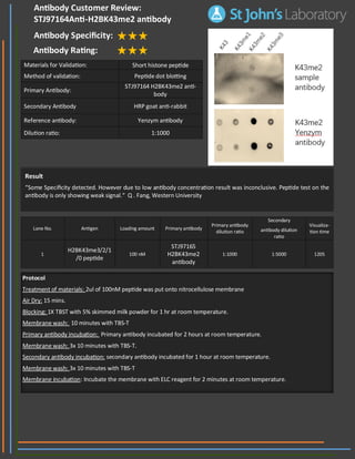
1. Result
“Some Specificity detected. However due to low antibody concentration result was inconclusive. Peptide test on the
antibody is only showing weak signal.“ Q . Fang, Western University
Lane No. Antigen Loading amount Primary antibody
Primary antibody
dilution ratio
Secondary
antibody dilution
ratio
Visualiza-
tion time
1
H2BK43me3/2/1
/0 peptide
100 nM
STJ97165
H2BK43me2
antibody
1:1000 1:5000 120S
Materials for Validation: Short histone peptide
Method of validation: Peptide dot blotting
Primary Antibody:
STJ97164 H2BK43me2 anti-
body
Secondary Antibody HRP goat anti-rabbit
Reference antibody: Yenzym antibody
Dilution ratio: 1:1000
Protocol
Treatment of materials: 2ul of 100nM peptide was put onto nitrocellulose membrane
Air Dry: 15 mins.
Blocking: 1X TBST with 5% skimmed milk powder for 1 hr at room temperature.
Membrane wash: 10 minutes with TBS-T
Primary antibody incubation: Primary antibody incubated for 2 hours at room temperature.
Membrane wash: 3x 10 minutes with TBS-T.
Secondary antibody incubation: secondary antibody incubated for 1 hour at room temperature.
Membrane wash: 3x 10 minutes with TBS-T
Membrane incubation: Incubate the membrane with ELC reagent for 2 minutes at room temperature.
Antibody Customer Review:
STJ97164Anti-H2BK43me2 antibody
Antibody Specificity:
Antibody Rating: