A Western blot was performed to validate the STJ92925 Anti-Endoplasmin Antibody using HeLa cell extracts. A band was observed around 92kD on the membrane, indicating the antibody recognized endoplasmin in the HeLa extracts as expected. The validation involved lysing HeLa cells, running protein samples on a 10% polyacrylamide gel, transferring to a PVDF membrane, blocking with milk, probing with the primary and secondary antibodies at specified dilutions, washing, and visualizing with ECL.
Western Blot Antibody Customer Review for Anti-RelB Polyclonal Antibody (STJ9...St John's Laboratory Ltd
The RelB protein is a transcription factor encoded by the RELB gene in humans. It is known to interact with NFKB2. The RelB polyclonal antibody detects endogenous levels of RelB protein.
Constitutively active protein kinase that acts as a negative regulator in the hormonal control of glucose homeostasis, Wnt signaling and regulation of transcription factors and microtubules, by phosphorylating and inactivating glycogen synthase (GYS1 or GYS2), EIF2B, CTNNB1/beta-catenin, APC, AXIN1, DPYSL2/CRMP2, JUN, NFATC1/NFATC, MAPT/TAU and MACF1. Requires primed phosphorylation of the majority of its substrates. In skeletal muscle, contributes to insulin regulation of glycogen synthesis by phosphorylating and inhibiting GYS1 activity and hence glycogen synthesis. May also mediate the development of insulin resistance by regulating activation of transcription factors. Regulates protein synthesis by controlling the activity of initiation factor 2B (EIF2BE/EIF2B5) in the same manner as glycogen synthase. In Wnt signaling, GSK3B forms a multimeric complex with APC, AXIN1 and CTNNB1/beta-catenin and phosphorylates the N-terminus of CTNNB1 leading to its degradation mediated by ubiquitin/proteasomes. Phosphorylates JUN at sites proximal to its DNA-binding domain, thereby reducing its affinity for DNA.
Anti-GSK3β-http://www.stjohnslabs.com/gsk3b-antibody-p-92568
Join our Antibody Validation Project - http://www.stjohnslabs.com/services/antibody-validation
Hypoxia-inducible factor 1-alpha, also known as HIF-1α, is a subunit of a heterodimeric transcription factor hypoxia-inducible factor 1 (HIF-1) that is encoded by the HIF1α gene. It is a basic helix-loop-helix PAS domain containing protein, and is considered as the master transcriptional regulator of cellular and developmental response to hypoxia. The dysregulation and overexpression of HIF1α by either hypoxia or genetic alternations have been heavily implicated in cancer biology, as well as a number of other pathophysiologies, specifically in areas of vascularization and angiogenesis, energy metabolism, cell survival and tumour invasion.
To purchase this antibody use the following link: http://www.stjohnslabs.com/hif-1a-antibody-p-92614?filter_name=STJ93498
Western Blot Antibody Customer Review for Anti-Peroxin 2 Antibody (STJ95040)St John's Laboratory Ltd
PEX2 is gene on chromosome 8q21.1 that encodes a peroxin, an integral membrane protein involved in peroxisome biosynthesis and integrity, which assembles membrane vesicles before translocation of matrix proteins.
Defects of PEX2 cause peroxisome biogenesis disorder complementation group 5, Zelleger syndrome and a form of Refsum disease.
Anti-Peroxin 2 - http://www.stjohnslabs.com/peroxin-2-antibody?filter_name=STJ95040
Join our Antibody Validation Project - http://www.stjohnslabs.com/services/antibody-validation
Western Blot Antibody Customer Review for p63 Polyclonal Antibody (STJ94912)St John's Laboratory Ltd
TP63 (tumour protein p63) or transformation-related protein 63 acts as a sequence specific DNA binding transcriptional activator or repressor. It is encoded by the TP63 gene in humans. The p63 polyclonal antibody detects endogenous levels of p63 protein.
Fukutin-related protein (FKRP) is targeted to the medial Golgi apparatus and is necessary for post-translational modification of dystroglycan. Mutations in the gene encoding this protein have been associated with congenital muscular dystrophy, mental retardation, and cerebellar cysts. Several alternatively spliced transcript variants of this gene have been described, but the full-length nature of some of these variants has not been determined.
To purchase this antibody use the following link: http://www.stjohnslabs.com/fkrp-antibody-p-95246?filter_name=FKRP
Facilitative glucose transporter. This isoform may be responsible for constitutive or basal glucose uptake. Has a very broad substrate specificity; can transport a wide range of aldoses including both pentoses and hexoses.
Anti-Glut1-http://www.stjohnslabs.com/glut1-antibody-p-92472
Join our Antibody Validation Project - http://www.stjohnslabs.com/services/antibody-validation
Molecular chaperone that promotes the maturation, structural maintenance and proper regulation of specific target proteins involved for instance in cell cycle control and signal transduction. Undergoes a functional cycle that is linked to its ATPase activity. This cycle probably induces conformational changes in the client proteins, thereby causing their activation. Interacts dynamically with various co-chaperones that modulate its substrate recognition, ATPase cycle and chaperone function.
Anti-HSP90β-http://www.stjohnslabs.com/hsp90b-antibody-p-98667
Join our Antibody Validation Project - http://www.stjohnslabs.com/services/antibody-validation
Involved in the transcriptional regulation of multiple genes expressed in the intestinal epithelium. Important in broad range of functions from early differentiation to maintenance of the intestinal epithelial lining of both the small and large intestine.
Anti-CDX2-http://www.stjohnslabs.com/cdx2-antibody-p-98594
Join our Antibody Validation Project - http://www.stjohnslabs.com/services/antibody-validation
Customer Review For Tubulin-beta Polyclonal Antibody- IF Application (STJ31562)St John's Laboratory Ltd
Tubulin is the major constituent of microtubules. It binds two moles of GTP, one at an exchangeable site on the beta chain and one at a non-exchangeable site on the alpha chain. TUBB3 plays a critical role in proper axon guidance and maintenance.
To purchase this antibody use the following link: http://www.stjohnslabs.com/tubulin-beta-antibody?filter_name=STJ31562
Western Blot Antibody Customer Review for Anti-PDHA1 Polyclonal Antibody (STJ...St John's Laboratory Ltd
Pyruvate dehydrogenase E1 component subunit alpha, somatic form, mitochondrial is an enzyme that in humans is encoded by the PDHA1 gene.
The pyruvate dehydrogenase complex is responsible for the oxidative decarboxylation of pyruvate, with the final product being Acetyl CoA.
Mutations in the PDHA1 gene have been known to cause one form of pyruvate dehydrogenase deficiency. Pyruvate dehydrogenase deficiency is characterized by the buildup of a chemical called lactic acid in the body and a variety of neurological problems.
The antibody was affinity-purified from rabbit antiserum by affinity-chromatography using epitope-specific immunogen.
Anti-PDHA1 - http://www.stjohnslabs.com/pdha1-antibody-p-93852?filter_name=STJ95006
Join our Antibody Validation Project - http://www.stjohnslabs.com/services/antibody-validation
Core component of nucleosome. Nucleosomes wrap and compact DNA into chromatin, limiting DNA accessibility to the cellular machineries which require DNA as a template. Histones thereby play a central role in transcription regulation, DNA repair, DNA replication and chromosomal stability. DNA accessibility is regulated via a complex set of post-translational modifications of histones, also called histone code, and nucleosome remodeling.
Anti-Histone H4 (Acetyl K16)-http://www.stjohnslabs.com/acetyl-histone-h4-k16-antibody
Join our Antibody Validation Project - http://www.stjohnslabs.com/services/antibody-validation
GSK3β Polyclonal Antibody is an active protein kinase that acts as a negative regulator in the hormonal control of glucose homeostasis, Wnt signaling and regulation of transcription factors and microtubules, by phosphorylating and inactivating glycogen synthase (GYS1 or GYS2), EIF2B, CTNNB1/beta-catenin, APC, AXIN1, DPYSL2/CRMP2, JUN, NFATC1/NFATC, MAPT/TAU and MACF1. Requires primed phosphorylation of the majority of its substrates.
To purchase this antibody, use the following link: http://www.stjohnslabs.com/gsk3b-antibody-p-92568?filter_name=STJ93447
Immunohistochemistry Antibody Validation Report for Anti-Phospho-GSK3α/β (Y27...St John's Laboratory Ltd
Constitutively active protein kinase that acts as a negative regulator in the hormonal control of glucose homeostasis, Wnt signaling and regulation of transcription factors and microtubules, by phosphorylating and inactivating glycogen synthase (GYS1 or GYS2), CTNNB1/beta-catenin, APC and AXIN1. Requires primed phosphorylation of the majority of its substrates. Contributes to insulin regulation of glycogen synthesis by phosphorylating and inhibiting GYS1 activity and hence glycogen synthesis. Regulates glycogen metabolism in liver, but not in muscle. May also mediate the development of insulin resistance by regulating activation of transcription factors. In Wnt signaling, regulates the level and transcriptional activity of nuclear CTNNB1/beta-catenin. Facilitates amyloid precursor protein (APP) processing and the generation of APP-derived amyloid plaques found in Alzheimer disease.
Anti-Phospho-GSK3α/β (Y279/216)-http://www.stjohnslabs.com/phospho-gsk3ab-y279216-antibody
Join our Antibody Validation Project - http://www.stjohnslabs.com/services/antibody-validation
Immunohistochemistry Antibody Validation Report for Anti-Phospho-NFκB-p65 (S2...St John's Laboratory Ltd
NF-kappa-B is a pleiotropic transcription factor present in almost all cell types and is the endpoint of a series of signal transduction events that are initiated by a vast array of stimuli related to many biological processes such as inflammation, immunity, differentiation, cell growth, tumorigenesis and apoptosis. NF-kappa-B is a homo- or heterodimeric complex formed by the Rel-like domain-containing proteins RELA/p65, RELB, NFKB1/p105, NFKB1/p50, REL and NFKB2/p52 and the heterodimeric p65-p50 complex appears to be most abundant one. The dimers bind at kappa-B sites in the DNA of their target genes and the individual dimers have distinct preferences for different kappa-B sites that they can bind with distinguishable affinity and specificity.
Anti-Phospho-NFκB-p65 (S276)-http://www.stjohnslabs.com/phospho-nfkb-p65-s276-antibody
Join our Antibody Validation Project - http://www.stjohnslabs.com/services/antibody-validation
Core component of nucleosome. Nucleosomes wrap and compact DNA into chromatin, limiting DNA accessibility to the cellular machineries which require DNA as a template. Histones thereby play a central role in transcription regulation, DNA repair, DNA replication and chromosomal stability. DNA accessibility is regulated via a complex set of post-translational modifications of histones, also called histone code, and nucleosome remodeling.
Anti-Histone H3-http://www.stjohnslabs.com/histone-h3-antibody-p-92641
Join our Antibody Validation Project - http://www.stjohnslabs.com/services/antibody-validation
Immunohistochemistry Antibody Validation Report for Anti-Phospho-CaMKIIβ/γ/δ ...St John's Laboratory Ltd
Calcium/calmodulin-dependent protein kinase that functions autonomously after Ca2+/calmodulin-binding and autophosphorylation, and is involved in dendritic spine and synapse formation, neuronal plasticity and regulation of sarcoplasmic reticulum Ca2+ transport in skeletal muscle. In neurons, plays an essential structural role in the reorganization of the actin cytoskeleton during plasticity by binding and bundling actin filaments in a kinase-independent manner. This structural function is required for correct targeting of CaMK2A, which acts downstream of NMDAR to promote dendritic spine and synapse formation and maintain synaptic plasticity which enables long-term potentiation (LTP) and hippocampus-dependent learning. In developing hippocampal neurons, promotes arborization of the dendritic tree and in mature neurons, promotes dendritic remodeling.
Anti-Phospho-CaMKIIβ/γ/δ (T287)-http://www.stjohnslabs.com/phospho-camkiibgd-t287-antibody
Join our Antibody Validation Project - http://www.stjohnslabs.com/services/antibody-validation
Receptor for the Fc region of IgG. Binds complexed or aggregated IgG and also monomeric IgG. Mediates antibody-dependent cellular cytotoxicity (ADCC) and other antibody-dependent responses, such as phagocytosis.
Anti-CD16-http://www.stjohnslabs.com/cd16-antibody-p-98627
Join our Antibody Validation Project - http://www.stjohnslabs.com/services/antibody-validation
CD1 (cluster of differentiation 1) is a family of glycoproteins expressed on the surface of various human antigen-presenting cells. They are related to the class I MHC molecules, and are involved in the presentation of lipid antigens to T cells. However their precise function is unknown.
Anti-CD1 -http://www.stjohnslabs.com/cd1-antibody-p-98605
Join our Antibody Validation Project - http://www.stjohnslabs.com/services/antibody-validation
Could play a role in phagocytic activities of tissue macrophages, both in intracellular lysosomal metabolism and extracellular cell-cell and cell-pathogen interactions. Binds to tissue- and organ-specific lectins or selectins, allowing homing of macrophage subsets to particular sites. Rapid recirculation of CD68 from endosomes and lysosomes to the plasma membrane may allow macrophages to crawl over selectin-bearing substrates or other cells.
Anti-CD68-http://www.stjohnslabs.com/cd68-antibody-p-98587
Join our Antibody Validation Project - http://www.stjohnslabs.com/services/antibody-validation
Core component of nucleosome. Nucleosomes wrap and compact DNA into chromatin, limiting DNA accessibility to the cellular machineries which require DNA as a template. Histones thereby play a central role in transcription regulation, DNA repair, DNA replication and chromosomal stability. DNA accessibility is regulated via a complex set of post-translational modifications of histones, also called histone code, and nucleosome remodeling.
Anti-Histone H3-http://www.stjohnslabs.com/histone-h3-antibody-p-98575
Join our Antibody Validation Project - http://www.stjohnslabs.com/services/antibody-validation
The Bcl-2-associated death promoter (BAD) protein is a pro-apoptotic member of the Bcl-2 gene family which is involved in initiating apoptosis. BAD is a member of the BH3-only family, a subfamily of the Bcl-2 family. After activation, it is able to form a heterodimer with anti-apoptotic proteins and prevent them from stopping apoptosis.
Anti-Bad - http://www.stjohnslabs.com/bad-antibody-p-91310?filter_name=STJ91797
Join our Antibody Validation Project - http://www.stjohnslabs.com/services/antibody-validation
Tau are stabilising proteins for axonal microtubules within the Central Nervous System, and as such are also recognised as Mictrotubule-associated Protein (MAP). Tau is predominantly active in the distal portions of axons to carry out its function.
Western Blot Antibody Customer Review for ADNP Polyclonal Antibody (STJ91502)St John's Laboratory Ltd
ADNP (Activity-Dependent Neuroprotector Homeobox) is expressed through the ADNP gene in humans. It is a possible transcription factor through having a homeobox and 9 zinc finger domains. It is said to have both stimulatory and inhibitory growth effects on different tumour cells. The ADNP Polyclonal Antibody detects endogenous levels of ADNP protein.
Western Blot Antibody Customer Review for Anti-RUNX1 Polyclonal Antibody (STJ...St John's Laboratory Ltd
RUNX1 protein (Runt-related transcription factor 1) also known as AML1 is a transcriptional corepressor encoded by the RUNX1 gene in humans. It regulates the differentiation of hematopoietic stem cells into mature blood cells via its association with DNA-binding transcription factors as well as its recruitment of other corepressors and histone-modifying enzymes.
Equilibrative nucleoside transporter 1 (ENT1) is a protein that in humans is encoded by the SLC29A1 gene. Multiple alternatively spliced variants, encoding the same protein, have been found for this gene.
This gene is a member of the equilibrative nucleoside transporter family. The gene encodes a transmembrane glycoprotein that localizes to the plasma and mitochondrial membranes and mediates the cellular uptake of nucleosides from the surrounding medium. The protein is categorized as an equilibrative (as opposed to concentrative) transporter that is sensitive to inhibition by nitrobenzylmercaptopurine ribonucleoside (NBMPR). Nucleoside transporters are required for nucleotide synthesis in cells that lack de novo nucleoside synthesis pathways, and are also necessary for the uptake of cytotoxic nucleosides used for cancer and viral chemotherapies.
To purchase this antibody, use the following link: http://www.stjohnslabs.com/ent1-antibody?filter_name=STJ96396
Western Blot Antibody Customer Review for Anti-Histone H3 (Acetyl Lys14) Ant...St John's Laboratory Ltd
Histone H3 is one of the five main histone proteins involved in the structure of chromatin in eukaryotic cells
Histones play a central role in transcription regulation, DNA repair, DNA replication and chromosomal stability. DNA accessibility is regulated via a complex set of post-translational modifications of histones, also called histone code, and nucleosome remodeling.
Anti-Histone H3 (Acetyl Lys14) Antibody - http://www.stjohnslabs.com/histone-h3-acetyl-lys14-antibody?filter_name=Stj97144
Join our Antibody Validation Project - http://www.stjohnslabs.com/services/antibody-validation
Vesicle-associated membrane protein-associated protein B (VAPB) is a type IV membrane protein found in plasma and intracellular vesicle membranes. The protein is found as a homodimer and as a heterodimer with VAPA. It also can interact with VAMP1 and VAMP2 and may be involved in vesicle trafficking. Like VAPA, VAPB binds to proteins that contain a FFAT motif. Considerable interest in VAPB has arisen because mutations in this protein are associated with rare, familial forms of Motor Neurone Disease (also called Amyotrophic Lateral Sclerosis and Lou Gehrig's disease).
To purchase this antibody use the following link: http://www.stjohnslabs.com/vapb-antibody-c-term-p-79486?filter_name=STJ47066
Has both glyceraldehyde-3-phosphate dehydrogenase and nitrosylase activities, thereby playing a role in glycolysis and nuclear functions, respectively. Participates in nuclear events including transcription, RNA transport, DNA replication and apoptosis. Nuclear functions are probably due to the nitrosylase activity that mediates cysteine S-nitrosylation of nuclear target proteins such as SIRT1, HDAC2 and PRKDC. Modulates the organization and assembly of the cytoskeleton. Facilitates the CHP1-dependent microtubule and membrane associations through its ability to stimulate the binding of CHP1 to microtubules (By similarity). Glyceraldehyde-3-phosphate dehydrogenase is a key enzyme in glycolysis that catalyzes the first step of the pathway by converting D-glyceraldehyde 3-phosphate (G3P) into 3-phospho-D-glyceroyl phosphate. Component of the GAIT (gamma interferon-activated inhibitor of translation) complex which mediates interferon-gamma-induced transcript-selective translation inhibition in inflammation processes. Upon interferon-gamma treatment assembles into the GAIT complex which binds to stem loop-containing GAIT elements in the 3'-UTR of diverse inflammatory mRNAs (such as ceruplasmin) and suppresses their translation.
To purchase this antibody, use the following link: http://www.stjohnslabs.com/gapdh-antibody-p-94882?filter_name=STJ96417
Histones are nuclear proteins that form octameric structures which bind DNA to form units of chromatin called nucleosomes. The family of histones—H2A, H2B, H3, and H4—are key players in gene regulation. They undergo a number of post-translational modifications (PTM) in response to various stimuli, including phosphorylation on serine and threonine residues and methylation on lysine residues. PTMs produce configural changes in histone proteins that may induce nucleosome remodeling and expose or hide DNA sequences from transcriptional complexes. Histone H4 lysine 20 (H4K20) may undergo mono-, di-, or trimethylation, which is catalyzed by the methyltransferase PR-Set7 (Set8 or KMT5a). Methylated H4K20 plays a role in regulating DNA damage responses, mitosis, DNA replication, and gene expression. Trimethylation of H4K20 contributes to gene silencing, and is a mark of the repressive heterochromatin state.
To purchase this antibody use the following link: http://www.stjohnslabs.com/histone-h4-tri-methyl-lys20-antibody?filter_name=STJ97154
Immunohistochemistry Antibody Validation Report for Anti-Cleaved-PARP-1 (D214...St John's Laboratory Ltd
Involved in the base excision repair (BER) pathway, by catalyzing the poly(ADP-ribosyl)ation of a limited number of acceptor proteins involved in chromatin architecture and in DNA metabolism. This modification follows DNA damages and appears as an obligatory step in a detection/signaling pathway leading to the reparation of DNA strand breaks. Mediates the poly(ADP-ribosyl)ation of APLF and CHFR. Positively regulates the transcription of MTUS1 and negatively regulates the transcription of MTUS2/TIP150. With EEF1A1 and TXK, forms a complex that acts as a T-helper 1 (Th1) cell-specific transcription factor and binds the promoter of IFN-gamma to directly regulate its transcription, and is thus involved importantly in Th1 cytokine production. Required for PARP9 and DTX3L recruitment to DNA damage sites.
Anti-Cleaved-PARP-1 (D214)-http://www.stjohnslabs.com/cleaved-parp-1-d214-antibody
Join our Antibody Validation Project - http://www.stjohnslabs.com/services/antibody-validation
Serine/threonine protein kinase which is a central regulator of cellular metabolism, growth and survival in response to hormones, growth factors, nutrients, energy and stress signals. MTOR directly or indirectly regulates the phosphorylation of at least 800 proteins. Functions as part of 2 structurally and functionally distinct signaling complexes mTORC1 and mTORC2 (mTOR complex 1 and 2). Activated mTORC1 up-regulates protein synthesis by phosphorylating key regulators of mRNA translation and ribosome synthesis. This includes phosphorylation of EIF4EBP1 and release of its inhibition toward the elongation initiation factor 4E (eiF4E). Moreover, phosphorylates and activates RPS6KB1 and RPS6KB2 that promote protein synthesis by modulating the activity of their downstream targets including ribosomal protein S6, eukaryotic translation initiation factor EIF4B, and the inhibitor of translation initiation PDCD4. Stimulates the pyrimidine biosynthesis pathway, both by acute regulation through RPS6KB1-mediated phosphorylation of the biosynthetic enzyme CAD, and delayed regulation, through transcriptional enhancement of the pentose phosphate pathway which produces 5-phosphoribosyl-1-pyrophosphate (PRPP), an allosteric activator of CAD at a later step in synthesis, this function is dependent on the mTORC1 complex. Regulates ribosome synthesis by activating RNA polymerase III-dependent transcription through phosphorylation and inhibition of MAF1 an RNA polymerase III-repressor.
To purchase this antibody, use the following link: http://www.stjohnslabs.com/mtor-antibody-p-93228?filter_name=STJ94280
Immunohistochemistry Antibody Validation Report for Anti-Cleaved-Caspase-3 p1...St John's Laboratory Ltd
Involved in the activation cascade of caspases responsible for apoptosis execution. At the onset of apoptosis it proteolytically cleaves poly(ADP-ribose) polymerase (PARP) at a '216-Asp-|-Gly-217' bond. Cleaves and activates sterol regulatory element binding proteins (SREBPs) between the basic helix-loop-helix leucine zipper domain and the membrane attachment domain. Cleaves and activates caspase-6, -7 and -9. Involved in the cleavage of huntingtin. Triggers cell adhesion in sympathetic neurons through RET cleavage. / Strict requirement for an Asp residue at positions P1 and P4. It has a preferred cleavage sequence of Asp-Xaa-Xaa-Asp-|- with a hydrophobic amino-acid residue at P2 and a hydrophilic amino-acid residue at P3, although Val or Ala are also accepted at this position. / Inhibited by isatin sulfonamides
Anti-Cleaved-Caspase-3 p12 (D175)-http://www.stjohnslabs.com/cleaved-caspase-3-p12-d175-
antibody
Join our Antibody Validation Project - http://www.stjohnslabs.com/services/antibody-validation
Acts as decoy receptor for TNFSF11/RANKL and thereby neutralizes its function in osteoclastogenesis. Inhibits the activation of osteoclasts and promotes osteoclast apoptosis in vitro. Bone homeostasis seems to depend on the local ratio between TNFSF11 and TNFRSF11B. May also play a role in preventing arterial calcification. May act as decoy receptor for TNFSF10/TRAIL and protect against apoptosis. TNFSF10/TRAIL binding blocks the inhibition of osteoclastogenesis.
Anti-OPG-http://www.stjohnslabs.com/opg-antibody
Join our Antibody Validation Project - http://www.stjohnslabs.com/services/antibody-validation
Probably plays a role in facilitating the assembly of multimeric protein complexes inside the endoplasmic reticulum. Involved in the correct folding of proteins and degradation of misfolded proteins via its interaction with DNAJC10, probably to facilitate the release of DNAJC10 from its substrate.
Anti-HSP A5-http://www.stjohnslabs.com/hsp-a5-antibody
Join our Antibody Validation Project - http://www.stjohnslabs.com/services/antibody-validation
Western Blot Antibody Customer Review for Anti-Phospho-ALK (Y1507) Polyclonal...St John's Laboratory Ltd
Anaplastic lymphoma kinase (ALK) also known as ALK tyrosine kinase receptor or CD246 (cluster of differentiation 246) is an enzyme that in humans is encoded by the ALK gene.
ALK plays an important role in the development of the brain and exerts its effects on specific neurons in the nervous system. Transduces signals from ligands at the cell surface, through specific activation of the mitogen-activated protein kinase (MAPK) pathway. Phospho-ALK (Y1507) Polyclonal Antibody detects endogenous levels of ALK protein only when phosphorylated at Y1507.
Anti-Phospho-ALK - http://www.stjohnslabs.com/phospho-alk-y1507-antibody?filter_name=STJ90845
Join our Antibody Validation Project - http://www.stjohnslabs.com/services/antibody-validation
Immunohistochemistry Antibody Validation Report for Anti-Cleaved-Caspase-1 (D...St John's Laboratory Ltd
Thiol protease that cleaves IL-1 beta between an Asp and an Ala, releasing the mature cytokine which is involved in a variety of inflammatory processes. Important for defense against pathogens. Cleaves and activates sterol regulatory element binding proteins (SREBPs). Can also promote apoptosis. / Strict requirement for an Asp residue at position P1 and has a preferred cleavage sequence of Tyr-Val-Ala-Asp-|-. / Specifically inhibited by the cowpox virus Crma protein.
Anti-Cleaved-Caspase-1 (D210)-
http://www.stjohnslabs.com/cleaved-caspase-1-d210-antibody
Join our Antibody Validation Project - http://www.stjohnslabs.com/services/antibody-validation
Histones are basic nuclear proteins that are responsible for the nucleosome structure of the chromosomal fiber in eukaryotes. Nucleosomes consist of approximately 146 bp of DNA wrapped around a histone octamer composed of pairs of each of the four core histones (H2A, H2B, H3, and H4). The chromatin fiber is further compacted through the interaction of a linker histone, H1, with the DNA between the nucleosomes to form higher order chromatin structures. This gene is intronless and encodes a member of the histone H3 family. Transcripts from this gene lack polyA tails; instead, they contain a palindromic termination element. This gene is located separately from the other H3 genes that are in the histone gene cluster on chromosome 6p22-p21.3.
To purchase this antibody use the following link: http://www.stjohnslabs.com/trimethyl-histone-h3-k56?filter_name=STJ29403
Functions as a master transcriptional regulator of the adaptive response to hypoxia. Under hypoxic conditions, activates the transcription of over 40 genes, including erythropoietin, glucose transporters, glycolytic enzymes, vascular endothelial growth factor, HILPDA, and other genes whose protein products increase oxygen delivery or facilitate metabolic adaptation to hypoxia. Plays an essential role in embryonic vascularization, tumor angiogenesis and pathophysiology of ischemic disease. Binds to core DNA sequence 5'-[AG]CGTG-3' within the hypoxia response element (HRE) of target gene promoters. Activation requires recruitment of transcriptional coactivators such as CREBPB and EP300. Activity is enhanced by interaction with both, NCOA1 or NCOA2. Interaction with redox regulatory protein APEX seems to activate CTAD and potentiates activation by NCOA1 and CREBBP. Involved in the axonal distribution and transport of mitochondria in neurons during hypoxia.
Anti-HIF-1α-http://www.stjohnslabs.com/hif-1a-antibody-p-92614
Join our Antibody Validation Project - http://www.stjohnslabs.com/services/antibody-validation
Immunohistochemistry Antibody Validation Report for Anti-Cleaved-Caspase-8 (D...St John's Laboratory Ltd
Most upstream protease of the activation cascade of caspases responsible for the TNFRSF6/FAS mediated and TNFRSF1A induced cell death. Binding to the adapter molecule FADD recruits it to either receptor. The resulting aggregate called death-inducing signalling complex (DISC) performs CASP8 proteolytic activation. The active dimeric enzyme is then liberated from the DISC and free to activate downstream apoptotic proteases. Proteolytic fragments of the N-terminal pro peptide (termed CAP3, CAP5 and CAP6) are likely retained in the DISC. Cleaves and activates CASP3, CASP4, CASP6, CASP7, CASP9 and CASP10. May participate in the GZMB apoptotic pathways. Cleaves ADPRT. Hydrolyzes the small-molecule substrate, Ac-Asp-Glu-Val-Asp-|-AMC. Likely target for the cowpox virus CRMA death inhibitory protein.
Anti-Cleaved-Caspase-8 (D384) -http://www.stjohnslabs.com/cleaved-caspase-8-d384-antibody
Join our Antibody Validation Project - http://www.stjohnslabs.com/services/antibody-validation
Metabolic-stress sensing protein kinase that phosphorylates the alpha subunit of eukaryotic translation initiation factor 2 (eIF-2-alpha/EIF2S1) on 'Ser-52' during the unfolded protein response (UPR) and in response to low amino acid availability. Converts phosphorylated eIF-2-alpha/EIF2S1 either in a global protein synthesis inhibitor, leading to a reduced overall utilization of amino acids, or to a translation initiation activator of specific mRNAs, such as the transcriptional activator ATF4, and hence allowing ATF4-mediated reprogramming of amino acid biosynthetic gene expression to alleviate nutrient depletion.
Anti-Phospho-PERK (T981)-http://www.stjohnslabs.com/phospho-perk-t981-antibody
Join our Antibody Validation Project - http://www.stjohnslabs.com/services/antibody-validation
Similar to Western Blot Antibody Customer Review for Anti-Endoplasmin Polyclonal Antibody (STJ92925) (20)
G protein-coupled receptor that probably associates with the patched protein (PTCH) to transduce the hedgehog's proteins signal. Binding of sonic hedgehog (SHH) to its receptor patched is thought to prevent normal inhibition by patched of smoothened (SMO). Required for the accumulation of KIF7 and GLI3 in the cilia.
Anti-Smo antibody (STJ95710): http://www.stjohnslabs.com/smo-antibody-p-94371?filter_name=STJ95710
Join our Antibody Validation Project - http://www.stjohnslabs.com/services/antibody-validation
Western Blot Customer Review Anti Glucocorticoid Receptor antibody (STJ97101)St John's Laboratory Ltd
Receptor for glucocorticoids (GC). Has a dual mode of action: as a transcription factor that binds to glucocorticoid response elements (GRE), both for nuclear and mitochondrial DNA, and as a modulator of other transcription factors. Affects inflammatory responses, cellular proliferation and differentiation in target tissues. Involved in chromatin remodeling . Plays a role in rapid mRNA degradation by binding to the 5' UTR of target mRNAs and interacting with PNRC2 in a ligand-dependent manner. Could act as a coactivator for STAT5-dependent transcription upon growth hormone (GH) stimulation and could reveal an essential role of hepatic GR in the control of body growth (By similarity). Has transcriptional activation and repression activity . Mediates glucocorticoid-induced apoptosis . Promotes accurate chromosome segregation during mitosis . May act as a tumor suppressor . May play a negative role in adipogenesis through the regulation of lipolytic and antilipogenic gene expression (By similarity). / Isoform Beta: Acts as a dominant negative inhibitor of isoform Alpha . Has intrinsic transcriptional activity independent of isoform Alpha when both isoforms are coexpressed.
Join our Antibody Validation Project: http://www.stjohnslabs.com/services/antibody-validation
Anti glucocorticoid receptor antibody (STJ97101):
http://www.stjohnslabs.com/glucocorticoid-receptor-antibody-p-98736?filter_name=STJ97101
Western Blot Customer Review Anti-Phospho-Cofilin (S3) Antibody (STJ90230)St John's Laboratory Ltd
Binds to F-actin and exhibits pH-sensitive F-actin depolymerizing activity. Regulates actin cytoskeleton dynamics. Important for normal progress through mitosis and normal cytokinesis. Plays a role in the regulation of cell morphology and cytoskeletal organization. Required for the up-regulation of atypical chemokine receptor ACKR2 from endosomal compartment to cell membrane, increasing its efficiency in chemokine uptake and degradation.
Anti-Phospho-Cofilin (S3) -http://www.stjohnslabs.com/phospho-cofilin-s3-antibody
Join our Antibody Validation Project - http://www.stjohnslabs.com/services/antibody-validation
This June, Dr. Byron Baron from the University of Malta, Malta, is our Scientist of the Month! He's shared with us his research highlights, his current projects and some comments on the biotechnology industry.
Want to be our Scientist of the Month? Contact info@stjohnslabs.com
Downstream effector molecule involved in the transmission of signals from tyrosine kinase receptors and small GTPases to the actin cytoskeleton. Promotes formation of actin filaments. Part of the WAVE complex that regulates lamellipodia formation. The WAVE complex regulates actin filament reorganization via its interaction with the Arp2/3 complex.
Anti-WAVE2 -http://www.stjohnslabs.com/wave2-antibody
Join our Antibody Validation Project - http://www.stjohnslabs.com/services/antibody-validation
Implicated in synaptic vesicle endocytosis. May recruit other proteins to membranes with high curvature.
Brain, mostly in frontal cortex. Expressed at high level in fetal cerebellum.
Anti-Endophilin I -http://www.stjohnslabs.com/endophilin-i-antibody
Join our Antibody Validation Project - http://www.stjohnslabs.com/services/antibody-validation
Tubulin is the major constituent of microtubules. It binds two moles of GTP, one at an exchangeable site on the beta chain and one at a non-exchangeable site on the alpha chain. TUBB3 plays a critical role in proper axon guidance and mantainance.
Anti-β-tubulin -http://www.stjohnslabs.com/b-tubulin-antibody-p-98672
Join our Antibody Validation Project - http://www.stjohnslabs.com/services/antibody-validation
Most upstream protease of the activation cascade of caspases responsible for the TNFRSF6/FAS mediated and TNFRSF1A induced cell death. Binding to the adapter molecule FADD recruits it to either receptor. The resulting aggregate called death-inducing signaling complex (DISC) performs CASP8 proteolytic activation. The active dimeric enzyme is then liberated from the DISC and free to activate downstream apoptotic proteases. Proteolytic fragments of the N-terminal propeptide (termed CAP3, CAP5 and CAP6) are likely retained in the DISC. Cleaves and activates CASP3, CASP4, CASP6, CASP7, CASP9 and CASP10. May participate in the GZMB apoptotic pathways. Cleaves ADPRT. Hydrolyzes the small-molecule substrate, Ac-Asp-Glu-Val-Asp-|-AMC. Likely target for the cowpox virus CRMA death inhibitory protein. Isoform 5, isoform 6, isoform 7 and isoform 8 lack the catalytic site and may interfere with the pro-apoptotic activity of the complex. / Strict requirement for Asp at position P1 and has a preferred cleavage sequence of (Leu/Asp/Val)-Glu-Thr-Asp-|-(Gly/Ser/Ala). / Inhibited by the effector protein NleF that is produced by pathogenic E.coli; this inhibits apoptosis.
Anti-Caspase-8-http://www.stjohnslabs.com/caspase-8-antibody-p-99045
Join our Antibody Validation Project - http://www.stjohnslabs.com/services/antibody-validation
Tubulin is the major constituent of microtubules. The gamma chain is found at microtubule organizing centers (MTOC) such as the spindle poles or the centrosome. Pericentriolar matrix component that regulates alpha/beta chain minus-end nucleation, centrosome duplication and spindle formation.
Anti-Gamma Tubulin-http://www.stjohnslabs.com/gamma-tubulin-antibody
Join our Antibody Validation Project - http://www.stjohnslabs.com/services/antibody-validation
Tubulin in molecular biology can refer either to the tubulin protein superfamily of globular proteins, or one of the member proteins of that superfamily. α- and β-tubulins polymerize into microtubules, a major component of the eukaryotic cytoskeleton. Microtubules function in many essential cellular processes, including mitosis. Tubulin-binding drugs kill cancerous cells by inhibiting microtubule dynamics, which are required for DNA segregation and therefore cell division. In eukaryotes there are six members of the tubulin superfamily, although not all are present in all species (see below). Both α and β tubulins have a mass of around 50 kDa and are thus in a similar range compared to actin with ~42 kDa. In contrast, tubulin polymers (microtubules) tend to be much bigger than actin filaments due to their cylindrical nature. Tubulin was long thought to be specific to eukaryotes. More recently, however, several prokaryotic proteins have been shown to be related to tubulin.
Anti-Epsilon Tubulin -http://www.stjohnslabs.com/epsilon-tubulin-antibody-2
Join our Antibody Validation Project - http://www.stjohnslabs.com/services/antibody-validation
Ubiquitin-like modifier involved in formation of autophagosomal vacuoles (autophagosomes) . Whereas LC3s are involved in elongation of the phagophore membrane, the GABARAP/GATE-16 subfamily is essential for a later stage in autophagosome maturation .
Anti-LC3A-http://www.stjohnslabs.com/lc3a-antibody
Join our Antibody Validation Project - http://www.stjohnslabs.com/services/antibody-validation
Multifunctional transcription factor in ER stress response. Plays an essential role in the response to a wide variety of cell stresses and induces cell cycle arrest and apoptosis in response to ER stress. Plays a dual role both as an inhibitor of CCAAT/enhancer-binding protein (C/EBP) function and as an activator of other genes. Acts as a dominant-negative regulator of C/EBP-induced transcription: dimerizes with members of the C/EBP family, impairs their association with C/EBP binding sites in the promoter regions, and inhibits the expression of C/EBP regulated genes. Positively regulates the transcription of TRIB3, IL6, IL8, IL23, TNFRSF10B/DR5, PPP1R15A/GADD34, BBC3/PUMA, BCL2L11/BIM and ERO1L. Negatively regulates; expression of BCL2 and MYOD1, ATF4-dependent transcriptional activation of asparagine synthetase (ASNS), CEBPA-dependent transcriptional activation of hepcidin (HAMP) and CEBPB-mediated expression of peroxisome proliferator-activated receptor gamma (PPARG).
Anti-CHOP-http://www.stjohnslabs.com/chop-antibody-2
Join our Antibody Validation Project - http://www.stjohnslabs.com/services/antibody-validation
Pseudokinase that plays a key role in TNF-induced necroptosis, a programmed cell death process. Activated following phosphorylation by RIPK3, leading to homotrimerization, localization to the plasma membrane and execution of programmed necrosis characterized by calcium influx and plasma membrane damage. Does not have protein kinase activity.
Anti-phospho-MLKL (S358)-http://www.stjohnslabs.com/phospho-mlkl-s358-antibody-1
Join our Antibody Validation Project - http://www.stjohnslabs.com/services/antibody-validation
Serine/threonine kinase which acts as an essential component of the MAP kinase signal transduction pathway. MAPK1/ERK2 and MAPK3/ERK1 are the 2 MAPKs which play an important role in the MAPK/ERK cascade. They participate also in a signaling cascade initiated by activated KIT and KITLG/SCF. Depending on the cellular context, the MAPK/ERK cascade mediates diverse biological functions such as cell growth, adhesion, survival and differentiation through the regulation of transcription, translation, cytoskeletal rearrangements. The MAPK/ERK cascade plays also a role in initiation and regulation of meiosis, mitosis, and postmitotic functions in differentiated cells by phosphorylating a number of transcription factors. About 160 substrates have already been discovered for ERKs. Many of these substrates are localized in the nucleus, and seem to participate in the regulation of transcription upon stimulation.
Anti-ERK1-http://www.stjohnslabs.com/erk1-antibody-3
Join our Antibody Validation Project - http://www.stjohnslabs.com/services/antibody-validation
Tyrosine-protein kinase that acts as a cell-surface receptor for PDGFA, PDGFB and PDGFC and plays an essential role in the regulation of embryonic development, cell proliferation, survival and chemotaxis. Depending on the context, promotes or inhibits cell proliferation and cell migration. Plays an important role in the differentiation of bone marrow-derived mesenchymal stem cells. Required for normal skeleton development and cephalic closure during embryonic development. Required for normal development of the mucosa lining the gastrointestinal tract, and for recruitment of mesenchymal cells and normal development of intestinal villi. Plays a role in cell migration and chemotaxis in wound healing. Plays a role in platelet activation, secretion of agonists from platelet granules, and in thrombin-induced platelet aggregation.
Anti-PDGFRα-http://www.stjohnslabs.com/pdgfra-antibody-2
Join our Antibody Validation Project - http://www.stjohnslabs.com/services/antibody-validation
Thiol protease that cleaves IL-1 beta between an Asp and an Ala, releasing the mature cytokine which is involved in a variety of inflammatory processes. Important for defense against pathogens. Cleaves and activates sterol regulatory element binding proteins (SREBPs). Can also promote apoptosis. / Strict requirement for an Asp residue at position P1 and has a preferred cleavage sequence of Tyr-Val-Ala-Asp-|-. / Specifically inhibited by the cowpox virus Crma protein.
Anti-Caspase-1-http://www.stjohnslabs.com/caspase-1-antibody-1
Join our Antibody Validation Project - http://www.stjohnslabs.com/services/antibody-validation
Functions as a cell surface receptor and performs physiological functions on the surface of neurons relevant to neurite growth, neuronal adhesion and axonogenesis. Involved in cell mobility and transcription regulation through protein-protein interactions. Can promote transcription activation through binding to APBB1-KAT5 and inhibits Notch signaling through interaction with Numb. Couples to apoptosis-inducing pathways such as those mediated by G(O) and JIP. Inhibits G(o) alpha ATPase activity (By similarity). Acts as a kinesin I membrane receptor, mediating the axonal transport of beta-secretase and presenilin 1. Involved in copper homeostasis/oxidative stress through copper ion reduction. In vitro, copper-metallated APP induces neuronal death directly or is potentiated through Cu2+-mediated low-density lipoprotein oxidation.
Anti-Amyloid-β-http://www.stjohnslabs.com/amyloid-v-antibody
Join our Antibody Validation Project - http://www.stjohnslabs.com/services/antibody-validation
Suppresses apoptosis in a variety of cell systems including factor-dependent lymphohematopoietic and neural cells. Regulates cell death by controlling the mitochondrial membrane permeability. Appears to function in a feedback loop system with caspases. Inhibits caspase activity either by preventing the release of cytochrome c from the mitochondria and/or by binding to the apoptosis-activating factor (APAF-1). May attenuate inflammation by impairing NLRP1-inflammasome activation, hence CASP1 activation and IL1B release .
Anti-Bcl-2-http://www.stjohnslabs.com/bcl-2-antibody-1
Join our Antibody Validation Project - http://www.stjohnslabs.com/services/antibody-validation
Alpha-actin-2 also known as actin, aortic smooth muscle or alpha smooth muscle actin (α-SMA, SMactin, alpha-SM-actin, ASMA) is a protein that in humans is encoded by the ACTA2 gene located on 10q22-q24. Actin alpha 2, the human aortic smooth muscle actin gene, is one of six different actin isoforms which have been identified. Actins are highly conserved proteins that are involved in cell motility, structure and integrity. Alpha actins are a major constituent of the contractile apparatus. Alpha-smooth muscle actin (α-SMA) is commonly used as a marker of myofibroblast formation.
Anti-α-SMA -http://www.stjohnslabs.com/a-sma-antibody-1
Join our Antibody Validation Project - http://www.stjohnslabs.com/services/antibody-validation
Multi-source connectivity as the driver of solar wind variability in the heli...Sérgio Sacani
The ambient solar wind that flls the heliosphere originates from multiple
sources in the solar corona and is highly structured. It is often described
as high-speed, relatively homogeneous, plasma streams from coronal
holes and slow-speed, highly variable, streams whose source regions are
under debate. A key goal of ESA/NASA’s Solar Orbiter mission is to identify
solar wind sources and understand what drives the complexity seen in the
heliosphere. By combining magnetic feld modelling and spectroscopic
techniques with high-resolution observations and measurements, we show
that the solar wind variability detected in situ by Solar Orbiter in March
2022 is driven by spatio-temporal changes in the magnetic connectivity to
multiple sources in the solar atmosphere. The magnetic feld footpoints
connected to the spacecraft moved from the boundaries of a coronal hole
to one active region (12961) and then across to another region (12957). This
is refected in the in situ measurements, which show the transition from fast
to highly Alfvénic then to slow solar wind that is disrupted by the arrival of
a coronal mass ejection. Our results describe solar wind variability at 0.5 au
but are applicable to near-Earth observatories.
This presentation explores a brief idea about the structural and functional attributes of nucleotides, the structure and function of genetic materials along with the impact of UV rays and pH upon them.
Nutraceutical market, scope and growth: Herbal drug technologyLokesh Patil
As consumer awareness of health and wellness rises, the nutraceutical market—which includes goods like functional meals, drinks, and dietary supplements that provide health advantages beyond basic nutrition—is growing significantly. As healthcare expenses rise, the population ages, and people want natural and preventative health solutions more and more, this industry is increasing quickly. Further driving market expansion are product formulation innovations and the use of cutting-edge technology for customized nutrition. With its worldwide reach, the nutraceutical industry is expected to keep growing and provide significant chances for research and investment in a number of categories, including vitamins, minerals, probiotics, and herbal supplements.
Professional air quality monitoring systems provide immediate, on-site data for analysis, compliance, and decision-making.
Monitor common gases, weather parameters, particulates.
Richard's entangled aventures in wonderlandRichard Gill
Since the loophole-free Bell experiments of 2020 and the Nobel prizes in physics of 2022, critics of Bell's work have retreated to the fortress of super-determinism. Now, super-determinism is a derogatory word - it just means "determinism". Palmer, Hance and Hossenfelder argue that quantum mechanics and determinism are not incompatible, using a sophisticated mathematical construction based on a subtle thinning of allowed states and measurements in quantum mechanics, such that what is left appears to make Bell's argument fail, without altering the empirical predictions of quantum mechanics. I think however that it is a smoke screen, and the slogan "lost in math" comes to my mind. I will discuss some other recent disproofs of Bell's theorem using the language of causality based on causal graphs. Causal thinking is also central to law and justice. I will mention surprising connections to my work on serial killer nurse cases, in particular the Dutch case of Lucia de Berk and the current UK case of Lucy Letby.
This pdf is about the Schizophrenia.
For more details visit on YouTube; @SELF-EXPLANATORY;
https://www.youtube.com/channel/UCAiarMZDNhe1A3Rnpr_WkzA/videos
Thanks...!
What is greenhouse gasses and how many gasses are there to affect the Earth.moosaasad1975
What are greenhouse gasses how they affect the earth and its environment what is the future of the environment and earth how the weather and the climate effects.
Western Blot Antibody Customer Review for Anti-Endoplasmin Polyclonal Antibody (STJ92925)
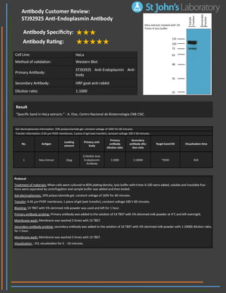
1. Result
“Specific band in HeLa extracts.” - A. Diaz, Centro Nacional de Biotecnologia CNB CSIC.
Cell Line: HeLa
Method of validation: Western Blot
Primary Antibody:
STJ92925 Anti-Endoplasmin Anti-
body
Secondary Antibody: HRP goat anti-rabbit
Dilution ratio: 1:1000
Protocol
Treatment of materials: When cells were cultured to 80% plating density, lysis buffer with triton X-100 were added, soluble and insoluble frac-
tions were separated by centrifugation and sample buffer was added and then boiled.
Gel electrophoresis: 10% polyacrylamide gel, constant voltage of 160V for 60 minutes.
Transfer: 0.45 µm PVDF membrane, 1 piece of gel (wet transfer), constant voltage 100 V 60 minutes.
Blocking: 1X TBST with 5% skimmed milk powder was used and left for 1 hour.
Primary antibody probing: Primary antibody was added to the solution of 1X TBST with 5% skimmed milk powder at 4˚C and left overnight.
Membrane wash: Membrane was washed 3 times with 1X TBST.
Secondary antibody probing: secondary antibody was added to the solution of 1X TBST with 5% skimmed milk powder with 1:10000 dilution ratio,
for 1 hour.
Membrane wash: Membrane was washed 3 times with 1X TBST.
Visualization: : ECL visualization for 5 - 10 minutes.
Antibody Customer Review:
STJ92925 Anti-Endoplasmin Antibody
Antibody Specificity:
Antibody Rating:
Gel electrophoresis information: 10% polyacrylamide gel, constant voltage of 160V for 60 minutes.
Transfer information: 0.45 µm PVDF membrane, 1 piece of gel (wet transfer), constant voltage 100 V 60 minutes.
No. Antigen
Loading
amount
Primary anti-
body
Primary
antibody
dilution ratio
Secondary
antibody dilu-
tion ratio
Target band KD Visualization time
1 HeLa Extract 10µg
STJ92925 Anti-
Endoplasmin
Antibody
1:1000 1:10000 ~92KD N/A