Acts as a tumor suppressor in many tumor types; induces growth arrest or apoptosis depending on the physiological circumstances and cell type. Involved in cell cycle regulation as a trans-activator that acts to negatively regulate cell division by controlling a set of genes required for this process. One of the activated genes is an inhibitor of cyclin-dependent kinases. Apoptosis induction seems to be mediated either by stimulation of BAX and FAS antigen expression, or by repression of Bcl-2 expression. In cooperation with mitochondrial PPIF is involved in activating oxidative stress-induced necrosis; the function is largely independent of transcription. Induces the transcription of long intergenic non-coding RNA p21 (lincRNA-p21) and lincRNA-Mkln1. LincRNA-p21 participates in TP53-dependent transcriptional repression leading to apoptosis and seem to have to effect on cell-cycle regulation. Implicated in Notch signaling cross-over. Prevents CDK7 kinase activity when associated to CAK complex in response to DNA damage, thus stopping cell cycle progression.
Anti-Phospho-p53 (T18)-http://www.stjohnslabs.com/phospho-p53-t18-antibody
Join our Antibody Validation Project - http://www.stjohnslabs.com/services/antibody-validation
Telomerase is a ribonucleoprotein enzyme essential for the replication of chromosome termini in most eukaryotes. Active in progenitor and cancer cells. Inactive, or very low activity, in normal somatic cells. Catalytic component of the teleromerase holoenzyme complex whose main activity is the elongation of telomeres by acting as a reverse transcriptase that adds simple sequence repeats to chromosome ends by copying a template sequence within the RNA component of the enzyme. Catalyzes the RNA-dependent extension of 3'-chromosomal termini with the 6-nucleotide telomeric repeat unit, 5'-TTAGGG-3'. The catalytic cycle involves primer binding, primer extension and release of product once the template boundary has been reached or nascent product translocation followed by further extension. More active on substrates containing 2 or 3 telomeric repeats. Telomerase activity is regulated by a number of factors including telomerase complex-associated proteins, chaperones and polypeptide modifiers. Modulates Wnt signaling. Plays important roles in aging and antiapoptosis.
Anti-TERT-http://www.stjohnslabs.com/anti-tert-antibody?filter_name=STJ98964
Join our Antibody Validation Project - http://www.stjohnslabs.com/services/antibody-validation
Ubiquitin: Exists either covalently attached to another protein, or free (unanchored). When covalently bound, it is conjugated to target proteins via an isopeptide bond either as a monomer (monoubiquitin), a polymer linked via different Lys residues of the ubiquitin (polyubiquitin chains) or a linear polymer linked via the initiator Met of the ubiquitin (linear polyubiquitin chains). Polyubiquitin chains, when attached to a target protein, have different functions depending on the Lys residue of the ubiquitin that is linked: Lys-6-linked may be involved in DNA repair; Lys-11-linked is involved in ERAD (endoplasmic reticulum-associated degradation) and in cell-cycle regulation; Lys-29-linked is involved in lysosomal degradation; Lys-33-linked is involved in kinase modification; Lys-48-linked is involved in protein degradation via the proteasome; Lys-63-linked is involved in endocytosis, DNA-damage responses as well as in signaling processes leading to activation of the transcription factor NF-kappa-B.
Anti-Ub -http://www.stjohnslabs.com/ub-antibody-p-98871
Join our Antibody Validation Project - http://www.stjohnslabs.com/services/antibody-validation
Immunohistochemistry Antibody Validation Report for Anti-CA IX Antibody (STJ9...St John's Laboratory Ltd
Reversible hydration of carbon dioxide. Participates in pH regulation. May be involved in the control of cell proliferation and transformation. Appears to be a novel specific biomarker for a cervical neoplasia.
Anti-CA IX -http://www.stjohnslabs.com/ca-ix-antibody-p-98613
Join our Antibody Validation Project - http://www.stjohnslabs.com/services/antibody-validation
Variant histone H2A which replaces conventional H2A in a subset of nucleosomes. Nucleosomes wrap and compact DNA into chromatin, limiting DNA accessibility to the cellular machineries which require DNA as a template. Histones thereby play a central role in transcription regulation, DNA repair, DNA replication and chromosomal stability. DNA accessibility is regulated via a complex set of post-translational modifications of histones, also called histone code, and nucleosome remodeling. Required for checkpoint-mediated arrest of cell cycle progression in response to low doses of ionizing radiation and for efficient repair of DNA double strand breaks (DSBs) specifically when modified by C-terminal phosphorylation.
Anti-Histone H2A.X-http://www.stjohnslabs.com/histone-h2ax-antibody-p-92631
Join our Antibody Validation Project - http://www.stjohnslabs.com/services/antibody-validation
Receptor for TNFSF6/FASLG. The adapter molecule FADD recruits caspase-8 to the activated receptor. The resulting death-inducing signaling complex (DISC) performs caspase-8 proteolytic activation which initiates the subsequent cascade of caspases (aspartate-specific cysteine proteases) mediating apoptosis. FAS-mediated apoptosis may have a role in the induction of peripheral tolerance, in the antigen-stimulated suicide of mature T-cells, or both. The secreted isoforms 2 to 6 block apoptosis (in vitro).
Anti-FAS-http://www.stjohnslabs.com/fas-antibody-p-92276
Join our Antibody Validation Project - http://www.stjohnslabs.com/services/antibody-validation
Nuclear phosphoprotein which forms a tight but non-covalently linked complex with the JUN/AP-1 transcription factor. In the heterodimer, FOS and JUN/AP-1 basic regions each seems to interact with symmetrical DNA half sites. On TGF-beta activation, forms a multimeric SMAD3/SMAD4/JUN/FOS complex at the AP1/SMAD-binding site to regulate TGF-beta-mediated signaling. Has a critical function in regulating the development of cells destined to form and maintain the skeleton. It is thought to have an important role in signal transduction, cell proliferation and differentiation. In growing cells, activates phospholipid synthesis, possibly by activating CDS1 and PI4K2A. This activity requires Tyr-dephosphorylation and association with the endoplasmic reticulum.
Anti-c-Fos-http://www.stjohnslabs.com/c-fos-antibody-p-91669
Join our Antibody Validation Project - http://www.stjohnslabs.com/services/antibody-validation
Protein tyrosine-protein phosphatase required for T-cell activation through the antigen receptor. Acts as a positive regulator of T-cell coactivation upon binding to DPP4. The first PTPase domain has enzymatic activity, while the second one seems to affect the substrate specificity of the first one. Upon T-cell activation, recruits and dephosphorylates SKAP1 and FYN. Dephosphorylates LYN, and thereby modulates LYN activity (By similarity).
Anti-CD45-http://www.stjohnslabs.com/cd45-antibody-p-98583
Join our Antibody Validation Project - http://www.stjohnslabs.com/services/antibody-validation
Receptor-regulated SMAD (R-SMAD) that is an intracellular signal transducer and transcriptional modulator activated by TGF-beta (transforming growth factor) and activin type 1 receptor kinases. Binds the TRE element in the promoter region of many genes that are regulated by TGF-beta and, on formation of the SMAD2/SMAD4 complex, activates transcription. May act as a tumor suppressor in colorectal carcinoma. Positively regulates PDPK1 kinase activity by stimulating its dissociation from the 14-3-3 protein YWHAQ which acts as a negative regulator.
Anti-Smad2/3-http://www.stjohnslabs.com/smad23-antibody-p-94359
Join our Antibody Validation Project - http://www.stjohnslabs.com/services/antibody-validation
NF-kappa-B is a pleiotropic transcription factor present in almost all cell types and is the endpoint of a series of signal transduction events that are initiated by a vast array of stimuli related to many biological processes such as inflammation, immunity, differentiation, cell growth, tumorigenesis and apoptosis. NF-kappa-B is a homo- or heterodimeric complex formed by the Rel-like domain-containing proteins RELA/p65, RELB, NFKB1/p105, NFKB1/p50, REL and NFKB2/p52 and the heterodimeric p65-p50 complex appears to be most abundant one. The dimers bind at kappa-B sites in the DNA of their target genes and the individual dimers have distinct preferences for different kappa-B sites that they can bind with distinguishable affinity and specificity. Different dimer combinations act as transcriptional activators or repressors, respectively. NF-kappa-B is controlled by various mechanisms of post-translational modification and subcellular compartmentalization as well as by interactions with other cofactors or corepressors.
Anti-NFkB p65-http://www.stjohnslabs.com/nfkb-p65-antibody-p-98648
Join our Antibody Validation Project - http://www.stjohnslabs.com/services/antibody-validation
Receptor for hyaluronic acid (HA). Mediates cell-cell and cell-matrix interactions through its affinity for HA, and possibly also through its affinity for other ligands such as osteopontin, collagens, and matrix metalloproteinases (MMPs). Adhesion with HA plays an important role in cell migration, tumor growth and progression. In cancer cells, may play an important role in invadopodia formation. Also involved in lymphocyte activation, recirculation and homing, and in hematopoiesis. Altered expression or dysfunction causes numerous pathogenic phenotypes. Great protein heterogeneity due to numerous alternative splicing and post-translational modification events. Receptor for LGALS9; the interaction enhances binding of SMAD3 to the FOXP3 promoter, leading to up-regulation of FOXP3 expression and increased induced regulatory T (iTreg) cell stability and suppressive function (By similarity).
Anti-CD44-http://www.stjohnslabs.com/cd44-antibody-p-91575
Join our Antibody Validation Project - http://www.stjohnslabs.com/services/antibody-validation
Antigen KI-67 also known as Ki-67 or MKI67 is a protein that in humans is encoded by the MKI67 gene (antigen identified by monoclonal antibody Ki-67). Antigen KI-67 is a nuclear protein that is associated with and may be necessary for cellular proliferation. Furthermore, it is associated with ribosomal RNA transcription.[5] Inactivation of antigen KI-67 leads to inhibition of ribosomal RNA synthesis.
Anti-Ki67 -http://www.stjohnslabs.com/ki-67-antibody-p-92897
Join our Antibody Validation Project - http://www.stjohnslabs.com/services/antibody-validation
Core component of nucleosome. Nucleosomes wrap and compact DNA into chromatin, limiting DNA accessibility to the cellular machineries which require DNA as a template. Histones thereby play a central role in transcription regulation, DNA repair, DNA replication and chromosomal stability. DNA accessibility is regulated via a complex set of post-translational modifications of histones, also called histone code, and nucleosome remodeling.
Anti-Histone H2A-http://www.stjohnslabs.com/histone-h2a-antibody-p-98500
Join our Antibody Validation Project - http://www.stjohnslabs.com/services/antibody-validation
Suppresses apoptosis in a variety of cell systems including factor-dependent lymphohematopoietic and neural cells. Regulates cell death by controlling the mitochondrial membrane permeability. Appears to function in a feedback loop system with caspases. Inhibits caspase activity either by preventing the release of cytochrome c from the mitochondria and/or by binding to the apoptosis-activating factor (APAF-1). May attenuate inflammation by impairing NLRP1-inflammasome activation, hence CASP1 activation and IL1B release .
Anti-Bcl-2-http://www.stjohnslabs.com/bcl-2-antibody-1
Join our Antibody Validation Project - http://www.stjohnslabs.com/services/antibody-validation
Receptor for the Fc region of IgG. Binds complexed or aggregated IgG and also monomeric IgG. Mediates antibody-dependent cellular cytotoxicity (ADCC) and other antibody-dependent responses, such as phagocytosis.
Anti-CD16-http://www.stjohnslabs.com/cd16-antibody-p-98627
Join our Antibody Validation Project - http://www.stjohnslabs.com/services/antibody-validation
Accelerates programmed cell death by binding to, and antagonizing the apoptosis repressor BCL2 or its adenovirus homolog E1B 19k protein. Under stress conditions, undergoes a conformation change that causes translocation to the mitochondrion membrane, leading to the release of cytochrome c that then triggers apoptosis. Promotes activation of CASP3, and thereby apoptosis.
Anti-Bax-http://www.stjohnslabs.com/bax-antibody-p-91328
Join our Antibody Validation Project - http://www.stjohnslabs.com/services/antibody-validation
Serine/threonine kinase which acts as an essential component of the MAP kinase signal transduction pathway. MAPK1/ERK2 and MAPK3/ERK1 are the 2 MAPKs which play an important role in the MAPK/ERK cascade. They participate also in a signaling cascade initiated by activated KIT and KITLG/SCF. Depending on the cellular context, the MAPK/ERK cascade mediates diverse biological functions such as cell growth, adhesion, survival and differentiation through the regulation of transcription, translation, cytoskeletal rearrangements. The MAPK/ERK cascade plays also a role in initiation and regulation of meiosis, mitosis, and postmitotic functions in differentiated cells by phosphorylating a number of transcription factors. About 160 substrates have already been discovered for ERKs. Many of these substrates are localized in the nucleus, and seem to participate in the regulation of transcription upon stimulation.
Anti-ERK1-http://www.stjohnslabs.com/erk1-antibody-3
Join our Antibody Validation Project - http://www.stjohnslabs.com/services/antibody-validation
Involved in redox regulation of the cell. Reduces peroxides with reducing equivalents provided through the thioredoxin system but not from glutaredoxin. May play an important role in eliminating peroxides generated during metabolism. Might participate in the signaling cascades of growth factors and tumor necrosis factor-alpha by regulating the intracellular concentrations of H2O2. Reduces an intramolecular disulfide bond in GDPD5 that gates the ability to GDPD5 to drive postmitotic motor neuron differentiation (By similarity). / 2 R'-SH + ROOH = R'-S-S-R' + H2O + ROH.
Anti-Peroxiredoxin 1-http://www.stjohnslabs.com/peroxiredoxin-1-antibody-p-98649
Join our Antibody Validation Project - http://www.stjohnslabs.com/services/antibody-validation
Integrin alpha-IIb/beta-3 is a receptor for fibronectin, fibrinogen, plasminogen, prothrombin, thrombospondin and vitronectin. It recognizes the sequence R-G-D in a wide array of ligands. It recognizes the sequence H-H-L-G-G-G-A-K-Q-A-G-D-V in fibrinogen gamma chain. Following activation integrin alpha-IIb/beta-3 brings about platelet/platelet interaction through binding of soluble fibrinogen. This step leads to rapid platelet aggregation which physically plugs ruptured endothelial cell surface.
Anti-CD41-http://www.stjohnslabs.com/cd41-antibody
Join our Antibody Validation Project - http://www.stjohnslabs.com/services/antibody-validation
Serine/threonine kinase which acts as an essential component of the MAP kinase signal transduction pathway. MAPK1/ERK2 and MAPK3/ERK1 are the 2 MAPKs which play an important role in the MAPK/ERK cascade. They participate also in a signaling cascade initiated by activated KIT and KITLG/SCF. Depending on the cellular context, the MAPK/ERK cascade mediates diverse biological functions such as cell growth, adhesion, survival and differentiation through the regulation of transcription, translation, cytoskeletal rearrangements.
Anti-Phospho-ERK 1/2 (T202/Y204)-http://www.stjohnslabs.com/phospho-erk-12-t202y204-antibody
Join our Antibody Validation Project - http://www.stjohnslabs.com/services/antibody-validation
Collagen alpha-2(I) chain is a protein that in humans is encoded by the COL1A2 gene.
This gene encodes one of the chains for type I collagen, the fibrillar collagen found in most connective tissues. Mutations in this gene are associated with osteogenesis imperfecta, Ehlers-Danlos syndrome, idiopathic osteoporosis, and atypical Marfan syndrome. Symptoms associated with mutations in this gene, however, tend to be less severe than mutations in the gene for alpha-1 type I collagen since alpha-2 is less abundant. Multiple messages for this gene result from multiple polyadenylation signals, a feature shared by most of the other collagen genes.
Anti-COL1A2 -http://www.stjohnslabs.com/col1a2-antibody
Join our Antibody Validation Project - http://www.stjohnslabs.com/services/antibody-validation
Tyrosine-protein kinase that acts as a cell-surface receptor for PDGFA, PDGFB and PDGFC and plays an essential role in the regulation of embryonic development, cell proliferation, survival and chemotaxis. Depending on the context, promotes or inhibits cell proliferation and cell migration. Plays an important role in the differentiation of bone marrow-derived mesenchymal stem cells. Required for normal skeleton development and cephalic closure during embryonic development. Required for normal development of the mucosa lining the gastrointestinal tract, and for recruitment of mesenchymal cells and normal development of intestinal villi. Plays a role in cell migration and chemotaxis in wound healing. Plays a role in platelet activation, secretion of agonists from platelet granules, and in thrombin-induced platelet aggregation.
Anti-PDGFRα-http://www.stjohnslabs.com/pdgfra-antibody-2
Join our Antibody Validation Project - http://www.stjohnslabs.com/services/antibody-validation
Transcription activator that binds DNA cooperatively with DP proteins through the E2 recognition site, 5'-TTTC[CG]CGC-3' found in the promoter region of a number of genes whose products are involved in cell cycle regulation or in DNA replication. The DRTF1/E2F complex functions in the control of cell-cycle progression from G1 to S phase. E2F1 binds preferentially RB1 in a cell-cycle dependent manner. It can mediate both cell proliferation and TP53/p53-dependent apoptosis. Blocks adipocyte differentiation by binding to specific promoters repressing CEBPA binding to its target gene promoters . / BIRC2/c-IAP1 stimulates its transcriptional activity.
Anti-E2F-1-http://www.stjohnslabs.com/e2f-1-antibody-1
Join our Antibody Validation Project - http://www.stjohnslabs.com/services/antibody-validation
Ligand for multiple Notch receptors and involved in the mediation of Notch signaling. May be involved in cell-fate decisions during hematopoiesis. Seems to be involved in early and late stages of mammalian cardiovascular development. Inhibits myoblast differentiation (By similarity). Enhances fibroblast growth factor-induced angiogenesis (in vitro).
Anti-Jagged1-http://www.stjohnslabs.com/jagged1-antibody
Join our Antibody Validation Project - http://www.stjohnslabs.com/services/antibody-validation
Transcription factor that recognizes and binds to the enhancer heptamer motif 5'-TGA[CG]TCA-3'. Promotes activity of NR5A1 when phosphorylated by HIPK3 leading to increased steroidogenic gene expression upon cAMP signaling pathway stimulation. Involved in activated KRAS-mediated transcriptional activation of USP28 in colorectal cancer (CRC) cells . Binds to the USP28 promoter in colorectal cancer (CRC) cells .
Anti-AP-1-http://www.stjohnslabs.com/ap-1-antibody-p-91169
Join our Antibody Validation Project - http://www.stjohnslabs.com/services/antibody-validation
Cytokine with a wide variety of biological functions. It is a potent inducer of the acute phase response. Plays an essential role in the final differentiation of B-cells into Ig-secreting cells Involved in lymphocyte and monocyte differentiation. Acts on B-cells, T-cells, hepatocytes, hematopoietic progenitor cells and cells of the CNS. Required for the generation of T(H)17 cells. Also acts as a myokine. It is discharged into the bloodstream after muscle contraction and acts to increase the breakdown of fats and to improve insulin resistance. It induces myeloma and plasmacytoma growth and induces nerve cells differentiation.
Anti-IL-6-http://www.stjohnslabs.com/il-6-antibody
Join our Antibody Validation Project - http://www.stjohnslabs.com/services/antibody-validation
IL-8 is a chemotactic factor that attracts neutrophils, basophils, and T-cells, but not monocytes. It is also involved in neutrophil activation. It is released from several cell types in response to an inflammatory stimulus. IL-8(6-77) has a 5-10-fold higher activity on neutrophil activation, IL-8(5-77) has increased activity on neutrophil activation and IL-8(7-77) has a higher affinity to receptors CXCR1 and CXCR2 as compared to IL-8(1-77), respectively.
Anti-IL8-http://www.stjohnslabs.com/il-8-antibody-p-94941
Join our Antibody Validation Project - http://www.stjohnslabs.com/services/antibody-validation
atalytic subunit of AMP-activated protein kinase (AMPK), an energy sensor protein kinase that plays a key role in regulating cellular energy metabolism. In response to reduction of intracellular ATP levels, AMPK activates energy-producing pathways and inhibits energy-consuming processes: inhibits protein, carbohydrate and lipid biosynthesis, as well as cell growth and proliferation. AMPK acts via direct phosphorylation of metabolic enzymes, and by longer-term effects via phosphorylation of transcription regulators. Also acts as a regulator of cellular polarity by remodeling the actin cytoskeleton; probably by indirectly activating myosin. Regulates lipid synthesis by phosphorylating and inactivating lipid metabolic enzymes such as ACACA, ACACB, GYS1, HMGCR and LIPE; regulates fatty acid and cholesterol synthesis by phosphorylating acetyl-CoA carboxylase (ACACA and ACACB) and hormone-sensitive lipase (LIPE) enzymes, respectively.
Anti-AMPKα1-http://www.stjohnslabs.com/ampka1-antibody
Join our Antibody Validation Project - http://www.stjohnslabs.com/services/antibody-validation
Functions as key mediator in apoptosis and inflammation. Promotes caspase-mediated apoptosis involving predominantly caspase-8 and also caspase-9 in a probable cell type-specific manner. Involved in activation of the mitochondrial apoptotic pathway, promotes caspase-8-dependent proteolytic maturation of BID independently of FADD in certain cell types and also mediates mitochondrial translocation of BAX and activates BAX-dependent apoptosis coupled to activation of caspase-9, -2 and -3. Involved in macrophage pyroptosis, a caspase-1-dependent inflammatory form of cell death and is the major constituent of the ASC pyroptosome which forms upon potassium depletion and rapidly recruits and activates caspase-1. In innate immune response believed to act as an integral adapter in the assembly of the inflammasome which activates caspase-1 leading to processing and secretion of proinflammatory cytokines.
Anti-ASC-http://www.stjohnslabs.com/asc-antibody-p-91257
Join our Antibody Validation Project - http://www.stjohnslabs.com/services/antibody-validation
Binds to activated (phosphorylated) protein-Tyr kinases, through its SH2 domain, and acts as an adapter, mediating the association of the p110 catalytic unit to the plasma membrane. Necessary for the insulin-stimulated increase in glucose uptake and glycogen synthesis in insulin-sensitive tissues. Plays an important role in signaling in response to FGFR1, FGFR2, FGFR3, FGFR4, KITLG/SCF, KIT, PDGFRA and PDGFRB. Likewise, plays a role in ITGB2 signaling . Modulates the cellular response to ER stress by promoting nuclear translocation of XBP1 isoform 2 in a ER stress-and/or insulin-dependent manner during metabolic overloading in the liver and hence plays a role in glucose tolerance improvement.
Anti-Phospho-PI 3-kinase p85/p55 (Y467/199) -http://www.stjohnslabs.com/phospho-pi-3-kinase-p85p55-y467199-antibody
Join our Antibody Validation Project - http://www.stjohnslabs.com/services/antibody-validation
IκBα (NF-kappa-B inhibitor alpha) is a cellular protein that inhibits the NF-κB transcription factor through masking nuclear localisation signals of NF-κB proteins and keeping them inactive in the cytoplasm. The Phospho-IκB-α (S32/S36) polyclonal antibody detects endogenous levels of IκB-α protein only when phosphorylated at S32/S36.
Immunofluorescence Antibody Validation Report for Anti-Phospho-NFκB-p65 (S529...St John's Laboratory Ltd
NF-kappa-B is a pleiotropic transcription factor present in almost all cell types and is the endpoint of a series of signal transduction events that are initiated by a vast array of stimuli related to many biological processes such as inflammation, immunity, differentiation, cell growth, tumorigenesis and apoptosis. NF-kappa-B is a homo- or heterodimeric complex formed by the Rel-like domain-containing proteins RELA/p65, RELB, NFKB1/p105, NFKB1/p50, REL and NFKB2/p52 and the heterodimeric p65-p50 complex appears to be most abundant one. The dimers bind at kappa-B sites in the DNA of their target genes and the individual dimers have distinct preferences for different kappa-B sites that they can bind with distinguishable affinity and specificity.
Anti-Phospho-NFκB-p65 (S529) -http://www.stjohnslabs.com/phospho-nfkb-p65-s529-antibody
Join our Antibody Validation Project - http://www.stjohnslabs.com/services/antibody-validation
Promotes cell death. Successfully competes for the binding to Bcl-X(L), Bcl-2 and Bcl-W, thereby affecting the level of heterodimerization of these proteins with BAX. Can reverse the death repressor activity of Bcl-X(L), but not that of Bcl-2 (By similarity). Appears to act as a link between growth factor receptor signaling and the apoptotic pathways.
Anti-Bad-http://www.stjohnslabs.com/bad-antibody-p-91313
Join our Antibody Validation Project - http://www.stjohnslabs.com/services/antibody-validation
Catalytic subunit of AMP-activated protein kinase (AMPK), an energy sensor protein kinase that plays a key role in regulating cellular energy metabolism. In response to reduction of intracellular ATP levels, AMPK activates energy-producing pathways and inhibits energy-consuming processes: inhibits protein, carbohydrate and lipid biosynthesis, as well as cell growth and proliferation. AMPK acts via direct phosphorylation of metabolic enzymes, and by longer-term effects via phosphorylation of transcription regulators. Also acts as a regulator of cellular polarity by remodeling the actin cytoskeleton; probably by indirectly activating myosin. Regulates lipid synthesis by phosphorylating and inactivating lipid metabolic enzymes such as ACACA, ACACB, GYS1, HMGCR and LIPE; regulates fatty acid and cholesterol synthesis by phosphorylating acetyl-CoA carboxylase (ACACA and ACACB) and hormone-sensitive lipase (LIPE) enzymes, respectively.
Anti-AMPKα1/2-http://www.stjohnslabs.com/ampka12-antibody
Join our Antibody Validation Project - http://www.stjohnslabs.com/services/antibody-validation
Serine/threonine kinase which acts as an essential component of the MAP kinase signal transduction pathway. MAPK14 is one of the four p38 MAPKs which play an important role in the cascades of cellular responses evoked by extracellular stimuli such as proinflammatory cytokines or physical stress leading to direct activation of transcription factors. Accordingly, p38 MAPKs phosphorylate a broad range of proteins and it has been estimated that they may have approximately 200 to 300 substrates each. Some of the targets are downstream kinases which are activated through phosphorylation and further phosphorylate additional targets. RPS6KA5/MSK1 and RPS6KA4/MSK2 can directly phosphorylate and activate transcription factors such as CREB1, ATF1, the NF-kappa-B isoform RELA/NFKB3, STAT1 and STAT3, but can also phosphorylate histone H3 and the nucleosomal protein HMGN1. RPS6KA5/MSK1 and RPS6KA4/MSK2 play important roles in the rapid induction of immediate-early genes in response to stress or mitogenic stimuli, either by inducing chromatin remodeling or by recruiting the transcription machinery.
Anti-Phospho-p38 (T180)-http://www.stjohnslabs.com/phospho-p38-t180-antibody
Join our Antibody Validation Project - http://www.stjohnslabs.com/services/antibody-validation
Non-receptor protein-tyrosine kinase that plays an essential role in regulating cell migration, adhesion, spreading, reorganization of the actin cytoskeleton, formation and disassembly of focal adhesions and cell protrusions, cell cycle progression, cell proliferation and apoptosis. Required for early embryonic development and placenta development. Required for embryonic angiogenesis, normal cardiomyocyte migration and proliferation, and normal heart development. Regulates axon growth and neuronal cell migration, axon branching and synapse formation; required for normal development of the nervous system. Plays a role in osteogenesis and differentiation of osteoblasts. Functions in integrin signal transduction, but also in signaling downstream of numerous growth factor receptors, G-protein coupled receptors (GPCR), EPHA2, netrin receptors and LDL receptors.
Anti-Phospho-FAK (Y397)-http://www.stjohnslabs.com/phospho-fak-y397-antibody-p-90511
Join our Antibody Validation Project - http://www.stjohnslabs.com/services/antibody-validation
Promotes microtubule assembly and stability, and might be involved in the establishment and maintenance of neuronal polarity. The C-terminus binds axonal microtubules while the N-terminus binds neural plasma membrane components, suggesting that tau functions as a linker protein between both.
Pathologies and dementias of the nervous system such as Alzheimer's disease and Parkinson's disease are associated with tau proteins that have become defective and no longer stabilize microtubules properly.
The tau proteins are the product of alternative splicing from a single gene that in humans is designated MAPT (microtubule-associated protein tau) and is located on chromosome 17.
Anti-Phospho-Tau (S396) -http://www.stjohnslabs.com/phospho-tau-s396-antibody
Join our Antibody Validation Project - http://www.stjohnslabs.com/services/antibody-validation
Immunohistochemistry Antibody Validation Report for Anti-Phospho-MEK-1/2 (S21...St John's Laboratory Ltd
Dual specificity protein kinase which acts as an essential component of the MAP kinase signal transduction pathway. Binding of extracellular ligands such as growth factors, cytokines and hormones to their cell-surface receptors activates RAS and this initiates RAF1 activation. RAF1 then further activates the dual-specificity protein kinases MAP2K1/MEK1 and MAP2K2/MEK2. Both MAP2K1/MEK1 and MAP2K2/MEK2 function specifically in the MAPK/ERK cascade, and catalyze the concomitant phosphorylation of a threonine and a tyrosine residue in a Thr-Glu-Tyr sequence located in the extracellular signal-regulated kinases MAPK3/ERK1 and MAPK1/ERK2, leading to their activation and further transduction of the signal within the MAPK/ERK cascade. Depending on the cellular context, this pathway mediates diverse biological functions such as cell growth, adhesion, survival and differentiation, predominantly through the regulation of transcription, metabolism and cytoskeletal rearrangements.
Anti-Phospho-MEK-1/2 (S218/222)-http://www.stjohnslabs.com/phospho-mek-12-s218222-antibody
Join our Antibody Validation Project - http://www.stjohnslabs.com/services/antibody-validation
Immunohistochemistry Antibody Validation Report for Anti-Phospho-JAK1 (Y1022)...St John's Laboratory Ltd
Tyrosine kinase of the non-receptor type, involved in the IFN-alpha/beta/gamma signal pathway . Kinase partner for the interleukin (IL)-2 receptor . / ATP + a [protein]-L-tyrosine = ADP + a [protein]-L-tyrosine phosphate. / Mg2+
Anti-Phospho-JAK1 (Y1022)-http://www.stjohnslabs.com/phospho-jak1-y1022-antibody
Join our Antibody Validation Project - http://www.stjohnslabs.com/services/antibody-validation
Immunohistochemistry Antibody Validation Report for Anti-Phospho-CaMKIIβ/γ/δ ...St John's Laboratory Ltd
Calcium/calmodulin-dependent protein kinase that functions autonomously after Ca2+/calmodulin-binding and autophosphorylation, and is involved in dendritic spine and synapse formation, neuronal plasticity and regulation of sarcoplasmic reticulum Ca2+ transport in skeletal muscle. In neurons, plays an essential structural role in the reorganization of the actin cytoskeleton during plasticity by binding and bundling actin filaments in a kinase-independent manner. This structural function is required for correct targeting of CaMK2A, which acts downstream of NMDAR to promote dendritic spine and synapse formation and maintain synaptic plasticity which enables long-term potentiation (LTP) and hippocampus-dependent learning. In developing hippocampal neurons, promotes arborization of the dendritic tree and in mature neurons, promotes dendritic remodeling.
Anti-Phospho-CaMKIIβ/γ/δ (T287)-http://www.stjohnslabs.com/phospho-camkiibgd-t287-antibody
Join our Antibody Validation Project - http://www.stjohnslabs.com/services/antibody-validation
Immunohistochemistry Antibody Validation Report for Anti-Phospho-NFκB-p65 (S2...St John's Laboratory Ltd
NF-kappa-B is a pleiotropic transcription factor present in almost all cell types and is the endpoint of a series of signal transduction events that are initiated by a vast array of stimuli related to many biological processes such as inflammation, immunity, differentiation, cell growth, tumorigenesis and apoptosis. NF-kappa-B is a homo- or heterodimeric complex formed by the Rel-like domain-containing proteins RELA/p65, RELB, NFKB1/p105, NFKB1/p50, REL and NFKB2/p52 and the heterodimeric p65-p50 complex appears to be most abundant one. The dimers bind at kappa-B sites in the DNA of their target genes and the individual dimers have distinct preferences for different kappa-B sites that they can bind with distinguishable affinity and specificity.
Anti-Phospho-NFκB-p65 (S276)-http://www.stjohnslabs.com/phospho-nfkb-p65-s276-antibody
Join our Antibody Validation Project - http://www.stjohnslabs.com/services/antibody-validation
Acts as a tumor suppressor in many tumor types; induces growth arrest or apoptosis depending on the physiological circumstances and cell type. Involved in cell cycle regulation as a trans-activator that acts to negatively regulate cell division by controlling a set of genes required for this process. One of the activated genes is an inhibitor of cyclin-dependent kinases. Apoptosis induction seems to be mediated either by stimulation of BAX and FAS antigen expression, or by repression of Bcl-2 expression. In cooperation with mitochondrial PPIF is involved in activating oxidative stress-induced necrosis; the function is largely independent of transcription. Induces the transcription of long intergenic non-coding RNA p21 (lincRNA-p21) and lincRNA-Mkln1. LincRNA-p21 participates in TP53-dependent transcriptional repression leading to apoptosis and seem to have to effect on cell-cycle regulation. Implicated in Notch signaling cross-over. Prevents CDK7 kinase activity when associated to CAK complex in response to DNA damage, thus stopping cell cycle progression.
Anti-Phospho-p53 (S15)-http://www.stjohnslabs.com/phospho-p53-s15-antibody
Join our Antibody Validation Project - http://www.stjohnslabs.com/services/antibody-validation
Immunohistochemistry Antibody Validation Report for Anti-Phospho-PTEN (S380/T...St John's Laboratory Ltd
Tumor suppressor. Acts as a dual-specificity protein phosphatase, dephosphorylating tyrosine-, serine- and threonine-phosphorylated proteins. Also acts as a lipid phosphatase, removing the phosphate in the D3 position of the inositol ring from phosphatidylinositol 3, 4, 5-trisphosphate, phosphatidylinositol 3, 4-diphosphate, phosphatidylinositol 3-phosphate and inositol 1, 3, 4, 5-tetrakisphosphate with order of substrate preference in vitro PtdIns(3, 4, 5)P3 > PtdIns(3, 4)P2 > PtdIns3P > Ins(1, 3, 4, 5)P4. The lipid phosphatase activity is critical for its tumor suppressor function. Antagonizes the PI3K-AKT/PKB signaling pathway by dephosphorylating phosphoinositides and thereby modulating cell cycle progression and cell survival. The unphosphorylated form cooperates with AIP1 to suppress AKT1 activation. Dephosphorylates tyrosine-phosphorylated focal adhesion kinase and inhibits cell migration and integrin-mediated cell spreading and focal adhesion formation. Plays a role as a key modulator of the AKT-mTOR signaling pathway controlling the tempo of the process of newborn neurons integration during adult neurogenesis, including correct neuron positioning, dendritic development and synapse formation. May be a negative regulator of insulin signaling and glucose metabolism in adipose tissue.
Anti-Phospho-PTEN (S380/T382/T383)-http://www.stjohnslabs.com/phospho-pten-s380t382t383-antibody
Join our Antibody Validation Project - http://www.stjohnslabs.com/services/antibody-validation
Immunohistochemistry Antibody Validation Report for Anti-Phospho-FoxO3A (S253...St John's Laboratory Ltd
Transcriptional activator which triggers apoptosis in the absence of survival factors, including neuronal cell death upon oxidative stress. Recognizes and binds to the DNA sequence 5'-[AG]TAAA[TC]A-3'. Participates in post-transcriptional regulation of MYC: following phosphorylation by MAPKAPK5, promotes induction of miR-34b and miR-34c expression, 2 post-transcriptional regulators of MYC that bind to the 3'UTR of MYC transcript and prevent its translation
Anti-Phospho-FoxO3A (S253)-http://www.stjohnslabs.com/phospho-foxo3a-s253-antibody
Join our Antibody Validation Project - http://www.stjohnslabs.com/services/antibody-validation
Immunohistochemistry Antibody Validation Report for Anti-Phospho-mTOR (S2448)...St John's Laboratory Ltd
Serine/threonine protein kinase which is a central regulator of cellular metabolism, growth and survival in response to hormones, growth factors, nutrients, energy and stress signals. MTOR directly or indirectly regulates the phosphorylation of at least 800 proteins. Functions as part of 2 structurally and functionally distinct signaling complexes mTORC1 and mTORC2 (mTOR complex 1 and 2). Activated mTORC1 up-regulates protein synthesis by phosphorylating key regulators of mRNA translation and ribosome synthesis. This includes phosphorylation of EIF4EBP1 and release of its inhibition toward the elongation initiation factor 4E (eiF4E). Moreover, phosphorylates and activates RPS6KB1 and RPS6KB2 that promote protein synthesis by modulating the activity of their downstream targets including ribosomal protein S6, eukaryotic translation initiation factor EIF4B, and the inhibitor of translation initiation PDCD4.
Anti-Phospho-mTOR (S2448)-http://www.stjohnslabs.com/phospho-mtor-s2448-antibody
Join our Antibody Validation Project - http://www.stjohnslabs.com/services/antibody-validation
Serine/threonine kinase which acts as an essential component of the MAP kinase signal transduction pathway. MAPK14 is one of the four p38 MAPKs which play an important role in the cascades of cellular responses evoked by extracellular stimuli such as proinflammatory cytokines or physical stress leading to direct activation of transcription factors. Accordingly, p38 MAPKs phosphorylate a broad range of proteins and it has been estimated that they may have approximately 200 to 300 substrates each. Some of the targets are downstream kinases which are activated through phosphorylation and further phosphorylate additional targets. RPS6KA5/MSK1 and RPS6KA4/MSK2 can directly phosphorylate and activate transcription factors such as CREB1, ATF1, the NF-kappa-B isoform RELA/NFKB3, STAT1 and STAT3, but can also phosphorylate histone H3 and the nucleosomal protein HMGN1. RPS6KA5/MSK1 and RPS6KA4/MSK2 play important roles in the rapid induction of immediate-early genes in response to stress or mitogenic stimuli, either by inducing chromatin remodeling or by recruiting the transcription machinery. On the other hand, two other kinase targets, MAPKAPK2/MK2 and MAPKAPK3/MK3, participate in the control of gene expression mostly at the post-transcriptional level, by phosphorylating ZFP36 (tristetraprolin) and ELAVL1, and by regulating EEF2K, which is important for the elongation of mRNA during translation.
Anti-Phospho-p38 (Y323)-http://www.stjohnslabs.com/phospho-p38-y323-antibody
Join our Antibody Validation Project - http://www.stjohnslabs.com/services/antibody-validation
Immunohistochemistry Antibody Validation Report for Anti-Phospho-TGFβ RI (S16...St John's Laboratory Ltd
Transmembrane serine/threonine kinase forming with the TGF-beta type II serine/threonine kinase receptor, TGFBR2, the non-promiscuous receptor for the TGF-beta cytokines TGFB1, TGFB2 and TGFB3. Transduces the TGFB1, TGFB2 and TGFB3 signal from the cell surface to the cytoplasm and is thus regulating a plethora of physiological and pathological processes including cell cycle arrest in epithelial and hematopoietic cells, control of mesenchymal cell proliferation and differentiation, wound healing, extracellular matrix production, immunosuppression and carcinogenesis. The formation of the receptor complex composed of 2 TGFBR1 and 2 TGFBR2 molecules symmetrically bound to the cytokine dimer results in the phosphorylation and the activation of TGFBR1 by the constitutively active TGFBR2. Activated TGFBR1 phosphorylates SMAD2 which dissociates from the receptor and interacts with SMAD4. The SMAD2-SMAD4 complex is subsequently translocated to the nucleus where it modulates the transcription of the TGF-beta-regulated genes.
Anti-Phospho-TGFβ RI (S165)-http://www.stjohnslabs.com/phospho-tgfb-ri-s165-antibody
Join our Antibody Validation Project - http://www.stjohnslabs.com/services/antibody-validation
Immunohistochemistry Antibody Validation Report for Anti-Phospho-IκB-α (S32/S...St John's Laboratory Ltd
Inhibits the activity of dimeric NF-kappa-B/REL complexes by trapping REL dimers in the cytoplasm through masking of their nuclear localization signals. On cellular stimulation by immune and proinflammatory responses, becomes phosphorylated promoting ubiquitination and degradation, enabling the dimeric RELA to translocate to the nucleus and activate transcription.
Anti-Phospho-IκB-α (S32/S36)-http://www.stjohnslabs.com/phospho-ikb-a-s32s36-antibody
Join our Antibody Validation Project - http://www.stjohnslabs.com/services/antibody-validation
Immunohistochemistry Antibody Validation Report for Anti-Phospho-Histone H2A....St John's Laboratory Ltd
Variant histone H2A which replaces conventional H2A in a subset of nucleosomes. Nucleosomes wrap and compact DNA into chromatin, limiting DNA accessibility to the cellular machineries which require DNA as a template. Histones thereby play a central role in transcription regulation, DNA repair, DNA replication and chromosomal stability. DNA accessibility is regulated via a complex set of post-translational modifications of histones, also called histone code, and nucleosome remodeling. Required for checkpoint-mediated arrest of cell cycle progression in response to low doses of ionizing radiation and for efficient repair of DNA double strand breaks (DSBs) specifically when modified by C-terminal phosphorylation.
Anti-Phospho-Histone H2A.X (S139)-http://www.stjohnslabs.com/phospho-histone-h2ax-s139-antibody
Join our Antibody Validation Project - http://www.stjohnslabs.com/services/antibody-validation
Transthyretin (TTR) is a transport protein in the serum and cerebrospinal fluid that carries the thyroid hormone thyroxine (T4) and retinol-binding protein bound to retinol. This is how transthyretin gained its name: transports thyroxine and retinol. The liver secretes transthyretin into the blood, and the choroid plexus secretes TTR into the cerebrospinal fluid.
TTR was originally called prealbumin(or thyroxine-binding prealbumin) because it ran faster than albumin on electrophoresis gels.
Anti-TTR - http://www.stjohnslabs.com/anti-ttr-antibody?filter_name=STJ98876
Join our Antibody Validation Project - http://www.stjohnslabs.com/services/antibody-validation
Involved in the base excision repair (BER) pathway, by catalyzing the poly(ADP-ribosyl)ation of a limited number of acceptor proteins involved in chromatin architecture and in DNA metabolism. This modification follows DNA damages and appears as an obligatory step in a detection/signaling pathway leading to the reparation of DNA strand breaks. Mediates the poly(ADP-ribosyl)ation of APLF and CHFR. Positively regulates the transcription of MTUS1 and negatively regulates the transcription of MTUS2/TIP150. With EEF1A1 and TXK, forms a complex that acts as a T-helper 1 (Th1) cell-specific transcription factor and binds the promoter of IFN-gamma to directly regulate its transcription, and is thus involved importantly in Th1 cytokine production. Required for PARP9 and DTX3L recruitment to DNA damage sites. PARP1-dependent PARP9-DTX3L-mediated ubiquitination promotes the rapid and specific recruitment of 53BP1/TP53BP1, UIMC1/RAP80, and BRCA1 to DNA damage sites.
Anti-Cleaved PARP-http://www.stjohnslabs.com/cleaved-parp-antibody-p-98682
Join our Antibody Validation Project - http://www.stjohnslabs.com/services/antibody-validation
Immunohistochemistry Antibody Validation Report for Anti-Phospho-Stat3 (S727)...St John's Laboratory Ltd
ignal transducer and transcription activator that mediates cellular responses to interleukins, KITLG/SCF, LEP and other growth factors. Once activated, recruits coactivators, such as NCOA1 or MED1, to the promoter region of the target gene . May mediate cellular responses to activated FGFR1, FGFR2, FGFR3 and FGFR4. Binds to the interleukin-6 (IL-6)-responsive elements identified in the promoters of various acute-phase protein genes. Activated by IL31 through IL31RA. Involved in cell cycle regulation by inducing the expression of key genes for the progression from G1 to S phase, such as CCND1 . Mediates the effects of LEP on melanocortin production, body energy homeostasis and lactation (By similarity). May play an apoptotic role by transctivating BIRC5 expression under LEP activation . Cytoplasmic STAT3 represses macroautophagy by inhibiting EIF2AK2/PKR activity.
Anti-Phospho-Stat3 (S727)-http://www.stjohnslabs.com/phospho-stat3-s727-antibody
Join our Antibody Validation Project - http://www.stjohnslabs.com/services/antibody-validation
Immunohistochemistry Antibody Validation Report for Anti-Phospho-Stat3 (Y705)...St John's Laboratory Ltd
Signal transducer and transcription activator that mediates cellular responses to interleukins, KITLG/SCF, LEP and other growth factors. Once activated, recruits coactivators, such as NCOA1 or MED1, to the promoter region of the target gene . May mediate cellular responses to activated FGFR1, FGFR2, FGFR3 and FGFR4. Binds to the interleukin-6 (IL-6)-responsive elements identified in the promoters of various acute-phase protein genes. Activated by IL31 through IL31RA. Involved in cell cycle regulation by inducing the expression of key genes for the progression from G1 to S phase, such as CCND1 . Mediates the effects of LEP on melanocortin production, body energy homeostasis and lactation (By similarity). May play an apoptotic role by transctivating BIRC5 expression under LEP activation . Cytoplasmic STAT3 represses macroautophagy by inhibiting EIF2AK2/PKR activity.
Anti-Phospho-Stat3 (Y705)-http://www.stjohnslabs.com/phospho-stat3-y705-antibody
Join our Antibody Validation Project - http://www.stjohnslabs.com/services/antibody-validation
Serine/threonine kinase which acts as an essential component of the MAP kinase signal transduction pathway. MAPK14 is one of the four p38 MAPKs which play an important role in the cascades of cellular responses evoked by extracellular stimuli such as proinflammatory cytokines or physical stress leading to direct activation of transcription factors. Accordingly, p38 MAPKs phosphorylate a broad range of proteins and it has been estimated that they may have approximately 200 to 300 substrates each. Some of the targets are downstream kinases which are activated through phosphorylation and further phosphorylate additional targets. RPS6KA5/MSK1 and RPS6KA4/MSK2 can directly phosphorylate and activate transcription factors such as CREB1, ATF1, the NF-kappa-B isoform RELA/NFKB3, STAT1 and STAT3, but can also phosphorylate histone H3 and the nucleosomal protein HMGN1. RPS6KA5/MSK1 and RPS6KA4/MSK2 play important roles in the rapid induction of immediate-early genes in response to stress or mitogenic stimuli, either by inducing chromatin remodeling or by recruiting the transcription machinery.
Anti-p38-http://www.stjohnslabs.com/p38-antibody-p-93734
Join our Antibody Validation Project - http://www.stjohnslabs.com/services/antibody-validation
Tyrosine-protein kinase that acts as a cell-surface receptor for VEGFA, VEGFC and VEGFD. Plays an essential role in the regulation of angiogenesis, vascular development, vascular permeability, and embryonic hematopoiesis. Promotes proliferation, survival, migration and differentiation of endothelial cells. Promotes reorganization of the actin cytoskeleton. Isoforms lacking a transmembrane domain, such as isoform 2 and isoform 3, may function as decoy receptors for VEGFA, VEGFC and/or VEGFD. Isoform 2 plays an important role as negative regulator of VEGFA- and VEGFC-mediated lymphangiogenesis by limiting the amount of free VEGFA and/or VEGFC and preventing their binding to FLT4.
Anti-Flk-1-http://www.stjohnslabs.com/flk-1-antibody-p-92317
Join our Antibody Validation Project - http://www.stjohnslabs.com/services/antibody-validation
Binds to activated (phosphorylated) protein-Tyr kinases, through its SH2 domain, and acts as an adapter, mediating the association of the p110 catalytic unit to the plasma membrane. Necessary for the insulin-stimulated increase in glucose uptake and glycogen synthesis in insulin-sensitive tissues. Plays an important role in signaling in response to FGFR1, FGFR2, FGFR3, FGFR4, KITLG/SCF, KIT, PDGFRA and PDGFRB. Likewise, plays a role in ITGB2 signaling . Modulates the cellular response to ER stress by promoting nuclear translocation of XBP1 isoform 2 in a ER stress-and/or insulin-dependent manner during metabolic overloading in the liver and hence plays a role in glucose tolerance improvement.
Anti-PI 3-kinase p85α-http://www.stjohnslabs.com/pi-3-kinase-p85a-antibody
Join our Antibody Validation Project - http://www.stjohnslabs.com/services/antibody-validation
The serine-threonine protein kinase AKT1 is catalytically inactive in serum-starved primary and immortalized fibroblasts. AKT1 and the related AKT2 are activated by platelet-derived growth factor. The activation is rapid and specific, and it is abrogated by mutations in the pleckstrin homology domain of AKT1. It was shown that the activation occurs through phosphatidylinositol 3-kinase. In the developing nervous system AKT is a critical mediator of growth factor-induced neuronal survival. Survival factors can suppress apoptosis in a transcription-independent manner by activating the serine/threonine kinase AKT1, which then phosphorylates and inactivates components of the apoptotic machinery. Mice lacking Akt1 display a 25% reduction in body mass, indicating that Akt1 is critical for transmitting growth-promoting signals, most likely via the IGF1 receptor. Mice lacking Akt1 are also resistant to cancer: They experience considerable delay in tumor growth initiated by the large T antigen or the Neu oncogene. A single-nucleotide polymorphism in this gene causes Proteus syndrome.
Anti-Phospho-Akt (T308) -http://www.stjohnslabs.com/phospho-akt-t308-antibody-p-90362
Join our Antibody Validation Project - http://www.stjohnslabs.com/services/antibody-validation
Important regulator of cell cycle progression. Involved in G1 arrest. Potent inhibitor of cyclin E- and cyclin A-CDK2 complexes. Forms a complex with cyclin type D-CDK4 complexes and is involved in the assembly, stability, and modulation of CCND1-CDK4 complex activation. Acts either as an inhibitor or an activator of cyclin type D-CDK4 complexes depending on its phosphorylation state and/or stoichometry.
Anti-p27-http://www.stjohnslabs.com/p27-antibody-p-93722
Join our Antibody Validation Project - http://www.stjohnslabs.com/services/antibody-validation
Immunohistochemistry Antibody Validation Report for Anti-Phospho-Smad3 (S425)...St John's Laboratory Ltd
Receptor-regulated SMAD (R-SMAD) that is an intracellular signal transducer and transcriptional modulator activated by TGF-beta (transforming growth factor) and activin type 1 receptor kinases. Binds the TRE element in the promoter region of many genes that are regulated by TGF-beta and, on formation of the SMAD3/SMAD4 complex, activates transcription. Also can form a SMAD3/SMAD4/JUN/FOS complex at the AP-1/SMAD site to regulate TGF-beta-mediated transcription. Has an inhibitory effect on wound healing probably by modulating both growth and migration of primary keratinocytes and by altering the TGF-mediated chemotaxis of monocytes. This effect on wound healing appears to be hormone-sensitive. Regulator of chondrogenesis and osteogenesis and inhibits early healing of bone fractures. Positively regulates PDPK1 kinase activity by stimulating its dissociation from the 14-3-3 protein YWHAQ which acts as a negative regulator.
Anti-Phospho-Smad3 (S425)-http://www.stjohnslabs.com/phospho-smad3-s425-antibody
Join our Antibody Validation Project - http://www.stjohnslabs.com/services/antibody-validation
Ligand-activated transcription factor. Receptor that binds peroxisome proliferators such as hypolipidemic drugs and fatty acids. Has a preference for poly-unsaturated fatty acids, such as gamma-linoleic acid and eicosapentanoic acid. Once activated by a ligand, the receptor binds to promoter elements of target genes. Regulates the peroxisomal beta-oxidation pathway of fatty acids. Functions as transcription activator for the acyl-CoA oxidase gene. Decreases expression of NPC1L1 once activated by a ligand.
Anti-PPAR Delta-http://www.stjohnslabs.com/ppar-delta-antibody-1
Join our Antibody Validation Project - http://www.stjohnslabs.com/services/antibody-validation
Nuclear hormone receptor. The steroid hormones and their receptors are involved in the regulation of eukaryotic gene expression and affect cellular proliferation and differentiation in target tissues. Ligand-dependent nuclear transactivation involves either direct homodimer binding to a palindromic estrogen response element (ERE) sequence or association with other DNA-binding transcription factors, such as AP-1/c-Jun, c-Fos, ATF-2, Sp1 and Sp3, to mediate ERE-independent signaling. Ligand binding induces a conformational change allowing subsequent or combinatorial association with multiprotein coactivator complexes through LXXLL motifs of their respective components. Mutual transrepression occurs between the estrogen receptor (ER) and NF-kappa-B in a cell-type specific manner. Decreases NF-kappa-B DNA-binding activity and inhibits NF-kappa-B-mediated transcription from the IL6 promoter and displace RELA/p65 and associated coregulators from the promoter. Recruited to the NF-kappa-B response element of the CCL2 and IL8 promoters and can displace CREBBP.
Anti-ERα-http://www.stjohnslabs.com/era-antibody-p-92238
Join our Antibody Validation Project - http://www.stjohnslabs.com/services/antibody-validation
Component of heterochromatin that recognizes and binds histone H3 tails methylated at 'Lys-9' (H3K9me), leading to epigenetic repression. In contrast, it is excluded from chromatin when 'Tyr-41' of histone H3 is phosphorylated (H3Y41ph). Can interact with lamin-B receptor (LBR). This interaction can contribute to the association of the heterochromatin with the inner nuclear membrane. Involved in the formation of functional kinetochore through interaction with MIS12 complex proteins.
Anti-HP-1α - http://www.stjohnslabs.com/anti-hp-1a-antibody?filter_name=STJ98895
Join our Antibody Validation Project - http://www.stjohnslabs.com/services/antibody-validation
Immunohistochemistry Antibody Validation Report for Anti-Phospho-CREB-1 (S133...St John's Laboratory Ltd
Phosphorylation-dependent transcription factor that stimulates transcription upon binding to the DNA cAMP response element (CRE), a sequence present in many viral and cellular promoters. Transcription activation is enhanced by the TORC coactivators which act independently of Ser-133 phosphorylation. Involved in different cellular processes including the synchronization of circadian rhythmicity and the differentiation of adipose cells.
Anti-Phospho-CREB-1 (S133) -http://www.stjohnslabs.com/phospho-creb-1-s133-antibody
Join our Antibody Validation Project - http://www.stjohnslabs.com/services/antibody-validation
May play an essential role in local proteolysis of the extracellular matrix and in leukocyte migration. Could play a role in bone osteoclastic resorption. Cleaves KiSS1 at a Gly-|-Leu bond. Cleaves type IV and type V collagen into large C-terminal three quarter fragments and shorter N-terminal one quarter fragments. Degrades fibronectin but not laminin or Pz-peptide. / Cleavage of gelatin types I and V and collagen types IV and V.
Anti-MMP-9-http://www.stjohnslabs.com/mmp-9-antibody-p-
95079
Join our Antibody Validation Project - http://www.stjohnslabs.com/services/antibody-validation
Plays a role in the cellular breakdown of insulin, IAPP, glucagon, bradykinin, kallidin and other peptides, and thereby plays a role in intercellular peptide signaling. Degrades amyloid formed by APP and IAPP. May play a role in the degradation and clearance of naturally secreted amyloid beta-protein by neurons and microglia. / (Microbial infection) The membrane-associated isoform acts as an entry receptor for varicella-zoster virus (VZV). / Degradation of insulin, glucagon and other polypeptides. No action on proteins. / Zn2+ / Activated by small peptides (By similarity). Activated by ATP and GTP, and to a lesser extent by CTP, TTP and PPPi. Inhibited by bacitracin. Inhibited by S-nitrosylation and oxidation agents.
Anti-IDE-http://www.stjohnslabs.com/ide-antibody-p-98620
Join our Antibody Validation Project - http://www.stjohnslabs.com/services/antibody-validation
Core component of nucleosome. Nucleosomes wrap and compact DNA into chromatin, limiting DNA accessibility to the cellular machineries which require DNA as a template. Histones thereby play a central role in transcription regulation, DNA repair, DNA replication and chromosomal stability. DNA accessibility is regulated via a complex set of post-translational modifications of histones, also called histone code, and nucleosome remodeling.
Anti-Histone H3-http://www.stjohnslabs.com/histone-h3-antibody-p-92641
Join our Antibody Validation Project - http://www.stjohnslabs.com/services/antibody-validation
Seems to be involved in transcriptional silencing in heterochromatin-like complexes. Recognizes and binds histone H3 tails methylated at 'Lys-9', leading to epigenetic repression. May contribute to the association of the heterochromatin with the inner nuclear membrane through its interaction with lamin B receptor (LBR). Involved in the formation of functional kinetochore through interaction with MIS12 complex proteins. Contributes to the conversion of local chromatin to a heterochromatin-like repressive state through H3 'Lys-9' trimethylation, mediates the recruitment of the methyltransferases SUV39H1 and/or SUV39H2 by the PER complex to the E-box elements of the circadian target genes such as PER2 itself or PER1.
Anti-HP-1γ-http://www.stjohnslabs.com/anti-hp-1g-antibody?filter_name=STJ98892
Join our Antibody Validation Project - http://www.stjohnslabs.com/services/antibody-validation
Immunohistochemistry Antibody Validation Report for Anti-Phospho-JAK2 (Y1007)...St John's Laboratory Ltd
Non-receptor tyrosine kinase involved in various processes such as cell growth, development, differentiation or histone modifications. Mediates essential signaling events in both innate and adaptive immunity. In the cytoplasm, plays a pivotal role in signal transduction via its association with type I receptors such as growth hormone (GHR), prolactin (PRLR), leptin (LEPR), erythropoietin (EPOR), thrombopoietin (THPO); or type II receptors including IFN-alpha, IFN-beta, IFN-gamma and multiple interleukins . Following ligand-binding to cell surface receptors, phosphorylates specific tyrosine residues on the cytoplasmic tails of the receptor, creating docking sites for STATs proteins . Subsequently, phosphorylates the STATs proteins once they are recruited to the receptor. Phosphorylated STATs then form homodimer or heterodimers and translocate to the nucleus to activate gene transcription. For example, cell stimulation with erythropoietin (EPO) during erythropoiesis leads to JAK2 autophosphorylation, activation, and its association with erythropoietin receptor (EPOR) that becomes phosphorylated in its cytoplasmic domain
Anti-Phospho-JAK2 (Y1007)-http://www.stjohnslabs.com/phospho-jak2-y1007-antibody
Join our Antibody Validation Project - http://www.stjohnslabs.com/services/antibody-validation
Serine/threonine protein kinase which is a central regulator of cellular metabolism, growth and survival in response to hormones, growth factors, nutrients, energy and stress signals. MTOR directly or indirectly regulates the phosphorylation of at least 800 proteins. Functions as part of 2 structurally and functionally distinct signaling complexes mTORC1 and mTORC2 (mTOR complex 1 and 2). Activated mTORC1 up-regulates protein synthesis by phosphorylating key regulators of mRNA translation and ribosome synthesis. This includes phosphorylation of EIF4EBP1 and release of its inhibition toward the elongation initiation factor 4E (eiF4E). Moreover, phosphorylates and activates RPS6KB1 and RPS6KB2 that promote protein synthesis by modulating the activity of their downstream targets including ribosomal protein S6, eukaryotic translation initiation factor EIF4B, and the inhibitor of translation initiation PDCD4. Stimulates the pyrimidine biosynthesis pathway, both by acute regulation through RPS6KB1-mediated phosphorylation of the biosynthetic enzyme CAD, and delayed regulation, through transcriptional enhancement of the pentose phosphate pathway which produces 5-phosphoribosyl-1-pyrophosphate (PRPP), an allosteric activator of CAD at a later step in synthesis, this function is dependent on the mTORC1 complex.
Anti-mTOR-http://www.stjohnslabs.com/mtor-antibody-p-93227
Join our Antibody Validation Project - http://www.stjohnslabs.com/services/antibody-validation
Similar to Immunohistochemistry Antibody Validation Report for Anti-Phospho-p53 (T18) Antibody (STJ90372) (20)
G protein-coupled receptor that probably associates with the patched protein (PTCH) to transduce the hedgehog's proteins signal. Binding of sonic hedgehog (SHH) to its receptor patched is thought to prevent normal inhibition by patched of smoothened (SMO). Required for the accumulation of KIF7 and GLI3 in the cilia.
Anti-Smo antibody (STJ95710): http://www.stjohnslabs.com/smo-antibody-p-94371?filter_name=STJ95710
Join our Antibody Validation Project - http://www.stjohnslabs.com/services/antibody-validation
Western Blot Customer Review Anti Glucocorticoid Receptor antibody (STJ97101)St John's Laboratory Ltd
Receptor for glucocorticoids (GC). Has a dual mode of action: as a transcription factor that binds to glucocorticoid response elements (GRE), both for nuclear and mitochondrial DNA, and as a modulator of other transcription factors. Affects inflammatory responses, cellular proliferation and differentiation in target tissues. Involved in chromatin remodeling . Plays a role in rapid mRNA degradation by binding to the 5' UTR of target mRNAs and interacting with PNRC2 in a ligand-dependent manner. Could act as a coactivator for STAT5-dependent transcription upon growth hormone (GH) stimulation and could reveal an essential role of hepatic GR in the control of body growth (By similarity). Has transcriptional activation and repression activity . Mediates glucocorticoid-induced apoptosis . Promotes accurate chromosome segregation during mitosis . May act as a tumor suppressor . May play a negative role in adipogenesis through the regulation of lipolytic and antilipogenic gene expression (By similarity). / Isoform Beta: Acts as a dominant negative inhibitor of isoform Alpha . Has intrinsic transcriptional activity independent of isoform Alpha when both isoforms are coexpressed.
Join our Antibody Validation Project: http://www.stjohnslabs.com/services/antibody-validation
Anti glucocorticoid receptor antibody (STJ97101):
http://www.stjohnslabs.com/glucocorticoid-receptor-antibody-p-98736?filter_name=STJ97101
Western Blot Customer Review Anti-Phospho-Cofilin (S3) Antibody (STJ90230)St John's Laboratory Ltd
Binds to F-actin and exhibits pH-sensitive F-actin depolymerizing activity. Regulates actin cytoskeleton dynamics. Important for normal progress through mitosis and normal cytokinesis. Plays a role in the regulation of cell morphology and cytoskeletal organization. Required for the up-regulation of atypical chemokine receptor ACKR2 from endosomal compartment to cell membrane, increasing its efficiency in chemokine uptake and degradation.
Anti-Phospho-Cofilin (S3) -http://www.stjohnslabs.com/phospho-cofilin-s3-antibody
Join our Antibody Validation Project - http://www.stjohnslabs.com/services/antibody-validation
This June, Dr. Byron Baron from the University of Malta, Malta, is our Scientist of the Month! He's shared with us his research highlights, his current projects and some comments on the biotechnology industry.
Want to be our Scientist of the Month? Contact info@stjohnslabs.com
Downstream effector molecule involved in the transmission of signals from tyrosine kinase receptors and small GTPases to the actin cytoskeleton. Promotes formation of actin filaments. Part of the WAVE complex that regulates lamellipodia formation. The WAVE complex regulates actin filament reorganization via its interaction with the Arp2/3 complex.
Anti-WAVE2 -http://www.stjohnslabs.com/wave2-antibody
Join our Antibody Validation Project - http://www.stjohnslabs.com/services/antibody-validation
Implicated in synaptic vesicle endocytosis. May recruit other proteins to membranes with high curvature.
Brain, mostly in frontal cortex. Expressed at high level in fetal cerebellum.
Anti-Endophilin I -http://www.stjohnslabs.com/endophilin-i-antibody
Join our Antibody Validation Project - http://www.stjohnslabs.com/services/antibody-validation
Tubulin is the major constituent of microtubules. It binds two moles of GTP, one at an exchangeable site on the beta chain and one at a non-exchangeable site on the alpha chain. TUBB3 plays a critical role in proper axon guidance and mantainance.
Anti-β-tubulin -http://www.stjohnslabs.com/b-tubulin-antibody-p-98672
Join our Antibody Validation Project - http://www.stjohnslabs.com/services/antibody-validation
Most upstream protease of the activation cascade of caspases responsible for the TNFRSF6/FAS mediated and TNFRSF1A induced cell death. Binding to the adapter molecule FADD recruits it to either receptor. The resulting aggregate called death-inducing signaling complex (DISC) performs CASP8 proteolytic activation. The active dimeric enzyme is then liberated from the DISC and free to activate downstream apoptotic proteases. Proteolytic fragments of the N-terminal propeptide (termed CAP3, CAP5 and CAP6) are likely retained in the DISC. Cleaves and activates CASP3, CASP4, CASP6, CASP7, CASP9 and CASP10. May participate in the GZMB apoptotic pathways. Cleaves ADPRT. Hydrolyzes the small-molecule substrate, Ac-Asp-Glu-Val-Asp-|-AMC. Likely target for the cowpox virus CRMA death inhibitory protein. Isoform 5, isoform 6, isoform 7 and isoform 8 lack the catalytic site and may interfere with the pro-apoptotic activity of the complex. / Strict requirement for Asp at position P1 and has a preferred cleavage sequence of (Leu/Asp/Val)-Glu-Thr-Asp-|-(Gly/Ser/Ala). / Inhibited by the effector protein NleF that is produced by pathogenic E.coli; this inhibits apoptosis.
Anti-Caspase-8-http://www.stjohnslabs.com/caspase-8-antibody-p-99045
Join our Antibody Validation Project - http://www.stjohnslabs.com/services/antibody-validation
Tubulin is the major constituent of microtubules. The gamma chain is found at microtubule organizing centers (MTOC) such as the spindle poles or the centrosome. Pericentriolar matrix component that regulates alpha/beta chain minus-end nucleation, centrosome duplication and spindle formation.
Anti-Gamma Tubulin-http://www.stjohnslabs.com/gamma-tubulin-antibody
Join our Antibody Validation Project - http://www.stjohnslabs.com/services/antibody-validation
Tubulin in molecular biology can refer either to the tubulin protein superfamily of globular proteins, or one of the member proteins of that superfamily. α- and β-tubulins polymerize into microtubules, a major component of the eukaryotic cytoskeleton. Microtubules function in many essential cellular processes, including mitosis. Tubulin-binding drugs kill cancerous cells by inhibiting microtubule dynamics, which are required for DNA segregation and therefore cell division. In eukaryotes there are six members of the tubulin superfamily, although not all are present in all species (see below). Both α and β tubulins have a mass of around 50 kDa and are thus in a similar range compared to actin with ~42 kDa. In contrast, tubulin polymers (microtubules) tend to be much bigger than actin filaments due to their cylindrical nature. Tubulin was long thought to be specific to eukaryotes. More recently, however, several prokaryotic proteins have been shown to be related to tubulin.
Anti-Epsilon Tubulin -http://www.stjohnslabs.com/epsilon-tubulin-antibody-2
Join our Antibody Validation Project - http://www.stjohnslabs.com/services/antibody-validation
Ubiquitin-like modifier involved in formation of autophagosomal vacuoles (autophagosomes) . Whereas LC3s are involved in elongation of the phagophore membrane, the GABARAP/GATE-16 subfamily is essential for a later stage in autophagosome maturation .
Anti-LC3A-http://www.stjohnslabs.com/lc3a-antibody
Join our Antibody Validation Project - http://www.stjohnslabs.com/services/antibody-validation
Multifunctional transcription factor in ER stress response. Plays an essential role in the response to a wide variety of cell stresses and induces cell cycle arrest and apoptosis in response to ER stress. Plays a dual role both as an inhibitor of CCAAT/enhancer-binding protein (C/EBP) function and as an activator of other genes. Acts as a dominant-negative regulator of C/EBP-induced transcription: dimerizes with members of the C/EBP family, impairs their association with C/EBP binding sites in the promoter regions, and inhibits the expression of C/EBP regulated genes. Positively regulates the transcription of TRIB3, IL6, IL8, IL23, TNFRSF10B/DR5, PPP1R15A/GADD34, BBC3/PUMA, BCL2L11/BIM and ERO1L. Negatively regulates; expression of BCL2 and MYOD1, ATF4-dependent transcriptional activation of asparagine synthetase (ASNS), CEBPA-dependent transcriptional activation of hepcidin (HAMP) and CEBPB-mediated expression of peroxisome proliferator-activated receptor gamma (PPARG).
Anti-CHOP-http://www.stjohnslabs.com/chop-antibody-2
Join our Antibody Validation Project - http://www.stjohnslabs.com/services/antibody-validation
Pseudokinase that plays a key role in TNF-induced necroptosis, a programmed cell death process. Activated following phosphorylation by RIPK3, leading to homotrimerization, localization to the plasma membrane and execution of programmed necrosis characterized by calcium influx and plasma membrane damage. Does not have protein kinase activity.
Anti-phospho-MLKL (S358)-http://www.stjohnslabs.com/phospho-mlkl-s358-antibody-1
Join our Antibody Validation Project - http://www.stjohnslabs.com/services/antibody-validation
Thiol protease that cleaves IL-1 beta between an Asp and an Ala, releasing the mature cytokine which is involved in a variety of inflammatory processes. Important for defense against pathogens. Cleaves and activates sterol regulatory element binding proteins (SREBPs). Can also promote apoptosis. / Strict requirement for an Asp residue at position P1 and has a preferred cleavage sequence of Tyr-Val-Ala-Asp-|-. / Specifically inhibited by the cowpox virus Crma protein.
Anti-Caspase-1-http://www.stjohnslabs.com/caspase-1-antibody-1
Join our Antibody Validation Project - http://www.stjohnslabs.com/services/antibody-validation
Functions as a cell surface receptor and performs physiological functions on the surface of neurons relevant to neurite growth, neuronal adhesion and axonogenesis. Involved in cell mobility and transcription regulation through protein-protein interactions. Can promote transcription activation through binding to APBB1-KAT5 and inhibits Notch signaling through interaction with Numb. Couples to apoptosis-inducing pathways such as those mediated by G(O) and JIP. Inhibits G(o) alpha ATPase activity (By similarity). Acts as a kinesin I membrane receptor, mediating the axonal transport of beta-secretase and presenilin 1. Involved in copper homeostasis/oxidative stress through copper ion reduction. In vitro, copper-metallated APP induces neuronal death directly or is potentiated through Cu2+-mediated low-density lipoprotein oxidation.
Anti-Amyloid-β-http://www.stjohnslabs.com/amyloid-v-antibody
Join our Antibody Validation Project - http://www.stjohnslabs.com/services/antibody-validation
Alpha-actin-2 also known as actin, aortic smooth muscle or alpha smooth muscle actin (α-SMA, SMactin, alpha-SM-actin, ASMA) is a protein that in humans is encoded by the ACTA2 gene located on 10q22-q24. Actin alpha 2, the human aortic smooth muscle actin gene, is one of six different actin isoforms which have been identified. Actins are highly conserved proteins that are involved in cell motility, structure and integrity. Alpha actins are a major constituent of the contractile apparatus. Alpha-smooth muscle actin (α-SMA) is commonly used as a marker of myofibroblast formation.
Anti-α-SMA -http://www.stjohnslabs.com/a-sma-antibody-1
Join our Antibody Validation Project - http://www.stjohnslabs.com/services/antibody-validation
Immunohistochemistry Antibody Validation Report for Anti-Annexin I Antibody (...St John's Laboratory Ltd
Plays important roles in the innate immune response as effector of glucocorticoid-mediated responses and regulator of the inflammatory process. Has anti-inflammatory activity. Plays a role in glucocorticoid-mediated down-regulation of the early phase of the inflammatory response (By similarity). Promotes resolution of inflammation and wound healing. Functions at least in part by activating the formyl peptide receptors and downstream signaling cascades. Promotes chemotaxis of granulocytes and monocytes via activation of the formyl peptide receptors.
Anti-Annexin I - http://www.stjohnslabs.com/anti-annexin-i-antibody?filter_name=STJ98699
Join our Antibody Validation Project - http://www.stjohnslabs.com/services/antibody-validation
Promotes microtubule assembly and stability, and might be involved in the establishment and maintenance of neuronal polarity. The C-terminus binds axonal microtubules while the N-terminus binds neural plasma membrane components, suggesting that tau functions as a linker protein between both. Axonal polarity is predetermined by TAU/MAPT localization (in the neuronal cell) in the domain of the cell body defined by the centrosome. The short isoforms allow plasticity of the cytoskeleton whereas the longer isoforms may preferentially play a role in its stabilization.
Anti-Tau- http://www.stjohnslabs.com/anti-tau-antibody?filter_name=STJ98827
Join our Antibody Validation Project - http://www.stjohnslabs.com/services/antibody-validation
The protein encoded by this gene was originally identified as an ovarian tumor antigen monitored in ovarian cancer. The encoded protein contains a B-box/coiled coil motif, which is present in many genes with transformation potential. This gene is located on a region of chromosome 17q21.1 that is in close proximity to tumor suppressor gene BRCA1. Three alternatively spliced variants encoding the same protein have been identified for this gene.[4] One implied function lies in autophagy, where it acts a cargo receptor in selective autophagy.
Anti-NBR1 - http://www.stjohnslabs.com/anti-nbr1-antibody?filter_name=STJ98902
Join our Antibody Validation Project - http://www.stjohnslabs.com/services/antibody-validation
What is greenhouse gasses and how many gasses are there to affect the Earth.moosaasad1975
What are greenhouse gasses how they affect the earth and its environment what is the future of the environment and earth how the weather and the climate effects.
Nutraceutical market, scope and growth: Herbal drug technologyLokesh Patil
As consumer awareness of health and wellness rises, the nutraceutical market—which includes goods like functional meals, drinks, and dietary supplements that provide health advantages beyond basic nutrition—is growing significantly. As healthcare expenses rise, the population ages, and people want natural and preventative health solutions more and more, this industry is increasing quickly. Further driving market expansion are product formulation innovations and the use of cutting-edge technology for customized nutrition. With its worldwide reach, the nutraceutical industry is expected to keep growing and provide significant chances for research and investment in a number of categories, including vitamins, minerals, probiotics, and herbal supplements.
Cancer cell metabolism: special Reference to Lactate PathwayAADYARAJPANDEY1
Normal Cell Metabolism:
Cellular respiration describes the series of steps that cells use to break down sugar and other chemicals to get the energy we need to function.
Energy is stored in the bonds of glucose and when glucose is broken down, much of that energy is released.
Cell utilize energy in the form of ATP.
The first step of respiration is called glycolysis. In a series of steps, glycolysis breaks glucose into two smaller molecules - a chemical called pyruvate. A small amount of ATP is formed during this process.
Most healthy cells continue the breakdown in a second process, called the Kreb's cycle. The Kreb's cycle allows cells to “burn” the pyruvates made in glycolysis to get more ATP.
The last step in the breakdown of glucose is called oxidative phosphorylation (Ox-Phos).
It takes place in specialized cell structures called mitochondria. This process produces a large amount of ATP. Importantly, cells need oxygen to complete oxidative phosphorylation.
If a cell completes only glycolysis, only 2 molecules of ATP are made per glucose. However, if the cell completes the entire respiration process (glycolysis - Kreb's - oxidative phosphorylation), about 36 molecules of ATP are created, giving it much more energy to use.
IN CANCER CELL:
Unlike healthy cells that "burn" the entire molecule of sugar to capture a large amount of energy as ATP, cancer cells are wasteful.
Cancer cells only partially break down sugar molecules. They overuse the first step of respiration, glycolysis. They frequently do not complete the second step, oxidative phosphorylation.
This results in only 2 molecules of ATP per each glucose molecule instead of the 36 or so ATPs healthy cells gain. As a result, cancer cells need to use a lot more sugar molecules to get enough energy to survive.
introduction to WARBERG PHENOMENA:
WARBURG EFFECT Usually, cancer cells are highly glycolytic (glucose addiction) and take up more glucose than do normal cells from outside.
Otto Heinrich Warburg (; 8 October 1883 – 1 August 1970) In 1931 was awarded the Nobel Prize in Physiology for his "discovery of the nature and mode of action of the respiratory enzyme.
WARNBURG EFFECT : cancer cells under aerobic (well-oxygenated) conditions to metabolize glucose to lactate (aerobic glycolysis) is known as the Warburg effect. Warburg made the observation that tumor slices consume glucose and secrete lactate at a higher rate than normal tissues.
Seminar of U.V. Spectroscopy by SAMIR PANDASAMIR PANDA
Spectroscopy is a branch of science dealing the study of interaction of electromagnetic radiation with matter.
Ultraviolet-visible spectroscopy refers to absorption spectroscopy or reflect spectroscopy in the UV-VIS spectral region.
Ultraviolet-visible spectroscopy is an analytical method that can measure the amount of light received by the analyte.
Introduction:
RNA interference (RNAi) or Post-Transcriptional Gene Silencing (PTGS) is an important biological process for modulating eukaryotic gene expression.
It is highly conserved process of posttranscriptional gene silencing by which double stranded RNA (dsRNA) causes sequence-specific degradation of mRNA sequences.
dsRNA-induced gene silencing (RNAi) is reported in a wide range of eukaryotes ranging from worms, insects, mammals and plants.
This process mediates resistance to both endogenous parasitic and exogenous pathogenic nucleic acids, and regulates the expression of protein-coding genes.
What are small ncRNAs?
micro RNA (miRNA)
short interfering RNA (siRNA)
Properties of small non-coding RNA:
Involved in silencing mRNA transcripts.
Called “small” because they are usually only about 21-24 nucleotides long.
Synthesized by first cutting up longer precursor sequences (like the 61nt one that Lee discovered).
Silence an mRNA by base pairing with some sequence on the mRNA.
Discovery of siRNA?
The first small RNA:
In 1993 Rosalind Lee (Victor Ambros lab) was studying a non- coding gene in C. elegans, lin-4, that was involved in silencing of another gene, lin-14, at the appropriate time in the
development of the worm C. elegans.
Two small transcripts of lin-4 (22nt and 61nt) were found to be complementary to a sequence in the 3' UTR of lin-14.
Because lin-4 encoded no protein, she deduced that it must be these transcripts that are causing the silencing by RNA-RNA interactions.
Types of RNAi ( non coding RNA)
MiRNA
Length (23-25 nt)
Trans acting
Binds with target MRNA in mismatch
Translation inhibition
Si RNA
Length 21 nt.
Cis acting
Bind with target Mrna in perfect complementary sequence
Piwi-RNA
Length ; 25 to 36 nt.
Expressed in Germ Cells
Regulates trnasposomes activity
MECHANISM OF RNAI:
First the double-stranded RNA teams up with a protein complex named Dicer, which cuts the long RNA into short pieces.
Then another protein complex called RISC (RNA-induced silencing complex) discards one of the two RNA strands.
The RISC-docked, single-stranded RNA then pairs with the homologous mRNA and destroys it.
THE RISC COMPLEX:
RISC is large(>500kD) RNA multi- protein Binding complex which triggers MRNA degradation in response to MRNA
Unwinding of double stranded Si RNA by ATP independent Helicase
Active component of RISC is Ago proteins( ENDONUCLEASE) which cleave target MRNA.
DICER: endonuclease (RNase Family III)
Argonaute: Central Component of the RNA-Induced Silencing Complex (RISC)
One strand of the dsRNA produced by Dicer is retained in the RISC complex in association with Argonaute
ARGONAUTE PROTEIN :
1.PAZ(PIWI/Argonaute/ Zwille)- Recognition of target MRNA
2.PIWI (p-element induced wimpy Testis)- breaks Phosphodiester bond of mRNA.)RNAse H activity.
MiRNA:
The Double-stranded RNAs are naturally produced in eukaryotic cells during development, and they have a key role in regulating gene expression .
A brief information about the SCOP protein database used in bioinformatics.
The Structural Classification of Proteins (SCOP) database is a comprehensive and authoritative resource for the structural and evolutionary relationships of proteins. It provides a detailed and curated classification of protein structures, grouping them into families, superfamilies, and folds based on their structural and sequence similarities.
Richard's aventures in two entangled wonderlandsRichard Gill
Since the loophole-free Bell experiments of 2020 and the Nobel prizes in physics of 2022, critics of Bell's work have retreated to the fortress of super-determinism. Now, super-determinism is a derogatory word - it just means "determinism". Palmer, Hance and Hossenfelder argue that quantum mechanics and determinism are not incompatible, using a sophisticated mathematical construction based on a subtle thinning of allowed states and measurements in quantum mechanics, such that what is left appears to make Bell's argument fail, without altering the empirical predictions of quantum mechanics. I think however that it is a smoke screen, and the slogan "lost in math" comes to my mind. I will discuss some other recent disproofs of Bell's theorem using the language of causality based on causal graphs. Causal thinking is also central to law and justice. I will mention surprising connections to my work on serial killer nurse cases, in particular the Dutch case of Lucia de Berk and the current UK case of Lucy Letby.
Observation of Io’s Resurfacing via Plume Deposition Using Ground-based Adapt...Sérgio Sacani
Since volcanic activity was first discovered on Io from Voyager images in 1979, changes
on Io’s surface have been monitored from both spacecraft and ground-based telescopes.
Here, we present the highest spatial resolution images of Io ever obtained from a groundbased telescope. These images, acquired by the SHARK-VIS instrument on the Large
Binocular Telescope, show evidence of a major resurfacing event on Io’s trailing hemisphere. When compared to the most recent spacecraft images, the SHARK-VIS images
show that a plume deposit from a powerful eruption at Pillan Patera has covered part
of the long-lived Pele plume deposit. Although this type of resurfacing event may be common on Io, few have been detected due to the rarity of spacecraft visits and the previously low spatial resolution available from Earth-based telescopes. The SHARK-VIS instrument ushers in a new era of high resolution imaging of Io’s surface using adaptive
optics at visible wavelengths.
This pdf is about the Schizophrenia.
For more details visit on YouTube; @SELF-EXPLANATORY;
https://www.youtube.com/channel/UCAiarMZDNhe1A3Rnpr_WkzA/videos
Thanks...!
Immunohistochemistry Antibody Validation Report for Anti-Phospho-p53 (T18) Antibody (STJ90372)
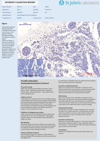
1. Figure:
Immunohistochemical
analysis of paraffin
embedded Human lung
cancer tissue. 1: p53
(phospho Thr18)
Polyclonal Antibody
was diluted at 1:200 (4
degree
Celsius,overnight). 2:
Sodium citrate pH 6.0
was used for antibody
retrieval (>98 degree
Celsius,20min). 3:
Secondary antibody was
diluted at 1:200 (room
temperature, 30min).
Negative control was
used by secondary
antibody only.
Report Number 90372-a Host Rabbit
Application IHC-P Clonality Polyclonal
Model Number STJ90372 Clone ID NA
Antibody Name Anti-Phospho-p53 (T18) antibody
Testing Species HUMAN Testing Tissue LUNG CANCER
ANTIBODY VALIDATION REPORT
b. (A small amount of distilled water was added into the incubation
box to prevent evaporation of antibody).
8. Secondary antibody incubation
a. Slides were washed 3 times, with PBS on a shaker for 5min.
Shortly after the slides were dried the corresponding secondary
antibody solution was added (HRP labelled), covering the
tissues, and incubated at room temperature for 30min.
b.
9. DAB staining
a. Slides were washed 3 times, with PBS on a shaker for 5min.
b. Shortly after, the slides were dried and fresh DAB staining buffer
was added inside the circles. The staining time was adjusted
under a microscope. Yellow-brown colour represented a positive
result. Slides were washed with water to stop the staining.
c.
10. Haematoxylin staining
a. Haematoxylin was used to counter-staining for 1min, and then
the slides were washed with water. 1% Hydrochloric acid and
alcohol was added for several seconds and then washed with
water. Ammonia was used to reveal blue colour, and then
flushed with water.
b.
11. Desolation and Clearing
i. Slides were incubated sequentially into: 75% alcohol 5min, 85%
alcohol 5min, Anhydrous ethanol - 5min, Anhydrous ethanol -
5min & Xylene - 5min. Shortly after slides were dried and neutral
gum was used to seal the slides.
ii.
12. Visualization
a. Results were validated with microscope, and the slides were
scanned.
Paraffin-Embedded
Immunohistochemistry Protocol
2.
3. Tissue processing
a. Slides were incubated sequentially into Xylene; 15min –
Xylene, 15min - Anhydrous ethanol, 5min - Anhydrous
ethanol, 5min - 85% alcohol, 5min - 75% alcohol & 5min –
wash in distilled water.
b.
4. Antigen retrieval
a. Tissue slides were incubated with citric acid (PH6.0) antigen
retrieval buffer and microwaved for antigen retrieval (heated
until boiled and then stopped heating) for 8min. Slides were
then heated with medium power for 7min. During this
process slides were kept from drying out. After cooling down
at room temperature, slides were washed with PBS on
shaker for 5min, repeated for 3 times.
b.
5. Inhibition of endogenous peroxidase
a. Slides were placed in 3% Hydrogen peroxide solution, and
incubated for 10 min at room temperature without light
exposure. Slides were then washed 3 times with PBS on a
shaker for 5mins.
b.
6. BSA Blocking
a. Shortly after slides were dried, a PAP pen was used to draw
circles around the tissue sections (and to prevent draining of
the antibody solution). Inside the circles, BSA was used to
cover the tissue evenly, blocking for 30min.
b.
7. Primary antibody incubation
After blocking solution was removed a 1:200 solution of
primary antibody/PBS was added on the slide, and incubated
overnight at 4°C.
St John's Laboratory Ltd.
www.stjohnslabs.com