Neutrophil cytosol factor 1, also known as p47phox, is a protein that in humans is encoded by the NCF1 gene.
The protein encoded by this gene is a 47 kDa cytosolic subunit of neutrophil NADPH oxidase. This oxidase is a multicomponent enzyme that is activated to produce superoxide anion. Mutations in this gene have been associated with chronic granulomatous disease. p47 is vital to the activation of NADPH oxidase. P47 becomes heavily phosphorylated.
Anti-p47-phox - http://www.stjohnslabs.com/p47-phox-antibody-p-93740?filter_name=stj94885
Join our Antibody Validation Project - http://www.stjohnslabs.com/services/antibody-validation
Hypoxia-inducible factor 1-alpha, also known as HIF-1α, is a subunit of a heterodimeric transcription factor hypoxia-inducible factor 1 (HIF-1) that is encoded by the HIF1α gene. It is a basic helix-loop-helix PAS domain containing protein, and is considered as the master transcriptional regulator of cellular and developmental response to hypoxia. The dysregulation and overexpression of HIF1α by either hypoxia or genetic alternations have been heavily implicated in cancer biology, as well as a number of other pathophysiologies, specifically in areas of vascularization and angiogenesis, energy metabolism, cell survival and tumour invasion.
To purchase this antibody use the following link: http://www.stjohnslabs.com/hif-1a-antibody-p-92614?filter_name=STJ93498
Functions as a master transcriptional regulator of the adaptive response to hypoxia. Under hypoxic conditions, activates the transcription of over 40 genes, including erythropoietin, glucose transporters, glycolytic enzymes, vascular endothelial growth factor, HILPDA, and other genes whose protein products increase oxygen delivery or facilitate metabolic adaptation to hypoxia. Plays an essential role in embryonic vascularization, tumor angiogenesis and pathophysiology of ischemic disease. Binds to core DNA sequence 5'-[AG]CGTG-3' within the hypoxia response element (HRE) of target gene promoters. Activation requires recruitment of transcriptional coactivators such as CREBPB and EP300. Activity is enhanced by interaction with both, NCOA1 or NCOA2. Interaction with redox regulatory protein APEX seems to activate CTAD and potentiates activation by NCOA1 and CREBBP. Involved in the axonal distribution and transport of mitochondria in neurons during hypoxia.
Anti-HIF-1α-http://www.stjohnslabs.com/hif-1a-antibody-p-92614
Join our Antibody Validation Project - http://www.stjohnslabs.com/services/antibody-validation
Fukutin-related protein (FKRP) is targeted to the medial Golgi apparatus and is necessary for post-translational modification of dystroglycan. Mutations in the gene encoding this protein have been associated with congenital muscular dystrophy, mental retardation, and cerebellar cysts. Several alternatively spliced transcript variants of this gene have been described, but the full-length nature of some of these variants has not been determined.
To purchase this antibody use the following link: http://www.stjohnslabs.com/fkrp-antibody-p-95246?filter_name=FKRP
Activity of CDK2 is maximal during S phase and G2; activated by interaction with cyclin E during the early stages of DNA synthesis to permit G1-S transition, and subsequently activated by cyclin A2 (cyclin A1 in germ cells) during the late stages of DNA replication to drive the transition from S phase to mitosis, the G2 phase. EZH2 phosphorylation promotes H3K27me3 maintenance and epigenetic gene silencing. Phosphorylates CABLES1 (By similarity). Cyclin E/CDK2 prevents oxidative stress-mediated Ras-induced senescence by phosphorylating MYC. Involved in G1-S phase DNA damage checkpoint that prevents cells with damaged DNA from initiating mitosis; regulates homologous recombination-dependent repair by phosphorylating BRCA2, this phosphorylation is low in S phase when recombination is active, but increases as cells progress towards mitosis. In response to DNA damage, double-strand break repair by homologous recombination a reduction of CDK2-mediated BRCA2 phosphorylation. Phosphorylation of RB1 disturbs its interaction with E2F1. NPM1 phosphorylation by cyclin E/CDK2 promotes its dissociates from unduplicated centrosomes, thus initiating centrosome duplication. Cyclin E/CDK2-mediated phosphorylation of NPAT at G1-S transition and until prophase stimulates the NPAT-mediated activation of histone gene transcription during S phase. Required for vitamin D-mediated growth inhibition by being itself inactivated. Involved in the nitric oxide- (NO) mediated signaling in a nitrosylation/activation-dependent manner. USP37 is activated by phosphorylation and thus triggers G1-S transition.
To purchase this antibody, use the following link: http://www.stjohnslabs.com/cdk2-antibody-p-91630?filter_name=STJ92197
Cdc2 Polyclonal Antibody plays a key role in the control of the eukaryotic cell cycle by modulating the centrosome cycle as well as mitotic onset; promotes G2-M transition, and regulates G1 progress and G1-S transition via association with multiple interphase cyclins.
To purchase this antibody, use the following link: http://www.stjohnslabs.com/cdc2-antibody-p-91597?filter_name=STJ92154
The Bcl-2-associated death promoter (BAD) protein is a pro-apoptotic member of the Bcl-2 gene family which is involved in initiating apoptosis. BAD is a member of the BH3-only family, a subfamily of the Bcl-2 family. After activation, it is able to form a heterodimer with anti-apoptotic proteins and prevent them from stopping apoptosis.
Anti-Bad - http://www.stjohnslabs.com/bad-antibody-p-91310?filter_name=STJ91797
Join our Antibody Validation Project - http://www.stjohnslabs.com/services/antibody-validation
Western Blot Antibody Customer Review for Anti-GPR18 antibody (STJ93370)St John's Laboratory Ltd
N-arachidonyl glycine receptor also known as G-protein coupled receptor 18 (GPR18) is a protein that in humans is encoded by the GPR18 gene. Along with the other previously "orphan" receptors GPR55 and GPR119, GPR18 has been found to be a receptor for endogenous lipid neurotransmitters, several of which also bind to cannabinoid receptors. Research supports the hypothesis that GPR18 is the abnormal cannabidiol receptor and N-arachidonoyl glycine, the endogenous lipid metabolite of anandamide, initiates directed microglial migration in the CNS through activation of GPR18.
Anti-GPR18 - http://www.stjohnslabs.com/gpr18-antibody-p-96212?filter_name=STJ93370
Join our Antibody Validation Project - http://www.stjohnslabs.com/services/antibody-validation
Western Blot Antibody Customer Review for Anti-Phospho-ALK (Y1507) Polyclonal...St John's Laboratory Ltd
Anaplastic lymphoma kinase (ALK) also known as ALK tyrosine kinase receptor or CD246 (cluster of differentiation 246) is an enzyme that in humans is encoded by the ALK gene.
ALK plays an important role in the development of the brain and exerts its effects on specific neurons in the nervous system. Transduces signals from ligands at the cell surface, through specific activation of the mitogen-activated protein kinase (MAPK) pathway. Phospho-ALK (Y1507) Polyclonal Antibody detects endogenous levels of ALK protein only when phosphorylated at Y1507.
Anti-Phospho-ALK - http://www.stjohnslabs.com/phospho-alk-y1507-antibody?filter_name=STJ90845
Join our Antibody Validation Project - http://www.stjohnslabs.com/services/antibody-validation
Western Blot Antibody Customer Review for p63 Polyclonal Antibody (STJ94912)St John's Laboratory Ltd
TP63 (tumour protein p63) or transformation-related protein 63 acts as a sequence specific DNA binding transcriptional activator or repressor. It is encoded by the TP63 gene in humans. The p63 polyclonal antibody detects endogenous levels of p63 protein.
Western Blot Antibody Customer Review for Anti-ALK Polyclonal Antibody (STJ91...St John's Laboratory Ltd
Anaplastic lymphoma kinase also known as ALK tyrosine kinase receptor or CD246 (cluster of differentiation 246) is an enzyme that in humans is encoded by the ALK gene.
ALK plays an important role in the development of the brain and exerts its effects on specific neurons in the nervous system. Transduces signals from ligands at the cell surface, through specific activation of the mitogen-activated protein kinase (MAPK) pathway.The antibody was affinity-purified from rabbit antiserum by affinity-chromatography using epitope-specific immunogen.
Anti-ALK - http://www.stjohnslabs.com/alk-antibody-p-91127?filter_name=STJ91562
Join our Antibody Validation Project - http://www.stjohnslabs.com/services/antibody-validation
Nucleic Acid Therapy Purity Methods by Capillary Gel ElectrophoresisCovance
The number of FDA-approved nucleic acid therapies including oligonucleotides and gene therapies has more than doubled since 2013. Safety, identity, purity and quality is of critical importance, so the characterization and routine testing of manufactured NAT drug substances and drug products is increasingly significant for public safety.
Explaining Biocide Tolerance of Gram Negative Bacteria Kate Barlow
Working on multiple organisms and constantly changing gene-targets requires use of an easily optimisable and cheap qPCR method, which is why we use SyBr Green qPCR. We have investigated chlorhexidine resistance in Klebsiella pneumoniae and are about to publish on biocide resistance in Acinetobacter baumannii and Pseudomonas aeruginosa. I will explain our approach and our optimisation and robustness strategies, as well as an overview of the hypotheses we developed and confirmed using our qPCR approach.
Lucy Bock, Senior Scientist/Project Team Leader, Technology Development Group, Public Health England, UK
2011 course on Molecular Diagnostic Automation - Part 2 - AmplificationPatrick Merel
2011 course on Molecular Diagnostic Automation - Part 2 - Amplification.
This is from early 2011. Prices and Specifications of instruments may have changed.
Part 2 of 3
ADCC Reporter Bioassay - V and F Variants: Novel, Bioluminescent Cell-Based A...Neal Cosby
The ADCC Reporter Bioassay from Promega is now available in both V158 and F158 variant formats. Use this functional, sister bioassay approach to better characterize Ab drugs with MOA directed through FcgammaRIIIa (CD16a) receptor. Not yet familiar with this novel bioassay that is becoming the industry standard? This slidedoc introduces the reporter-based ADCC technology. Contact me for any questions.
Next generation biotherapeutics production system Trichoderma reeseiChristopher Landowski
The filamentous fungus Trichoderma reesei is an important production organism used by industrial enzyme companies world-wide. It is a low cost production system that secretes its native enzymes at levels exceeding 100 g/L of culture medium. Several T. reesei produced enzymes have obtained the generally recognized as safe status by the U.S. Food and Drug Administration. T. reesei has tremendous prospects to be a cost efficient and high yield system for producing therapeutic proteins. We have adapted the fungus to become more suitable for biotherapeutic production by reducing secreted protease activity and altering glycosylation pathways needed for adding mammalian glycoforms.
Expression strains for monoclonal antibodies, Fab antibody fragments, interferon alpha-2b, insulin-like growth factor 1, and fibroblast growth factor 21 were constructed, cultivated in bioreactors, and expression levels were measured from the culture medium. After deleting 13 of the most critical protease genes, the general secreted protease activity was reduced over 30-fold. Monoclonal antibodies could be produced up to 7.6 g/L, Fab antibody fragments up to 8.2 g/L, interferon alpha-2b at 7.9 g/L, and insulin-like growth factor fusion protein at 8 g/L. With protease inhibitor treatment interferon alpha-2b could be produced at over 10 g/L, insulin-like growth factor fusion protein at 19 g/L, and full length fibroblast growth factor 21 at 200 mg/L in addition to a shorter form at 3.5 g/L. Human glycoforms such as G0 and FG0 were produced on monoclonal antibodies.
Expression levels and product quality improved dramatically after multiple protease deletions and optimization of culture conditions. While the production levels achieved are already relatively high, the strains could be developed further to reach the 100 g/L potential of the organism. This study demonstrates the excellent prospects of T. reesei as a host for therapeutic protein production.
Immunocytochemistry Antibody Customer Review for Anti β-actin Antibody (STJ96...St John's Laboratory Ltd
Beta-actin (human gene and protein symbol ACTB/ACTB) is one of six different actin isoforms which have been identified in humans. This is one of the two nonmuscle cytoskeletal actins. Actins are highly conserved proteins that are involved in cell motility, structure and integrity. Alpha actins are a major constituent of the contractile apparatus.
Beta actin is usually used as a loading control, for among others, the integrity of cells, protein degradation, in PCR and Western blotting. Its molecular weight is approximately 42 kDa.
Anti β-actin - http://www.stjohnslabs.com/b-actin-antibody-p-98565?filter_name=STJ96930
Join our Antibody Validation Project - http://www.stjohnslabs.com/services/antibody-validation
Beta-actin (human gene and protein symbol ACTB/ACTB) is one of six different actin isoforms which have been identified in humans. This is one of the two nonmuscle cytoskeletal actins. Actins are highly conserved proteins[3][4] that are involved in cell motility, structure and integrity. Alpha actins are a major constituent of the contractile apparatus.
Beta actin is usually used as a loading control, for among others, the integrity of cells, protein degradation, in PCR and Western blotting. Its molecular weight is approximately 42 kDa.
Anti-β-actin -http://www.stjohnslabs.com/b-actin-antibody-p-98565
Join our Antibody Validation Project - http://www.stjohnslabs.com/services/antibody-validation
Involved in the activation cascade of caspases responsible for apoptosis execution. At the onset of apoptosis it proteolytically cleaves poly(ADP-ribose) polymerase (PARP) at a '216-Asp-|-Gly-217' bond. Cleaves and activates sterol regulatory element binding proteins (SREBPs) between the basic helix-loop-helix leucine zipper domain and the membrane attachment domain. Cleaves and activates caspase-6, -7 and -9. Involved in the cleavage of huntingtin. Triggers cell adhesion in sympathetic neurons through RET cleavage.
Anti-Active Caspase-3 -http://www.stjohnslabs.com/active-caspase-3-antibody-p-99099
Join our Antibody Validation Project - http://www.stjohnslabs.com/services/antibody-validation
Western Blot Antibody Customer Review for Anti-ChemR23 Antibody (STJ92262)St John's Laboratory Ltd
Chemokine like receptor 1 also known as ChemR23 (Chemerin Receptor 23) is a protein that in humans is encoded by the CMKLR1 gene. Chemokine receptor-like 1 is a G protein-coupled receptor for the chemoattractant adipokine chemerin. Activating CMKLR1 by an agonist mobilizes intracellular calcium and causes the activation of several other signaling cascades like the ERK1 and NF-κB. Initial studies of CMKLR1 suggested that it might have a role in the inflammatory pathways. Its cognate ligand, chemerin was found in joint aspirate from rheumatoid arthritis and absent in aspirate from degenerative arthritis.
Anti-ChemR23 - http://www.stjohnslabs.com/chemr23-antibody?filter_name=STJ92262
Join our Antibody Validation Project - http://www.stjohnslabs.com/services/antibody-validation
Western Blot Antibody Customer Review for Anti-Histone H3 (Phospho-Tyr41) An...St John's Laboratory Ltd
Core component of nucleosome. Nucleosomes wrap and compact DNA into chromatin, limiting DNA accessibility to the cellular machineries which require DNA as a template. Histones thereby play a central role in transcription regulation, DNA repair, DNA replication and chromosomal stability. DNA accessibility is regulated via a complex set of post-translational modifications of histones, also called histone code, and nucleosome remodeling.
Anti-Histone H3 (Phospho-Tyr41) - http://www.stjohnslabs.com/histone-h3-phospho-tyr41-antibody?filter_name=STJ97138
Join our Antibody Validation Project - http://www.stjohnslabs.com/services/antibody-validation
Involved in the activation cascade of caspases responsible for apoptosis execution. At the onset of apoptosis it proteolytically cleaves poly(ADP-ribose) polymerase (PARP) at a '216-Asp-|-Gly-217' bond. Cleaves and activates sterol regulatory element binding proteins (SREBPs) between the basic helix-loop-helix leucine zipper domain and the membrane attachment domain. Cleaves and activates caspase-6, -7 and -9. Involved in the cleavage of huntingtin. Triggers cell adhesion in sympathetic neurons through RET cleavage. / Strict requirement for an Asp residue at positions P1 and P4. It has a preferred cleavage sequence of Asp-Xaa-Xaa-Asp-|- with a hydrophobic amino-acid residue at P2 and a hydrophilic amino-acid residue at P3, although Val or Ala are also accepted at this position. / Inhibited by isatin sulfonamides.
Anti-Active Caspase-3-http://www.stjohnslabs.com/active-caspase-3-antibody-p-99099
Join our Antibody Validation Project - http://www.stjohnslabs.com/services/antibody-validation
Equilibrative nucleoside transporter 1 (ENT1) is a protein that in humans is encoded by the SLC29A1 gene. Multiple alternatively spliced variants, encoding the same protein, have been found for this gene.
This gene is a member of the equilibrative nucleoside transporter family. The gene encodes a transmembrane glycoprotein that localizes to the plasma and mitochondrial membranes and mediates the cellular uptake of nucleosides from the surrounding medium. The protein is categorized as an equilibrative (as opposed to concentrative) transporter that is sensitive to inhibition by nitrobenzylmercaptopurine ribonucleoside (NBMPR). Nucleoside transporters are required for nucleotide synthesis in cells that lack de novo nucleoside synthesis pathways, and are also necessary for the uptake of cytotoxic nucleosides used for cancer and viral chemotherapies.
To purchase this antibody, use the following link: http://www.stjohnslabs.com/ent1-antibody?filter_name=STJ96396
Has both glyceraldehyde-3-phosphate dehydrogenase and nitrosylase activities, thereby playing a role in glycolysis and nuclear functions, respectively. Participates in nuclear events including transcription, RNA transport, DNA replication and apoptosis. Nuclear functions are probably due to the nitrosylase activity that mediates cysteine S-nitrosylation of nuclear target proteins such as SIRT1, HDAC2 and PRKDC. Modulates the organization and assembly of the cytoskeleton. Facilitates the CHP1-dependent microtubule and membrane associations through its ability to stimulate the binding of CHP1 to microtubules (By similarity). Glyceraldehyde-3-phosphate dehydrogenase is a key enzyme in glycolysis that catalyzes the first step of the pathway by converting D-glyceraldehyde 3-phosphate (G3P) into 3-phospho-D-glyceroyl phosphate. Component of the GAIT (gamma interferon-activated inhibitor of translation) complex which mediates interferon-gamma-induced transcript-selective translation inhibition in inflammation processes. Upon interferon-gamma treatment assembles into the GAIT complex which binds to stem loop-containing GAIT elements in the 3'-UTR of diverse inflammatory mRNAs (such as ceruplasmin) and suppresses their translation.
To purchase this antibody, use the following link: http://www.stjohnslabs.com/gapdh-antibody-p-94882?filter_name=STJ96417
Western Blot Antibody Customer Review for Anti-PDHA1 Polyclonal Antibody (STJ...St John's Laboratory Ltd
Pyruvate dehydrogenase E1 component subunit alpha, somatic form, mitochondrial is an enzyme that in humans is encoded by the PDHA1 gene.
The pyruvate dehydrogenase complex is responsible for the oxidative decarboxylation of pyruvate, with the final product being Acetyl CoA.
Mutations in the PDHA1 gene have been known to cause one form of pyruvate dehydrogenase deficiency. Pyruvate dehydrogenase deficiency is characterized by the buildup of a chemical called lactic acid in the body and a variety of neurological problems.
The antibody was affinity-purified from rabbit antiserum by affinity-chromatography using epitope-specific immunogen.
Anti-PDHA1 - http://www.stjohnslabs.com/pdha1-antibody-p-93852?filter_name=STJ95006
Join our Antibody Validation Project - http://www.stjohnslabs.com/services/antibody-validation
NF-kappa-B is a pleiotropic transcription factor present in almost all cell types and is the endpoint of a series of signal transduction events that are initiated by a vast array of stimuli related to many biological processes such as inflammation, immunity, differentiation, cell growth, tumorigenesis and apoptosis. NF-kappa-B is a homo- or heterodimeric complex formed by the Rel-like domain-containing proteins RELA/p65, RELB, NFKB1/p105, NFKB1/p50, REL and NFKB2/p52 and the heterodimeric p65-p50 complex appears to be most abundant one. The dimers bind at kappa-B sites in the DNA of their target genes and the individual dimers have distinct preferences for different kappa-B sites that they can bind with distinguishable affinity and specificity. Different dimer combinations act as transcriptional activators or repressors, respectively.
To purchase this antibody, use the following link: http://www.stjohnslabs.com/nfkb-p65-antibody-p-93367?filter_name=STJ94468
Western Blot Antibody Customer Review for ADNP Polyclonal Antibody (STJ91502)St John's Laboratory Ltd
ADNP (Activity-Dependent Neuroprotector Homeobox) is expressed through the ADNP gene in humans. It is a possible transcription factor through having a homeobox and 9 zinc finger domains. It is said to have both stimulatory and inhibitory growth effects on different tumour cells. The ADNP Polyclonal Antibody detects endogenous levels of ADNP protein.
Histones are nuclear proteins that form octameric structures which bind DNA to form units of chromatin called nucleosomes. The family of histones—H2A, H2B, H3, and H4—are key players in gene regulation. They undergo a number of post-translational modifications (PTM) in response to various stimuli, including phosphorylation on serine and threonine residues and methylation on lysine residues. PTMs produce configural changes in histone proteins that may induce nucleosome remodeling and expose or hide DNA sequences from transcriptional complexes. Histone H4 lysine 20 (H4K20) may undergo mono-, di-, or trimethylation, which is catalyzed by the methyltransferase PR-Set7 (Set8 or KMT5a). Methylated H4K20 plays a role in regulating DNA damage responses, mitosis, DNA replication, and gene expression. Trimethylation of H4K20 contributes to gene silencing, and is a mark of the repressive heterochromatin state.
To purchase this antibody use the following link: http://www.stjohnslabs.com/histone-h4-tri-methyl-lys20-antibody?filter_name=STJ97154
GSK3β Polyclonal Antibody is an active protein kinase that acts as a negative regulator in the hormonal control of glucose homeostasis, Wnt signaling and regulation of transcription factors and microtubules, by phosphorylating and inactivating glycogen synthase (GYS1 or GYS2), EIF2B, CTNNB1/beta-catenin, APC, AXIN1, DPYSL2/CRMP2, JUN, NFATC1/NFATC, MAPT/TAU and MACF1. Requires primed phosphorylation of the majority of its substrates.
To purchase this antibody, use the following link: http://www.stjohnslabs.com/gsk3b-antibody-p-92568?filter_name=STJ93447
Beta-actin (human gene and protein symbol ACTB/ACTB) is one of six different actin isoforms which have been identified in humans. This is one of the two nonmuscle cytoskeletal actins. Actins are highly conserved proteins that are involved in cell motility, structure and integrity. Alpha actins are a major constituent of the contractile apparatus.
To purchase this antibody, use the following link: http://www.stjohnslabs.com/actin-b-antibody?filter_name=STJ91464
Tau are stabilising proteins for axonal microtubules within the Central Nervous System, and as such are also recognised as Mictrotubule-associated Protein (MAP). Tau is predominantly active in the distal portions of axons to carry out its function.
Western Blot Antibody Customer Review for Anti-PKM2 Polyclonal Antibody (STJ9...St John's Laboratory Ltd
Pyruvate kinase isozymes M1/M2 (PKM1/M2), also known as pyruvate kinase muscle isozyme (PKM), pyruvate kinase type K, cytosolic thyroid hormone-binding protein (CTHBP), thyroid hormone-binding protein 1 (THBP1), or opa-interacting protein 3 (OIP3), is an enzyme that in humans is encoded by the PKM2 gene.
Is a glycolytic enzyme that catalyzes the transfer of a phosphoryl group from phosphoenolpyruvate (PEP) to ADP, generating ATP. It plays a general role in caspase independent cell death of tumor cells.
The antibody was affinity-purified from rabbit antiserum by affinity-chromatography using epitope-specific immunogen.
Anti-PMK2 - http://www.stjohnslabs.com/pkm2-antibody-p-93954?filter_name=Stj95142
Join our Antibody Validation Project - http://www.stjohnslabs.com/services/antibody-validation
Component of the epithelial apical junction complex that may function as an homophilic cell adhesion molecule and is essential for tight junction integrity. Also involved in transepithelial migration of leukocytes through adhesive interactions with AMICA1/JAML a transmembrane protein of the plasma membrane of leukocytes. The interaction between both receptors also mediates the activation of gamma-delta T-cells, a subpopulation of T-cells residing in epithelia and involved in tissue homeostasis and repair. Upon epithelial CXADR-binding, AMICA1 induces downstream cell signaling events in gamma-delta T-cells through PI3-kinase and MAP kinases. It results in proliferation and production of cytokines and growth factors by T-cells that in turn stimulate epithelial tissues repair.
To purchase this antibody, use the following link: http://www.stjohnslabs.com/car-antibody?filter_name=STJ91996
Serine/threonine protein kinase which is a central regulator of cellular metabolism, growth and survival in response to hormones, growth factors, nutrients, energy and stress signals. MTOR directly or indirectly regulates the phosphorylation of at least 800 proteins. Functions as part of 2 structurally and functionally distinct signaling complexes mTORC1 and mTORC2 (mTOR complex 1 and 2). Activated mTORC1 up-regulates protein synthesis by phosphorylating key regulators of mRNA translation and ribosome synthesis. This includes phosphorylation of EIF4EBP1 and release of its inhibition toward the elongation initiation factor 4E (eiF4E). Moreover, phosphorylates and activates RPS6KB1 and RPS6KB2 that promote protein synthesis by modulating the activity of their downstream targets including ribosomal protein S6, eukaryotic translation initiation factor EIF4B, and the inhibitor of translation initiation PDCD4. Stimulates the pyrimidine biosynthesis pathway, both by acute regulation through RPS6KB1-mediated phosphorylation of the biosynthetic enzyme CAD, and delayed regulation, through transcriptional enhancement of the pentose phosphate pathway which produces 5-phosphoribosyl-1-pyrophosphate (PRPP), an allosteric activator of CAD at a later step in synthesis, this function is dependent on the mTORC1 complex. Regulates ribosome synthesis by activating RNA polymerase III-dependent transcription through phosphorylation and inhibition of MAF1 an RNA polymerase III-repressor.
To purchase this antibody, use the following link: http://www.stjohnslabs.com/mtor-antibody-p-93228?filter_name=STJ94280
Histones are basic nuclear proteins that are responsible for the nucleosome structure of the chromosomal fiber in eukaryotes. Nucleosomes consist of approximately 146 bp of DNA wrapped around a histone octamer composed of pairs of each of the four core histones (H2A, H2B, H3, and H4). The chromatin fiber is further compacted through the interaction of a linker histone, H1, with the DNA between the nucleosomes to form higher order chromatin structures. This gene is intronless and encodes a member of the histone H3 family. Transcripts from this gene lack polyA tails; instead, they contain a palindromic termination element. This gene is located separately from the other H3 genes that are in the histone gene cluster on chromosome 6p22-p21.3.
To purchase this antibody use the following link: http://www.stjohnslabs.com/trimethyl-histone-h3-k56?filter_name=STJ29403
Tyrosine-protein kinase that plays an essential role as cell surface receptor for neuregulins and EGF family members and regulates development of the heart, the central nervous system and the mammary gland, gene transcription, cell proliferation, differentiation, migration and apoptosis. Required for normal cardiac muscle differentiation during embryonic development, and for postnatal cardiomyocyte proliferation. Required for normal development of the embryonic central nervous system, especially for normal neural crest cell migration and normal axon guidance. Required for mammary gland differentiation, induction of milk proteins and lactation. Acts as cell-surface receptor for the neuregulins NRG1, NRG2, NRG3 and NRG4 and the EGF family members BTC, EREG and HBEGF. Ligand binding triggers receptor dimerization and autophosphorylation at specific tyrosine residues that then serve as binding sites for scaffold proteins and effectors. Ligand specificity and signaling is modulated by alternative splicing, proteolytic processing, and by the formation of heterodimers with other ERBB family members, thereby creating multiple combinations of intracellular phosphotyrosines that trigger ligand- and context-specific cellular responses. Mediates phosphorylation of SHC1 and activation of the MAP kinases MAPK1/ERK2 and MAPK3/ERK1. Isoform JM-A CYT-1 and isoform JM-B CYT-1 phosphorylate PIK3R1, leading to the activation of phosphatidylinositol 3-kinase and AKT1 and protect cells against apoptosis. Isoform JM-A CYT-1 and isoform JM-B CYT-1 mediate reorganization of the actin cytoskeleton and promote cell migration in response to NRG1. Isoform JM-A CYT-2 and isoform JM-B CYT-2 lack the phosphotyrosine that mediates interaction with PIK3R1, and hence do not phosphorylate PIK3R1, do not protect cells against apoptosis, and do not promote reorganization of the actin cytoskeleton and cell migration. Proteolytic processing of isoform JM-A CYT-1 and isoform JM-A CYT-2 gives rise to the corresponding soluble intracellular domains (4ICD) that translocate to the nucleus, promote nuclear import of STAT5A, activation of STAT5A, mammary epithelium differentiation, cell proliferation and activation of gene expression. The ERBB4 soluble intracellular domains (4ICD) colocalize with STAT5A at the CSN2 promoter to regulate transcription of milk proteins during lactation. The ERBB4 soluble intracellular domains can also translocate to mitochondria and promote apoptosis.
To purchase this antibody, use the following link: http://www.stjohnslabs.com/erbb4?filter_name=STJ27886
A polyhistidine-tag is an amino acid motif in proteins that consists of at least six histidine (His) residues, often at the N- or C-terminus of the protein. It is also known as hexa histidine-tag, 6xHis-tag, His6 tag and by the trademarked name His-tag. Polyhistidine-tags are often used for affinity purification of polyhistidine-tagged recombinant proteins expressed in Escherichia coli and other prokaryotic expression systems.
To purchase this antibody, use the following link: http://www.stjohnslabs.com/his-tag-antibody-p-95792?filter_name=STJ90106
Western Blot Antibody Customer Review for Anti-Peroxin 2 Antibody (STJ95040)St John's Laboratory Ltd
PEX2 is gene on chromosome 8q21.1 that encodes a peroxin, an integral membrane protein involved in peroxisome biosynthesis and integrity, which assembles membrane vesicles before translocation of matrix proteins.
Defects of PEX2 cause peroxisome biogenesis disorder complementation group 5, Zelleger syndrome and a form of Refsum disease.
Anti-Peroxin 2 - http://www.stjohnslabs.com/peroxin-2-antibody?filter_name=STJ95040
Join our Antibody Validation Project - http://www.stjohnslabs.com/services/antibody-validation
Antibody Customer Review For Phospho-eIF2α (S51) Polyclonal Antibody (STJ90253) St John's Laboratory Ltd
eIF2 is an initiation factor found within eukaryotes. It mediates binding of tRNA to the ribosome in a GTP dependent manner. The alpha subunit is the main target for phosphorylation at Serine-51, and is considered the regulatory subunit of this trimeric (formed of eIF2a, eIF2b and eIF2y).
Visit www.stjohnslabs.com
Western Blot Antibody Customer Review for Anti-RelB Polyclonal Antibody (STJ9...St John's Laboratory Ltd
The RelB protein is a transcription factor encoded by the RELB gene in humans. It is known to interact with NFKB2. The RelB polyclonal antibody detects endogenous levels of RelB protein.
Western Blot Antibody Customer Review for Anti-Endoplasmin Polyclonal Antibod...St John's Laboratory Ltd
The Endoplasmin protein (also known as HSP90B1) is a molecular chaperone encoded by the HSP90B1 gene in humans. It plays a role in the processing and transport of proteins such as integrins and Toll-like receptors. It is located in the endoplasmic reticulum. The Endoplasmin polyclonal antibody detects endogenous levels of Endoplasmin protein.
Similar to Western Blot Antibody Customer Review for Anti-p47-phox Antibody (STJ94885) (20)
G protein-coupled receptor that probably associates with the patched protein (PTCH) to transduce the hedgehog's proteins signal. Binding of sonic hedgehog (SHH) to its receptor patched is thought to prevent normal inhibition by patched of smoothened (SMO). Required for the accumulation of KIF7 and GLI3 in the cilia.
Anti-Smo antibody (STJ95710): http://www.stjohnslabs.com/smo-antibody-p-94371?filter_name=STJ95710
Join our Antibody Validation Project - http://www.stjohnslabs.com/services/antibody-validation
Western Blot Customer Review Anti Glucocorticoid Receptor antibody (STJ97101)St John's Laboratory Ltd
Receptor for glucocorticoids (GC). Has a dual mode of action: as a transcription factor that binds to glucocorticoid response elements (GRE), both for nuclear and mitochondrial DNA, and as a modulator of other transcription factors. Affects inflammatory responses, cellular proliferation and differentiation in target tissues. Involved in chromatin remodeling . Plays a role in rapid mRNA degradation by binding to the 5' UTR of target mRNAs and interacting with PNRC2 in a ligand-dependent manner. Could act as a coactivator for STAT5-dependent transcription upon growth hormone (GH) stimulation and could reveal an essential role of hepatic GR in the control of body growth (By similarity). Has transcriptional activation and repression activity . Mediates glucocorticoid-induced apoptosis . Promotes accurate chromosome segregation during mitosis . May act as a tumor suppressor . May play a negative role in adipogenesis through the regulation of lipolytic and antilipogenic gene expression (By similarity). / Isoform Beta: Acts as a dominant negative inhibitor of isoform Alpha . Has intrinsic transcriptional activity independent of isoform Alpha when both isoforms are coexpressed.
Join our Antibody Validation Project: http://www.stjohnslabs.com/services/antibody-validation
Anti glucocorticoid receptor antibody (STJ97101):
http://www.stjohnslabs.com/glucocorticoid-receptor-antibody-p-98736?filter_name=STJ97101
Western Blot Customer Review Anti-Phospho-Cofilin (S3) Antibody (STJ90230)St John's Laboratory Ltd
Binds to F-actin and exhibits pH-sensitive F-actin depolymerizing activity. Regulates actin cytoskeleton dynamics. Important for normal progress through mitosis and normal cytokinesis. Plays a role in the regulation of cell morphology and cytoskeletal organization. Required for the up-regulation of atypical chemokine receptor ACKR2 from endosomal compartment to cell membrane, increasing its efficiency in chemokine uptake and degradation.
Anti-Phospho-Cofilin (S3) -http://www.stjohnslabs.com/phospho-cofilin-s3-antibody
Join our Antibody Validation Project - http://www.stjohnslabs.com/services/antibody-validation
This June, Dr. Byron Baron from the University of Malta, Malta, is our Scientist of the Month! He's shared with us his research highlights, his current projects and some comments on the biotechnology industry.
Want to be our Scientist of the Month? Contact info@stjohnslabs.com
Downstream effector molecule involved in the transmission of signals from tyrosine kinase receptors and small GTPases to the actin cytoskeleton. Promotes formation of actin filaments. Part of the WAVE complex that regulates lamellipodia formation. The WAVE complex regulates actin filament reorganization via its interaction with the Arp2/3 complex.
Anti-WAVE2 -http://www.stjohnslabs.com/wave2-antibody
Join our Antibody Validation Project - http://www.stjohnslabs.com/services/antibody-validation
Implicated in synaptic vesicle endocytosis. May recruit other proteins to membranes with high curvature.
Brain, mostly in frontal cortex. Expressed at high level in fetal cerebellum.
Anti-Endophilin I -http://www.stjohnslabs.com/endophilin-i-antibody
Join our Antibody Validation Project - http://www.stjohnslabs.com/services/antibody-validation
Tubulin is the major constituent of microtubules. It binds two moles of GTP, one at an exchangeable site on the beta chain and one at a non-exchangeable site on the alpha chain. TUBB3 plays a critical role in proper axon guidance and mantainance.
Anti-β-tubulin -http://www.stjohnslabs.com/b-tubulin-antibody-p-98672
Join our Antibody Validation Project - http://www.stjohnslabs.com/services/antibody-validation
Most upstream protease of the activation cascade of caspases responsible for the TNFRSF6/FAS mediated and TNFRSF1A induced cell death. Binding to the adapter molecule FADD recruits it to either receptor. The resulting aggregate called death-inducing signaling complex (DISC) performs CASP8 proteolytic activation. The active dimeric enzyme is then liberated from the DISC and free to activate downstream apoptotic proteases. Proteolytic fragments of the N-terminal propeptide (termed CAP3, CAP5 and CAP6) are likely retained in the DISC. Cleaves and activates CASP3, CASP4, CASP6, CASP7, CASP9 and CASP10. May participate in the GZMB apoptotic pathways. Cleaves ADPRT. Hydrolyzes the small-molecule substrate, Ac-Asp-Glu-Val-Asp-|-AMC. Likely target for the cowpox virus CRMA death inhibitory protein. Isoform 5, isoform 6, isoform 7 and isoform 8 lack the catalytic site and may interfere with the pro-apoptotic activity of the complex. / Strict requirement for Asp at position P1 and has a preferred cleavage sequence of (Leu/Asp/Val)-Glu-Thr-Asp-|-(Gly/Ser/Ala). / Inhibited by the effector protein NleF that is produced by pathogenic E.coli; this inhibits apoptosis.
Anti-Caspase-8-http://www.stjohnslabs.com/caspase-8-antibody-p-99045
Join our Antibody Validation Project - http://www.stjohnslabs.com/services/antibody-validation
Tubulin is the major constituent of microtubules. The gamma chain is found at microtubule organizing centers (MTOC) such as the spindle poles or the centrosome. Pericentriolar matrix component that regulates alpha/beta chain minus-end nucleation, centrosome duplication and spindle formation.
Anti-Gamma Tubulin-http://www.stjohnslabs.com/gamma-tubulin-antibody
Join our Antibody Validation Project - http://www.stjohnslabs.com/services/antibody-validation
Tubulin in molecular biology can refer either to the tubulin protein superfamily of globular proteins, or one of the member proteins of that superfamily. α- and β-tubulins polymerize into microtubules, a major component of the eukaryotic cytoskeleton. Microtubules function in many essential cellular processes, including mitosis. Tubulin-binding drugs kill cancerous cells by inhibiting microtubule dynamics, which are required for DNA segregation and therefore cell division. In eukaryotes there are six members of the tubulin superfamily, although not all are present in all species (see below). Both α and β tubulins have a mass of around 50 kDa and are thus in a similar range compared to actin with ~42 kDa. In contrast, tubulin polymers (microtubules) tend to be much bigger than actin filaments due to their cylindrical nature. Tubulin was long thought to be specific to eukaryotes. More recently, however, several prokaryotic proteins have been shown to be related to tubulin.
Anti-Epsilon Tubulin -http://www.stjohnslabs.com/epsilon-tubulin-antibody-2
Join our Antibody Validation Project - http://www.stjohnslabs.com/services/antibody-validation
Ubiquitin-like modifier involved in formation of autophagosomal vacuoles (autophagosomes) . Whereas LC3s are involved in elongation of the phagophore membrane, the GABARAP/GATE-16 subfamily is essential for a later stage in autophagosome maturation .
Anti-LC3A-http://www.stjohnslabs.com/lc3a-antibody
Join our Antibody Validation Project - http://www.stjohnslabs.com/services/antibody-validation
Multifunctional transcription factor in ER stress response. Plays an essential role in the response to a wide variety of cell stresses and induces cell cycle arrest and apoptosis in response to ER stress. Plays a dual role both as an inhibitor of CCAAT/enhancer-binding protein (C/EBP) function and as an activator of other genes. Acts as a dominant-negative regulator of C/EBP-induced transcription: dimerizes with members of the C/EBP family, impairs their association with C/EBP binding sites in the promoter regions, and inhibits the expression of C/EBP regulated genes. Positively regulates the transcription of TRIB3, IL6, IL8, IL23, TNFRSF10B/DR5, PPP1R15A/GADD34, BBC3/PUMA, BCL2L11/BIM and ERO1L. Negatively regulates; expression of BCL2 and MYOD1, ATF4-dependent transcriptional activation of asparagine synthetase (ASNS), CEBPA-dependent transcriptional activation of hepcidin (HAMP) and CEBPB-mediated expression of peroxisome proliferator-activated receptor gamma (PPARG).
Anti-CHOP-http://www.stjohnslabs.com/chop-antibody-2
Join our Antibody Validation Project - http://www.stjohnslabs.com/services/antibody-validation
Pseudokinase that plays a key role in TNF-induced necroptosis, a programmed cell death process. Activated following phosphorylation by RIPK3, leading to homotrimerization, localization to the plasma membrane and execution of programmed necrosis characterized by calcium influx and plasma membrane damage. Does not have protein kinase activity.
Anti-phospho-MLKL (S358)-http://www.stjohnslabs.com/phospho-mlkl-s358-antibody-1
Join our Antibody Validation Project - http://www.stjohnslabs.com/services/antibody-validation
Serine/threonine kinase which acts as an essential component of the MAP kinase signal transduction pathway. MAPK1/ERK2 and MAPK3/ERK1 are the 2 MAPKs which play an important role in the MAPK/ERK cascade. They participate also in a signaling cascade initiated by activated KIT and KITLG/SCF. Depending on the cellular context, the MAPK/ERK cascade mediates diverse biological functions such as cell growth, adhesion, survival and differentiation through the regulation of transcription, translation, cytoskeletal rearrangements. The MAPK/ERK cascade plays also a role in initiation and regulation of meiosis, mitosis, and postmitotic functions in differentiated cells by phosphorylating a number of transcription factors. About 160 substrates have already been discovered for ERKs. Many of these substrates are localized in the nucleus, and seem to participate in the regulation of transcription upon stimulation.
Anti-ERK1-http://www.stjohnslabs.com/erk1-antibody-3
Join our Antibody Validation Project - http://www.stjohnslabs.com/services/antibody-validation
Tyrosine-protein kinase that acts as a cell-surface receptor for PDGFA, PDGFB and PDGFC and plays an essential role in the regulation of embryonic development, cell proliferation, survival and chemotaxis. Depending on the context, promotes or inhibits cell proliferation and cell migration. Plays an important role in the differentiation of bone marrow-derived mesenchymal stem cells. Required for normal skeleton development and cephalic closure during embryonic development. Required for normal development of the mucosa lining the gastrointestinal tract, and for recruitment of mesenchymal cells and normal development of intestinal villi. Plays a role in cell migration and chemotaxis in wound healing. Plays a role in platelet activation, secretion of agonists from platelet granules, and in thrombin-induced platelet aggregation.
Anti-PDGFRα-http://www.stjohnslabs.com/pdgfra-antibody-2
Join our Antibody Validation Project - http://www.stjohnslabs.com/services/antibody-validation
Thiol protease that cleaves IL-1 beta between an Asp and an Ala, releasing the mature cytokine which is involved in a variety of inflammatory processes. Important for defense against pathogens. Cleaves and activates sterol regulatory element binding proteins (SREBPs). Can also promote apoptosis. / Strict requirement for an Asp residue at position P1 and has a preferred cleavage sequence of Tyr-Val-Ala-Asp-|-. / Specifically inhibited by the cowpox virus Crma protein.
Anti-Caspase-1-http://www.stjohnslabs.com/caspase-1-antibody-1
Join our Antibody Validation Project - http://www.stjohnslabs.com/services/antibody-validation
Functions as a cell surface receptor and performs physiological functions on the surface of neurons relevant to neurite growth, neuronal adhesion and axonogenesis. Involved in cell mobility and transcription regulation through protein-protein interactions. Can promote transcription activation through binding to APBB1-KAT5 and inhibits Notch signaling through interaction with Numb. Couples to apoptosis-inducing pathways such as those mediated by G(O) and JIP. Inhibits G(o) alpha ATPase activity (By similarity). Acts as a kinesin I membrane receptor, mediating the axonal transport of beta-secretase and presenilin 1. Involved in copper homeostasis/oxidative stress through copper ion reduction. In vitro, copper-metallated APP induces neuronal death directly or is potentiated through Cu2+-mediated low-density lipoprotein oxidation.
Anti-Amyloid-β-http://www.stjohnslabs.com/amyloid-v-antibody
Join our Antibody Validation Project - http://www.stjohnslabs.com/services/antibody-validation
Suppresses apoptosis in a variety of cell systems including factor-dependent lymphohematopoietic and neural cells. Regulates cell death by controlling the mitochondrial membrane permeability. Appears to function in a feedback loop system with caspases. Inhibits caspase activity either by preventing the release of cytochrome c from the mitochondria and/or by binding to the apoptosis-activating factor (APAF-1). May attenuate inflammation by impairing NLRP1-inflammasome activation, hence CASP1 activation and IL1B release .
Anti-Bcl-2-http://www.stjohnslabs.com/bcl-2-antibody-1
Join our Antibody Validation Project - http://www.stjohnslabs.com/services/antibody-validation
Alpha-actin-2 also known as actin, aortic smooth muscle or alpha smooth muscle actin (α-SMA, SMactin, alpha-SM-actin, ASMA) is a protein that in humans is encoded by the ACTA2 gene located on 10q22-q24. Actin alpha 2, the human aortic smooth muscle actin gene, is one of six different actin isoforms which have been identified. Actins are highly conserved proteins that are involved in cell motility, structure and integrity. Alpha actins are a major constituent of the contractile apparatus. Alpha-smooth muscle actin (α-SMA) is commonly used as a marker of myofibroblast formation.
Anti-α-SMA -http://www.stjohnslabs.com/a-sma-antibody-1
Join our Antibody Validation Project - http://www.stjohnslabs.com/services/antibody-validation
Slide 1: Title Slide
Extrachromosomal Inheritance
Slide 2: Introduction to Extrachromosomal Inheritance
Definition: Extrachromosomal inheritance refers to the transmission of genetic material that is not found within the nucleus.
Key Components: Involves genes located in mitochondria, chloroplasts, and plasmids.
Slide 3: Mitochondrial Inheritance
Mitochondria: Organelles responsible for energy production.
Mitochondrial DNA (mtDNA): Circular DNA molecule found in mitochondria.
Inheritance Pattern: Maternally inherited, meaning it is passed from mothers to all their offspring.
Diseases: Examples include Leber’s hereditary optic neuropathy (LHON) and mitochondrial myopathy.
Slide 4: Chloroplast Inheritance
Chloroplasts: Organelles responsible for photosynthesis in plants.
Chloroplast DNA (cpDNA): Circular DNA molecule found in chloroplasts.
Inheritance Pattern: Often maternally inherited in most plants, but can vary in some species.
Examples: Variegation in plants, where leaf color patterns are determined by chloroplast DNA.
Slide 5: Plasmid Inheritance
Plasmids: Small, circular DNA molecules found in bacteria and some eukaryotes.
Features: Can carry antibiotic resistance genes and can be transferred between cells through processes like conjugation.
Significance: Important in biotechnology for gene cloning and genetic engineering.
Slide 6: Mechanisms of Extrachromosomal Inheritance
Non-Mendelian Patterns: Do not follow Mendel’s laws of inheritance.
Cytoplasmic Segregation: During cell division, organelles like mitochondria and chloroplasts are randomly distributed to daughter cells.
Heteroplasmy: Presence of more than one type of organellar genome within a cell, leading to variation in expression.
Slide 7: Examples of Extrachromosomal Inheritance
Four O’clock Plant (Mirabilis jalapa): Shows variegated leaves due to different cpDNA in leaf cells.
Petite Mutants in Yeast: Result from mutations in mitochondrial DNA affecting respiration.
Slide 8: Importance of Extrachromosomal Inheritance
Evolution: Provides insight into the evolution of eukaryotic cells.
Medicine: Understanding mitochondrial inheritance helps in diagnosing and treating mitochondrial diseases.
Agriculture: Chloroplast inheritance can be used in plant breeding and genetic modification.
Slide 9: Recent Research and Advances
Gene Editing: Techniques like CRISPR-Cas9 are being used to edit mitochondrial and chloroplast DNA.
Therapies: Development of mitochondrial replacement therapy (MRT) for preventing mitochondrial diseases.
Slide 10: Conclusion
Summary: Extrachromosomal inheritance involves the transmission of genetic material outside the nucleus and plays a crucial role in genetics, medicine, and biotechnology.
Future Directions: Continued research and technological advancements hold promise for new treatments and applications.
Slide 11: Questions and Discussion
Invite Audience: Open the floor for any questions or further discussion on the topic.
Observation of Io’s Resurfacing via Plume Deposition Using Ground-based Adapt...Sérgio Sacani
Since volcanic activity was first discovered on Io from Voyager images in 1979, changes
on Io’s surface have been monitored from both spacecraft and ground-based telescopes.
Here, we present the highest spatial resolution images of Io ever obtained from a groundbased telescope. These images, acquired by the SHARK-VIS instrument on the Large
Binocular Telescope, show evidence of a major resurfacing event on Io’s trailing hemisphere. When compared to the most recent spacecraft images, the SHARK-VIS images
show that a plume deposit from a powerful eruption at Pillan Patera has covered part
of the long-lived Pele plume deposit. Although this type of resurfacing event may be common on Io, few have been detected due to the rarity of spacecraft visits and the previously low spatial resolution available from Earth-based telescopes. The SHARK-VIS instrument ushers in a new era of high resolution imaging of Io’s surface using adaptive
optics at visible wavelengths.
Multi-source connectivity as the driver of solar wind variability in the heli...Sérgio Sacani
The ambient solar wind that flls the heliosphere originates from multiple
sources in the solar corona and is highly structured. It is often described
as high-speed, relatively homogeneous, plasma streams from coronal
holes and slow-speed, highly variable, streams whose source regions are
under debate. A key goal of ESA/NASA’s Solar Orbiter mission is to identify
solar wind sources and understand what drives the complexity seen in the
heliosphere. By combining magnetic feld modelling and spectroscopic
techniques with high-resolution observations and measurements, we show
that the solar wind variability detected in situ by Solar Orbiter in March
2022 is driven by spatio-temporal changes in the magnetic connectivity to
multiple sources in the solar atmosphere. The magnetic feld footpoints
connected to the spacecraft moved from the boundaries of a coronal hole
to one active region (12961) and then across to another region (12957). This
is refected in the in situ measurements, which show the transition from fast
to highly Alfvénic then to slow solar wind that is disrupted by the arrival of
a coronal mass ejection. Our results describe solar wind variability at 0.5 au
but are applicable to near-Earth observatories.
Professional air quality monitoring systems provide immediate, on-site data for analysis, compliance, and decision-making.
Monitor common gases, weather parameters, particulates.
Introduction:
RNA interference (RNAi) or Post-Transcriptional Gene Silencing (PTGS) is an important biological process for modulating eukaryotic gene expression.
It is highly conserved process of posttranscriptional gene silencing by which double stranded RNA (dsRNA) causes sequence-specific degradation of mRNA sequences.
dsRNA-induced gene silencing (RNAi) is reported in a wide range of eukaryotes ranging from worms, insects, mammals and plants.
This process mediates resistance to both endogenous parasitic and exogenous pathogenic nucleic acids, and regulates the expression of protein-coding genes.
What are small ncRNAs?
micro RNA (miRNA)
short interfering RNA (siRNA)
Properties of small non-coding RNA:
Involved in silencing mRNA transcripts.
Called “small” because they are usually only about 21-24 nucleotides long.
Synthesized by first cutting up longer precursor sequences (like the 61nt one that Lee discovered).
Silence an mRNA by base pairing with some sequence on the mRNA.
Discovery of siRNA?
The first small RNA:
In 1993 Rosalind Lee (Victor Ambros lab) was studying a non- coding gene in C. elegans, lin-4, that was involved in silencing of another gene, lin-14, at the appropriate time in the
development of the worm C. elegans.
Two small transcripts of lin-4 (22nt and 61nt) were found to be complementary to a sequence in the 3' UTR of lin-14.
Because lin-4 encoded no protein, she deduced that it must be these transcripts that are causing the silencing by RNA-RNA interactions.
Types of RNAi ( non coding RNA)
MiRNA
Length (23-25 nt)
Trans acting
Binds with target MRNA in mismatch
Translation inhibition
Si RNA
Length 21 nt.
Cis acting
Bind with target Mrna in perfect complementary sequence
Piwi-RNA
Length ; 25 to 36 nt.
Expressed in Germ Cells
Regulates trnasposomes activity
MECHANISM OF RNAI:
First the double-stranded RNA teams up with a protein complex named Dicer, which cuts the long RNA into short pieces.
Then another protein complex called RISC (RNA-induced silencing complex) discards one of the two RNA strands.
The RISC-docked, single-stranded RNA then pairs with the homologous mRNA and destroys it.
THE RISC COMPLEX:
RISC is large(>500kD) RNA multi- protein Binding complex which triggers MRNA degradation in response to MRNA
Unwinding of double stranded Si RNA by ATP independent Helicase
Active component of RISC is Ago proteins( ENDONUCLEASE) which cleave target MRNA.
DICER: endonuclease (RNase Family III)
Argonaute: Central Component of the RNA-Induced Silencing Complex (RISC)
One strand of the dsRNA produced by Dicer is retained in the RISC complex in association with Argonaute
ARGONAUTE PROTEIN :
1.PAZ(PIWI/Argonaute/ Zwille)- Recognition of target MRNA
2.PIWI (p-element induced wimpy Testis)- breaks Phosphodiester bond of mRNA.)RNAse H activity.
MiRNA:
The Double-stranded RNAs are naturally produced in eukaryotic cells during development, and they have a key role in regulating gene expression .
Earliest Galaxies in the JADES Origins Field: Luminosity Function and Cosmic ...Sérgio Sacani
We characterize the earliest galaxy population in the JADES Origins Field (JOF), the deepest
imaging field observed with JWST. We make use of the ancillary Hubble optical images (5 filters
spanning 0.4−0.9µm) and novel JWST images with 14 filters spanning 0.8−5µm, including 7 mediumband filters, and reaching total exposure times of up to 46 hours per filter. We combine all our data
at > 2.3µm to construct an ultradeep image, reaching as deep as ≈ 31.4 AB mag in the stack and
30.3-31.0 AB mag (5σ, r = 0.1” circular aperture) in individual filters. We measure photometric
redshifts and use robust selection criteria to identify a sample of eight galaxy candidates at redshifts
z = 11.5 − 15. These objects show compact half-light radii of R1/2 ∼ 50 − 200pc, stellar masses of
M⋆ ∼ 107−108M⊙, and star-formation rates of SFR ∼ 0.1−1 M⊙ yr−1
. Our search finds no candidates
at 15 < z < 20, placing upper limits at these redshifts. We develop a forward modeling approach to
infer the properties of the evolving luminosity function without binning in redshift or luminosity that
marginalizes over the photometric redshift uncertainty of our candidate galaxies and incorporates the
impact of non-detections. We find a z = 12 luminosity function in good agreement with prior results,
and that the luminosity function normalization and UV luminosity density decline by a factor of ∼ 2.5
from z = 12 to z = 14. We discuss the possible implications of our results in the context of theoretical
models for evolution of the dark matter halo mass function.
Cancer cell metabolism: special Reference to Lactate PathwayAADYARAJPANDEY1
Normal Cell Metabolism:
Cellular respiration describes the series of steps that cells use to break down sugar and other chemicals to get the energy we need to function.
Energy is stored in the bonds of glucose and when glucose is broken down, much of that energy is released.
Cell utilize energy in the form of ATP.
The first step of respiration is called glycolysis. In a series of steps, glycolysis breaks glucose into two smaller molecules - a chemical called pyruvate. A small amount of ATP is formed during this process.
Most healthy cells continue the breakdown in a second process, called the Kreb's cycle. The Kreb's cycle allows cells to “burn” the pyruvates made in glycolysis to get more ATP.
The last step in the breakdown of glucose is called oxidative phosphorylation (Ox-Phos).
It takes place in specialized cell structures called mitochondria. This process produces a large amount of ATP. Importantly, cells need oxygen to complete oxidative phosphorylation.
If a cell completes only glycolysis, only 2 molecules of ATP are made per glucose. However, if the cell completes the entire respiration process (glycolysis - Kreb's - oxidative phosphorylation), about 36 molecules of ATP are created, giving it much more energy to use.
IN CANCER CELL:
Unlike healthy cells that "burn" the entire molecule of sugar to capture a large amount of energy as ATP, cancer cells are wasteful.
Cancer cells only partially break down sugar molecules. They overuse the first step of respiration, glycolysis. They frequently do not complete the second step, oxidative phosphorylation.
This results in only 2 molecules of ATP per each glucose molecule instead of the 36 or so ATPs healthy cells gain. As a result, cancer cells need to use a lot more sugar molecules to get enough energy to survive.
introduction to WARBERG PHENOMENA:
WARBURG EFFECT Usually, cancer cells are highly glycolytic (glucose addiction) and take up more glucose than do normal cells from outside.
Otto Heinrich Warburg (; 8 October 1883 – 1 August 1970) In 1931 was awarded the Nobel Prize in Physiology for his "discovery of the nature and mode of action of the respiratory enzyme.
WARNBURG EFFECT : cancer cells under aerobic (well-oxygenated) conditions to metabolize glucose to lactate (aerobic glycolysis) is known as the Warburg effect. Warburg made the observation that tumor slices consume glucose and secrete lactate at a higher rate than normal tissues.
THE IMPORTANCE OF MARTIAN ATMOSPHERE SAMPLE RETURN.Sérgio Sacani
The return of a sample of near-surface atmosphere from Mars would facilitate answers to several first-order science questions surrounding the formation and evolution of the planet. One of the important aspects of terrestrial planet formation in general is the role that primary atmospheres played in influencing the chemistry and structure of the planets and their antecedents. Studies of the martian atmosphere can be used to investigate the role of a primary atmosphere in its history. Atmosphere samples would also inform our understanding of the near-surface chemistry of the planet, and ultimately the prospects for life. High-precision isotopic analyses of constituent gases are needed to address these questions, requiring that the analyses are made on returned samples rather than in situ.
(May 29th, 2024) Advancements in Intravital Microscopy- Insights for Preclini...Scintica Instrumentation
Intravital microscopy (IVM) is a powerful tool utilized to study cellular behavior over time and space in vivo. Much of our understanding of cell biology has been accomplished using various in vitro and ex vivo methods; however, these studies do not necessarily reflect the natural dynamics of biological processes. Unlike traditional cell culture or fixed tissue imaging, IVM allows for the ultra-fast high-resolution imaging of cellular processes over time and space and were studied in its natural environment. Real-time visualization of biological processes in the context of an intact organism helps maintain physiological relevance and provide insights into the progression of disease, response to treatments or developmental processes.
In this webinar we give an overview of advanced applications of the IVM system in preclinical research. IVIM technology is a provider of all-in-one intravital microscopy systems and solutions optimized for in vivo imaging of live animal models at sub-micron resolution. The system’s unique features and user-friendly software enables researchers to probe fast dynamic biological processes such as immune cell tracking, cell-cell interaction as well as vascularization and tumor metastasis with exceptional detail. This webinar will also give an overview of IVM being utilized in drug development, offering a view into the intricate interaction between drugs/nanoparticles and tissues in vivo and allows for the evaluation of therapeutic intervention in a variety of tissues and organs. This interdisciplinary collaboration continues to drive the advancements of novel therapeutic strategies.
(May 29th, 2024) Advancements in Intravital Microscopy- Insights for Preclini...
Western Blot Antibody Customer Review for Anti-p47-phox Antibody (STJ94885)
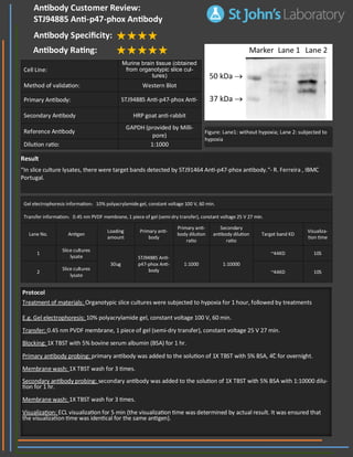
1. Result
“In slice culture lysates, there were target bands detected by STJ91464 Anti-p47-phox antibody.“- R. Ferreira , IBMC
Portugal.
Gel electrophoresis information:10% polyacrylamide gel, constant voltage 100 V, 60 min.
Transfer information:0.45 nm PVDF membrane, 1 piece of gel (semi-dry transfer), constant voltage 25 V 27 min.
Lane No. Antigen
Loading
amount
Primary anti-
body
Primary anti-
body dilution
ratio
Secondary
antibody dilution
ratio
Target band KD
Visualiza-
tion time
1
Slice cultures
lysate
30ug
STJ94885 Anti-
p47-phox Anti-
body
1:1000
~44KD 10S
1:10000
2
Slice cultures
lysate
~44KD 10S
Cell Line:
Murine brain tissue (obtained
from organotypic slice cul-
tures)
Method of validation: Western Blot
Primary Antibody: STJ94885 Anti-p47-phox Anti-
Secondary Antibody HRP goat anti-rabbit
Reference Antibody
GAPDH (provided by Milli-
pore)
Dilution ratio: 1:1000
Antibody Customer Review:
STJ94885 Anti-p47-phox Antibody
Antibody Specificity:
Antibody Rating:
Protocol
Treatment of materials: Organotypic slice cultures were subjected to hypoxia for 1 hour, followed by treatments
E.g. Gel electrophoresis: 10% polyacrylamide gel, constant voltage 100 V, 60 min.
Transfer: 0.45 nm PVDF membrane, 1 piece of gel (semi-dry transfer), constant voltage 25 V 27 min.
Blocking: 1X TBST with 5% bovine serum albumin (BSA) for 1 hr.
Primary antibody probing: primary antibody was added to the solution of 1X TBST with 5% BSA, 4˚C for overnight.
Membrane wash: 1X TBST wash for 3 times.
Secondary antibody probing: secondary antibody was added to the solution of 1X TBST with 5% BSA with 1:10000 dilu-
tion for 1 hr.
Membrane wash: 1X TBST wash for 3 times.
Visualization: ECL visualization for 5 min (the visualization time was determined by actual result. It was ensured that
the visualization time was identical for the same antigen).
Figure: Lane1: without hypoxia; Lane 2: subjected to
hypoxia