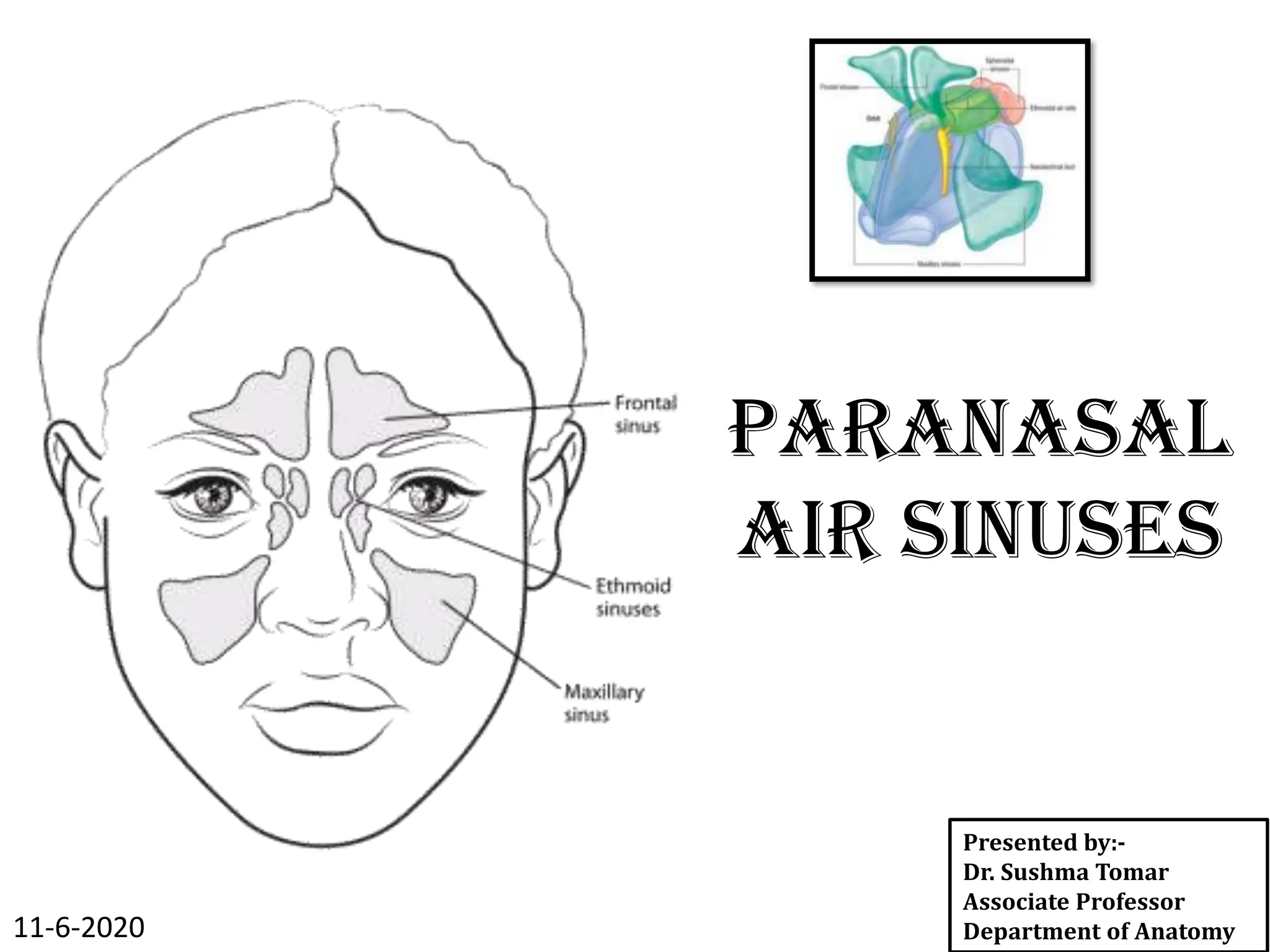
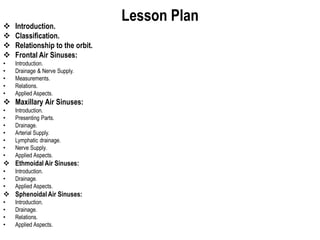
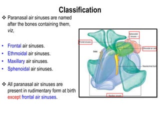
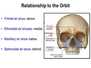
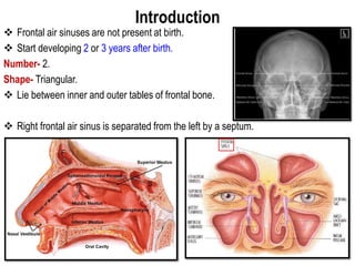
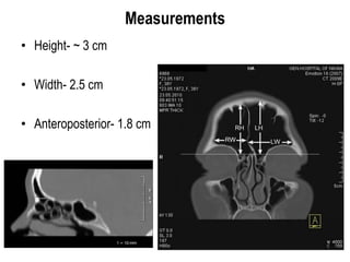
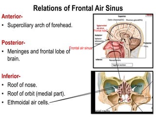
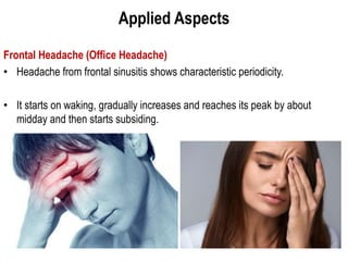
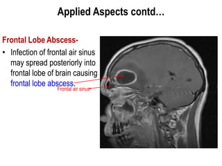
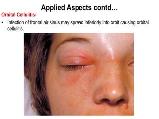
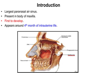
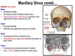
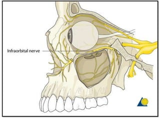
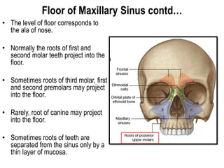
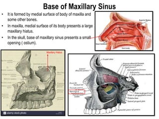
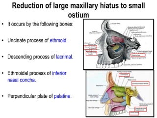
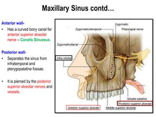
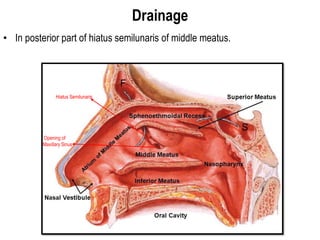
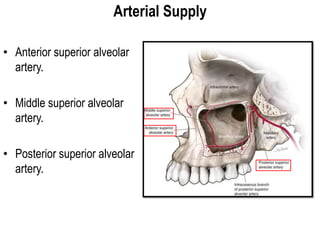
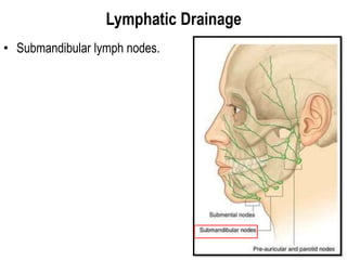
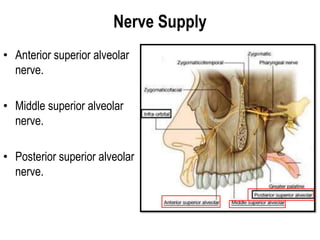
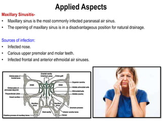
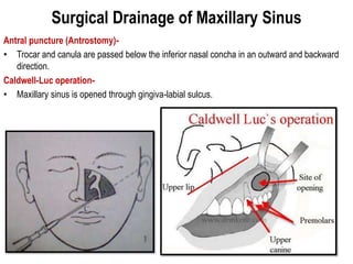
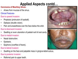
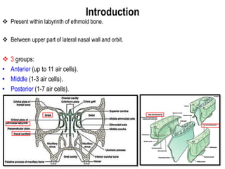
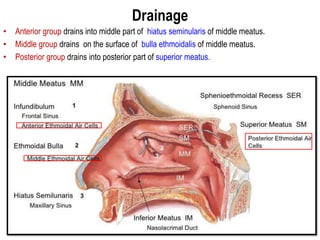
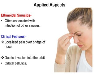
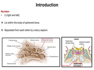
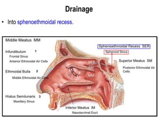
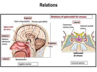
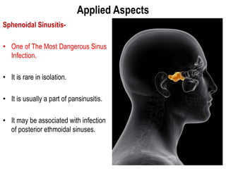
PARANASAL
AIR SINUSES
Presented by:-
Dr. Sushma Tomar
Associate Professor
Department of Anatomy
11-6-2020
Lesson Plan
 Introduction.
 Classification.
 Relationship to the orbit.
 Frontal Air Sinuses:
• Introduction.
• Drainage & Nerve Supply.
• Measurements.
• Relations.
• Applied Aspects.
 Maxillary Air Sinuses:
• Introduction.
• Presenting Parts.
• Drainage.
• Arterial Supply.
• Lymphatic drainage.
• Nerve Supply.
• Applied Aspects.
 Ethmoidal Air Sinuses:
• Introduction.
• Drainage.
• Applied Aspects.
 SphenoidalAir Sinuses:
• Introduction.
• Drainage.
• Relations.
• Applied Aspects.
Introduction
 Air-containing cavities in the bones
around nasal cavity.
 Paranasal air sinuses develop as
mucosal diverticulae of nasal cavity,
invading the adjacent bones.
 Paranasal air sinuses perform the
following functions:
• Make the skull lighter.
• Add resonance to the voice.
• Act as air conditioning chambers by
making the inspired air moist and warm.
• Aid in growth of facial skeleton.
 Paranasal air sinuses are lined by respiratory epithelium and
respiratory mucosa is highly vascular and contains a large
number of cavernous spaces and sinusoids to warm the air.
 Respiratory mucosa also contains a large number of serous
glands and secretion of these glands makes the air moist.
Classification
 Paranasal air sinuses are named
after the bones containing them,
viz,
• Frontal air sinuses.
• Ethmoidal air sinuses.
• Maxillary air sinuses.
• Sphenoidal air sinuses.
 All paranasal air sinuses are
present in rudimentary form at birth
except frontal air sinuses.
Relationship to the Orbit
• Frontal air sinus- above.
• Ethmoidal air sinuses- medial.
• Maxillary air sinus- below.
• Sphenoidal air sinus- behind.
Frontal Air Sinuses
Introduction
 Frontal air sinuses are not present at birth.
 Start developing 2 or 3 years after birth.
Number- 2.
Shape- Triangular.
 Lie between inner and outer tables of frontal bone.
 Right frontal air sinus is separated from the left by a septum.
Drainage & Nerve Supply
• Drainage- Drains into anterior part of hiatus semilunaris of middle meatus
through frontonasal duct.
• Nerve Supply- Supraorbital nerve.
Hiatus
Semilunaris
Measurements
• Height- ~ 3 cm
• Width- 2.5 cm
• Anteroposterior- 1.8 cm
Relations of Frontal Air Sinus
Anterior-
• Superciliary arch of forehead.
Posterior-
• Meninges and frontal lobe of
brain.
Inferior-
• Roof of nose.
• Roof of orbit (medial part).
• Ethmoidal air cells.
Frontal air sinus
Frontal lobe
Applied Aspects
Frontal Headache (Office Headache)
• Headache from frontal sinusitis shows characteristic periodicity.
• It starts on waking, gradually increases and reaches its peak by about
midday and then starts subsiding.
Applied Aspects contd…
Frontal Lobe Abscess-
• Infection of frontal air sinus
may spread posteriorly into
frontal lobe of brain causing
frontal lobe abscess.
Frontal air sinus
Applied Aspects contd…
Orbital Cellulitis-
• Infection of frontal air sinus may spread inferiorly into orbit causing orbital
cellulitis.
Maxillary Sinus
(Antrum of Highmore)
Introduction
• Largest paranasal air sinus.
• Present in body of maxilla.
• First to develop.
• Appears around 4th month of intrauterine life.
Maxillary Sinus contd…
SHAPE- Pyramidal.
Base-
• Directed medially.
• Formed by a part of lateral wall of nose.
• Opening or ostium of the sinus is present in the
upper part of base, close to the roof.
Apex-
• Directed laterally.
• Extends into zygomatic process of maxilla.
Roof-
• Formed by the floor of orbital cavity.
• Infraorbital nerve and artery traverse the roof in a
bony canal.
Floor-
• Formed by the alveolar process of maxilla.
• Lies ~1.25 cm below the floor of nasal cavity.
Zygomatic bone
Ostium of Maxillary air sinus
Floor of Maxillary Sinus contd…
• The level of floor corresponds to
the ala of nose.
• Normally the roots of first and
second molar teeth project into the
floor.
• Sometimes roots of third molar, first
and second premolars may project
into the floor.
• Rarely, root of canine may project
into the floor.
• Sometimes roots of teeth are
separated from the sinus only by a
thin layer of mucosa.
Base of Maxillary Sinus
• It is formed by medial surface of body of maxilla and
some other bones.
• In maxilla, medial surface of its body presents a large
maxillary hiatus.
• In the skull, base of maxillary sinus presents a small
opening ( ostium).
Maxillary Hiatus
Reduction of large maxillary hiatus to small
ostium
• It occurs by the following bones:
• Uncinate process of ethmoid.
• Descending process of lacrimal.
• Ethmoidal process of inferior
nasal concha.
• Perpendicular plate of palatine.
Maxillary Sinus contd…
Anterior wall-
• Has a curved bony canal for
anterior superior alveolar
nerve – Canalis Sinuosus.
Posterior wall-
• Separates the sinus from
infratemporal and
pterygopalatine fossae.
• It is pierced by the posterior
superior alveolar nerves and
vessels.
Drainage
• In posterior part of hiatus semilunaris of middle meatus.
Hiatus Semilunaris
Opening of
Maxillary Sinus
Arterial Supply
• Anterior superior alveolar
artery.
• Middle superior alveolar
artery.
• Posterior superior alveolar
artery.
Lymphatic Drainage
• Submandibular lymph nodes.
Nerve Supply
• Anterior superior alveolar
nerve.
• Middle superior alveolar
nerve.
• Posterior superior alveolar
nerve.
Applied Aspects
Maxillary Sinusitis-
• Maxillary sinus is the most commonly infected paranasal air sinus.
• The opening of maxillary sinus is in a disadvantageous position for natural drainage.
Sources of infection:
• Infected nose.
• Carious upper premolar and molar teeth.
• Infected frontal and anterior ethmoidal air sinuses.
Surgical Drainage of Maxillary Sinus
Antral puncture (Antrostomy)-
• Trocar and canula are passed below the inferior nasal concha in an outward and backward
direction.
Caldwell-Luc operation-
• Maxillary sinus is opened through gingiva-labial sulcus.
Applied Aspects contd…
Carcinoma of Maxillary Sinus-
• Arises from mucosa of the sinus.
Clinical Features-
Due to upward invasion:
• Proptosis (protrusion of eyeball).
• Diplopia (double vision).
• Pain and anaesthesia over the face below the orbit.
Due to downward invasion:
• Swelling or even ulceration of palatal roof of oral cavity.
Due to medial invasion:
• Nasal obstruction.
• Epistaxis.
• Epiphora (overflow of tears).
Due to lateral invasion:
• Swelling on the face and palpable mass in gingiva-labial sulcus.
Due to posterior invasion:
• Referred pain to upper teeth.
Ethmoidal Sinuses
Introduction
 Present within labyrinth of ethmoid bone.
 Between upper part of lateral nasal wall and orbit.
 3 groups:
• Anterior (up to 11 air cells).
• Middle (1-3 air cells).
• Posterior (1-7 air cells).
Drainage
• Anterior group drains into middle part of hiatus seminularis of middle meatus.
• Middle group drains on the surface of bulla ethmoidalis of middle meatus.
• Posterior group drains into posterior part of superior meatus.
Applied Aspects
Ethmoidal Sinusitis-
• Often asoociated with
infection of other sinuses.
Clinical Features-
Localized pain over bridge of
nose.
Due to invasion into the orbit-
• Orbital cellulitis.
Sphenoidal Sinuses
Introduction
Number-
• 2 (right and left)
 Lie within the body of sphenoid bone.
 Separated from each other by a bony septum.
Bony Septum
Drainage
• Into sphenoethmoidal recess.
Relations
Applied Aspects
Sphenoidal Sinusitis-
• One of The Most Dangerous Sinus
Infection.
• It is rare in isolation.
• It is usually a part of pansinusitis.
• It may be associated with infection
of posterior ethmoidal sinuses.
PARANASAL-AIR-SINUSES ethmoid, mastoidpdf

PARANASAL-AIR-SINUSES ethmoid, mastoidpdf

  • 1.
    PARANASAL AIR SINUSES Presented by:- Dr.Sushma Tomar Associate Professor Department of Anatomy 11-6-2020
  • 2.
    Lesson Plan  Introduction. Classification.  Relationship to the orbit.  Frontal Air Sinuses: • Introduction. • Drainage & Nerve Supply. • Measurements. • Relations. • Applied Aspects.  Maxillary Air Sinuses: • Introduction. • Presenting Parts. • Drainage. • Arterial Supply. • Lymphatic drainage. • Nerve Supply. • Applied Aspects.  Ethmoidal Air Sinuses: • Introduction. • Drainage. • Applied Aspects.  SphenoidalAir Sinuses: • Introduction. • Drainage. • Relations. • Applied Aspects.
  • 3.
    Introduction  Air-containing cavitiesin the bones around nasal cavity.  Paranasal air sinuses develop as mucosal diverticulae of nasal cavity, invading the adjacent bones.  Paranasal air sinuses perform the following functions: • Make the skull lighter. • Add resonance to the voice. • Act as air conditioning chambers by making the inspired air moist and warm. • Aid in growth of facial skeleton.  Paranasal air sinuses are lined by respiratory epithelium and respiratory mucosa is highly vascular and contains a large number of cavernous spaces and sinusoids to warm the air.  Respiratory mucosa also contains a large number of serous glands and secretion of these glands makes the air moist.
  • 4.
    Classification  Paranasal airsinuses are named after the bones containing them, viz, • Frontal air sinuses. • Ethmoidal air sinuses. • Maxillary air sinuses. • Sphenoidal air sinuses.  All paranasal air sinuses are present in rudimentary form at birth except frontal air sinuses.
  • 5.
    Relationship to theOrbit • Frontal air sinus- above. • Ethmoidal air sinuses- medial. • Maxillary air sinus- below. • Sphenoidal air sinus- behind.
  • 6.
  • 7.
    Introduction  Frontal airsinuses are not present at birth.  Start developing 2 or 3 years after birth. Number- 2. Shape- Triangular.  Lie between inner and outer tables of frontal bone.  Right frontal air sinus is separated from the left by a septum.
  • 8.
    Drainage & NerveSupply • Drainage- Drains into anterior part of hiatus semilunaris of middle meatus through frontonasal duct. • Nerve Supply- Supraorbital nerve. Hiatus Semilunaris
  • 9.
    Measurements • Height- ~3 cm • Width- 2.5 cm • Anteroposterior- 1.8 cm
  • 10.
    Relations of FrontalAir Sinus Anterior- • Superciliary arch of forehead. Posterior- • Meninges and frontal lobe of brain. Inferior- • Roof of nose. • Roof of orbit (medial part). • Ethmoidal air cells. Frontal air sinus Frontal lobe
  • 11.
    Applied Aspects Frontal Headache(Office Headache) • Headache from frontal sinusitis shows characteristic periodicity. • It starts on waking, gradually increases and reaches its peak by about midday and then starts subsiding.
  • 12.
    Applied Aspects contd… FrontalLobe Abscess- • Infection of frontal air sinus may spread posteriorly into frontal lobe of brain causing frontal lobe abscess. Frontal air sinus
  • 13.
    Applied Aspects contd… OrbitalCellulitis- • Infection of frontal air sinus may spread inferiorly into orbit causing orbital cellulitis.
  • 14.
  • 15.
    Introduction • Largest paranasalair sinus. • Present in body of maxilla. • First to develop. • Appears around 4th month of intrauterine life.
  • 16.
    Maxillary Sinus contd… SHAPE-Pyramidal. Base- • Directed medially. • Formed by a part of lateral wall of nose. • Opening or ostium of the sinus is present in the upper part of base, close to the roof. Apex- • Directed laterally. • Extends into zygomatic process of maxilla. Roof- • Formed by the floor of orbital cavity. • Infraorbital nerve and artery traverse the roof in a bony canal. Floor- • Formed by the alveolar process of maxilla. • Lies ~1.25 cm below the floor of nasal cavity. Zygomatic bone Ostium of Maxillary air sinus
  • 18.
    Floor of MaxillarySinus contd… • The level of floor corresponds to the ala of nose. • Normally the roots of first and second molar teeth project into the floor. • Sometimes roots of third molar, first and second premolars may project into the floor. • Rarely, root of canine may project into the floor. • Sometimes roots of teeth are separated from the sinus only by a thin layer of mucosa.
  • 19.
    Base of MaxillarySinus • It is formed by medial surface of body of maxilla and some other bones. • In maxilla, medial surface of its body presents a large maxillary hiatus. • In the skull, base of maxillary sinus presents a small opening ( ostium). Maxillary Hiatus
  • 20.
    Reduction of largemaxillary hiatus to small ostium • It occurs by the following bones: • Uncinate process of ethmoid. • Descending process of lacrimal. • Ethmoidal process of inferior nasal concha. • Perpendicular plate of palatine.
  • 21.
    Maxillary Sinus contd… Anteriorwall- • Has a curved bony canal for anterior superior alveolar nerve – Canalis Sinuosus. Posterior wall- • Separates the sinus from infratemporal and pterygopalatine fossae. • It is pierced by the posterior superior alveolar nerves and vessels.
  • 22.
    Drainage • In posteriorpart of hiatus semilunaris of middle meatus. Hiatus Semilunaris Opening of Maxillary Sinus
  • 23.
    Arterial Supply • Anteriorsuperior alveolar artery. • Middle superior alveolar artery. • Posterior superior alveolar artery.
  • 24.
  • 25.
    Nerve Supply • Anteriorsuperior alveolar nerve. • Middle superior alveolar nerve. • Posterior superior alveolar nerve.
  • 26.
    Applied Aspects Maxillary Sinusitis- •Maxillary sinus is the most commonly infected paranasal air sinus. • The opening of maxillary sinus is in a disadvantageous position for natural drainage. Sources of infection: • Infected nose. • Carious upper premolar and molar teeth. • Infected frontal and anterior ethmoidal air sinuses.
  • 27.
    Surgical Drainage ofMaxillary Sinus Antral puncture (Antrostomy)- • Trocar and canula are passed below the inferior nasal concha in an outward and backward direction. Caldwell-Luc operation- • Maxillary sinus is opened through gingiva-labial sulcus.
  • 28.
    Applied Aspects contd… Carcinomaof Maxillary Sinus- • Arises from mucosa of the sinus. Clinical Features- Due to upward invasion: • Proptosis (protrusion of eyeball). • Diplopia (double vision). • Pain and anaesthesia over the face below the orbit. Due to downward invasion: • Swelling or even ulceration of palatal roof of oral cavity. Due to medial invasion: • Nasal obstruction. • Epistaxis. • Epiphora (overflow of tears). Due to lateral invasion: • Swelling on the face and palpable mass in gingiva-labial sulcus. Due to posterior invasion: • Referred pain to upper teeth.
  • 29.
  • 30.
    Introduction  Present withinlabyrinth of ethmoid bone.  Between upper part of lateral nasal wall and orbit.  3 groups: • Anterior (up to 11 air cells). • Middle (1-3 air cells). • Posterior (1-7 air cells).
  • 31.
    Drainage • Anterior groupdrains into middle part of hiatus seminularis of middle meatus. • Middle group drains on the surface of bulla ethmoidalis of middle meatus. • Posterior group drains into posterior part of superior meatus.
  • 32.
    Applied Aspects Ethmoidal Sinusitis- •Often asoociated with infection of other sinuses. Clinical Features- Localized pain over bridge of nose. Due to invasion into the orbit- • Orbital cellulitis.
  • 33.
  • 34.
    Introduction Number- • 2 (rightand left)  Lie within the body of sphenoid bone.  Separated from each other by a bony septum. Bony Septum
  • 35.
  • 36.
  • 37.
    Applied Aspects Sphenoidal Sinusitis- •One of The Most Dangerous Sinus Infection. • It is rare in isolation. • It is usually a part of pansinusitis. • It may be associated with infection of posterior ethmoidal sinuses.