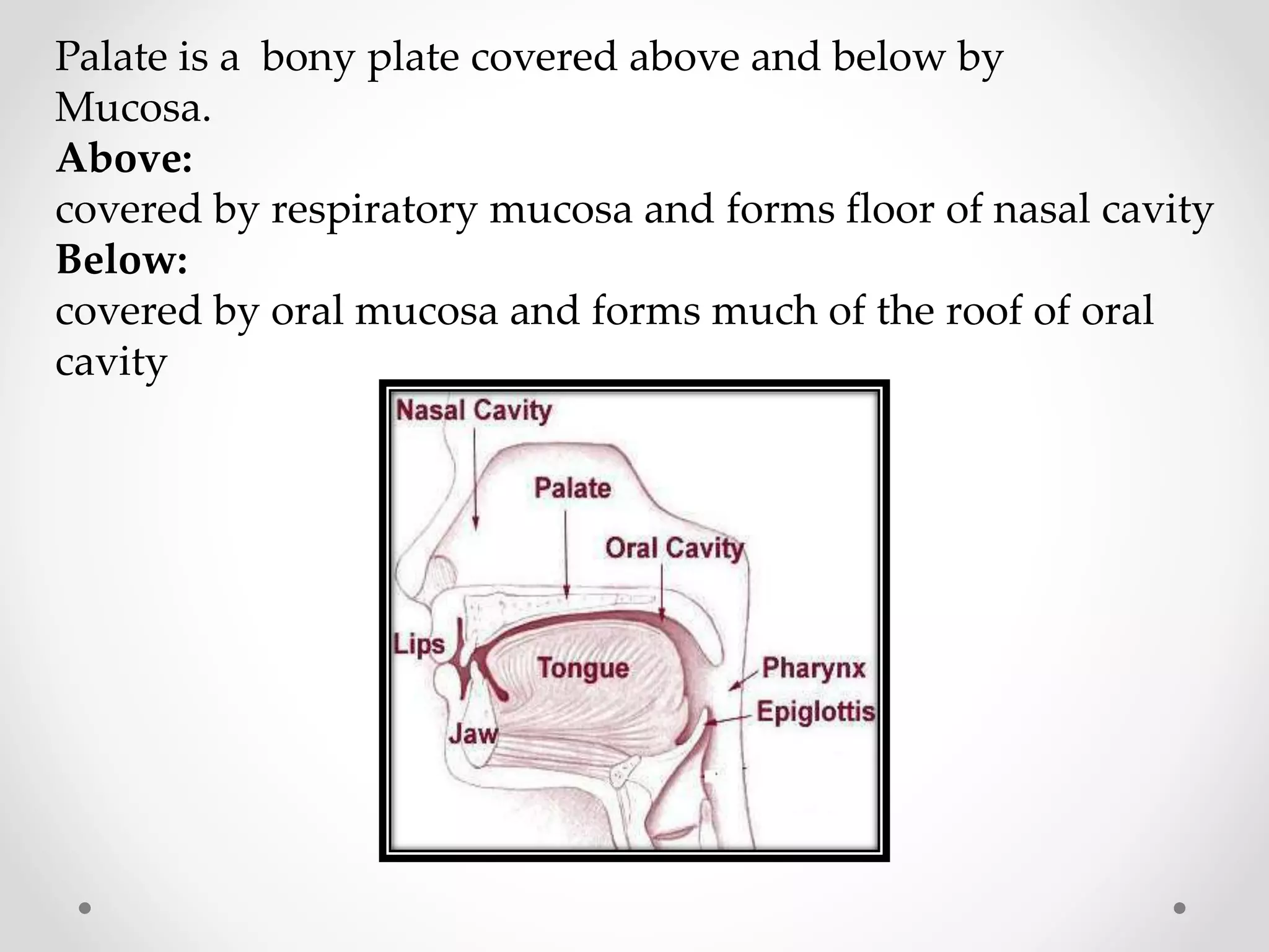
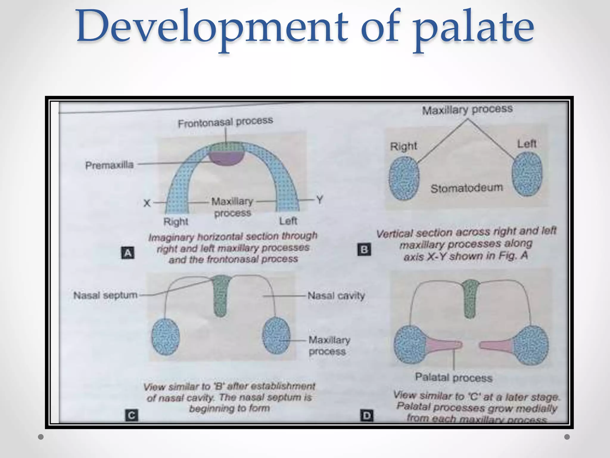
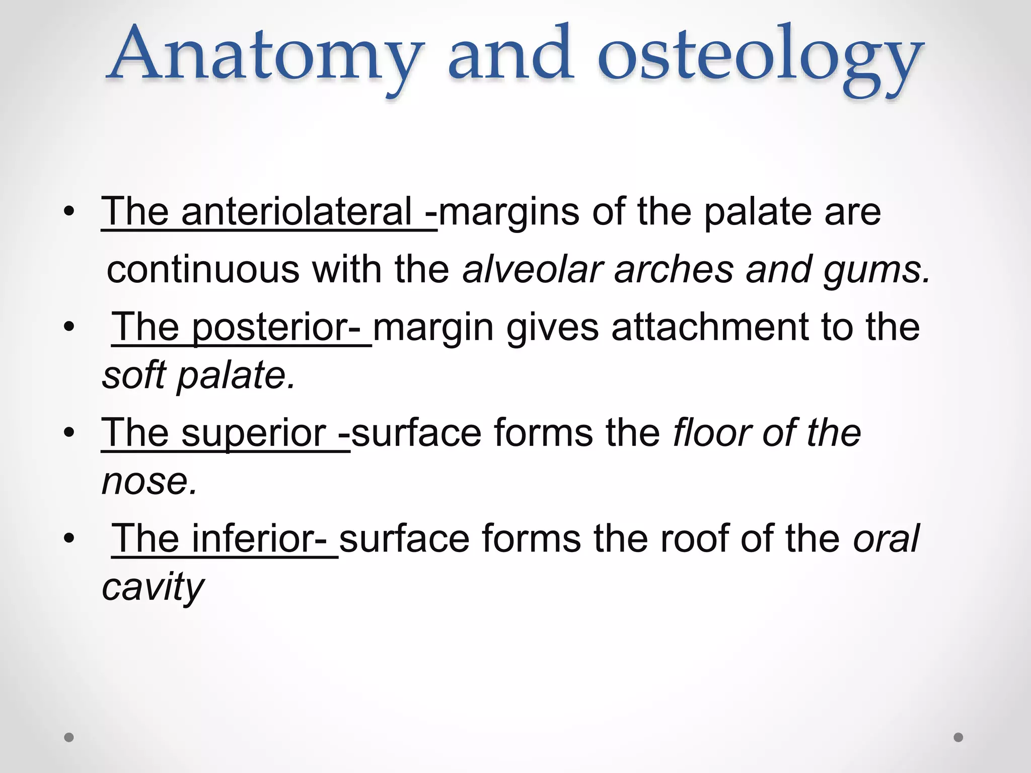
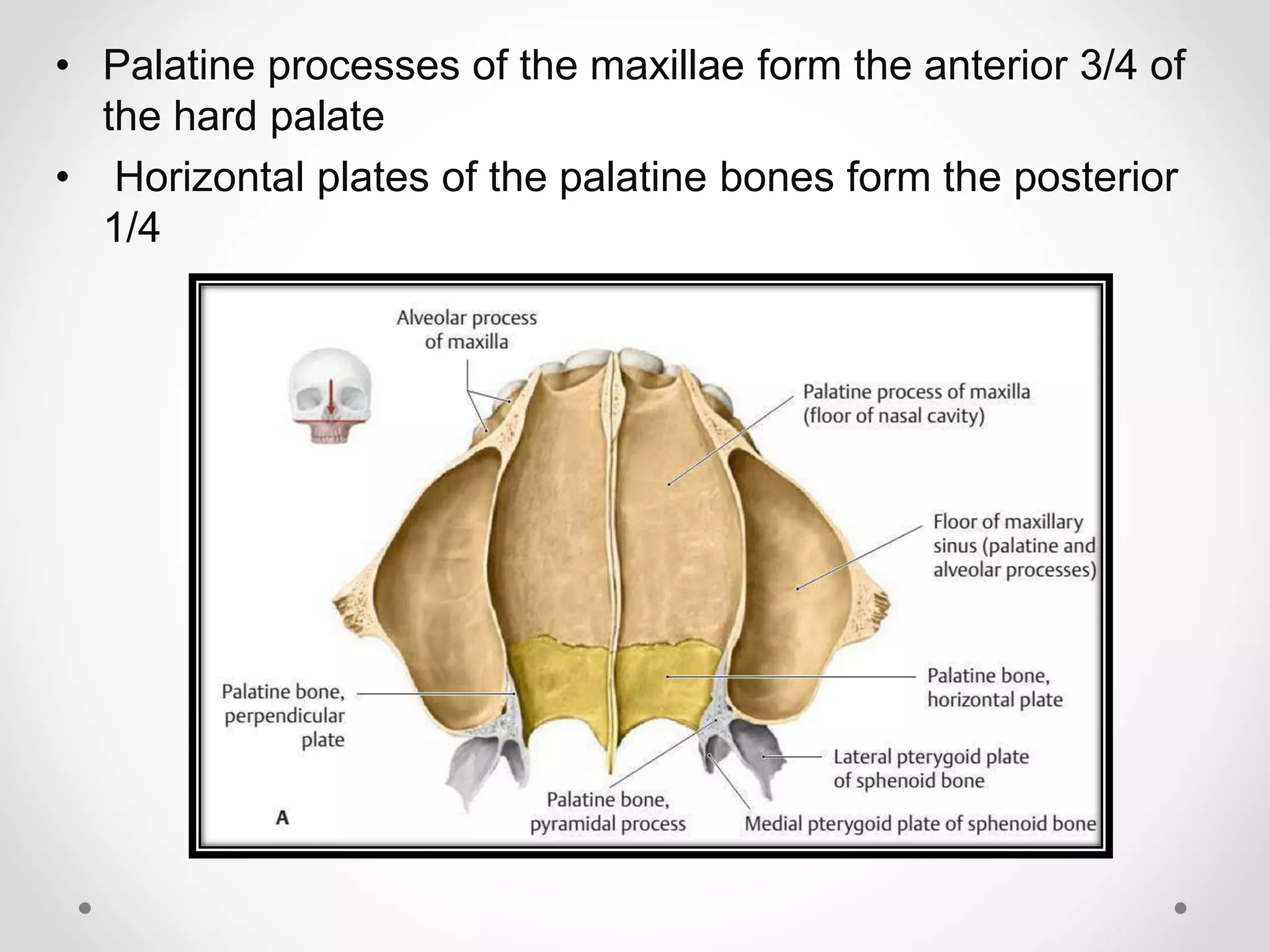
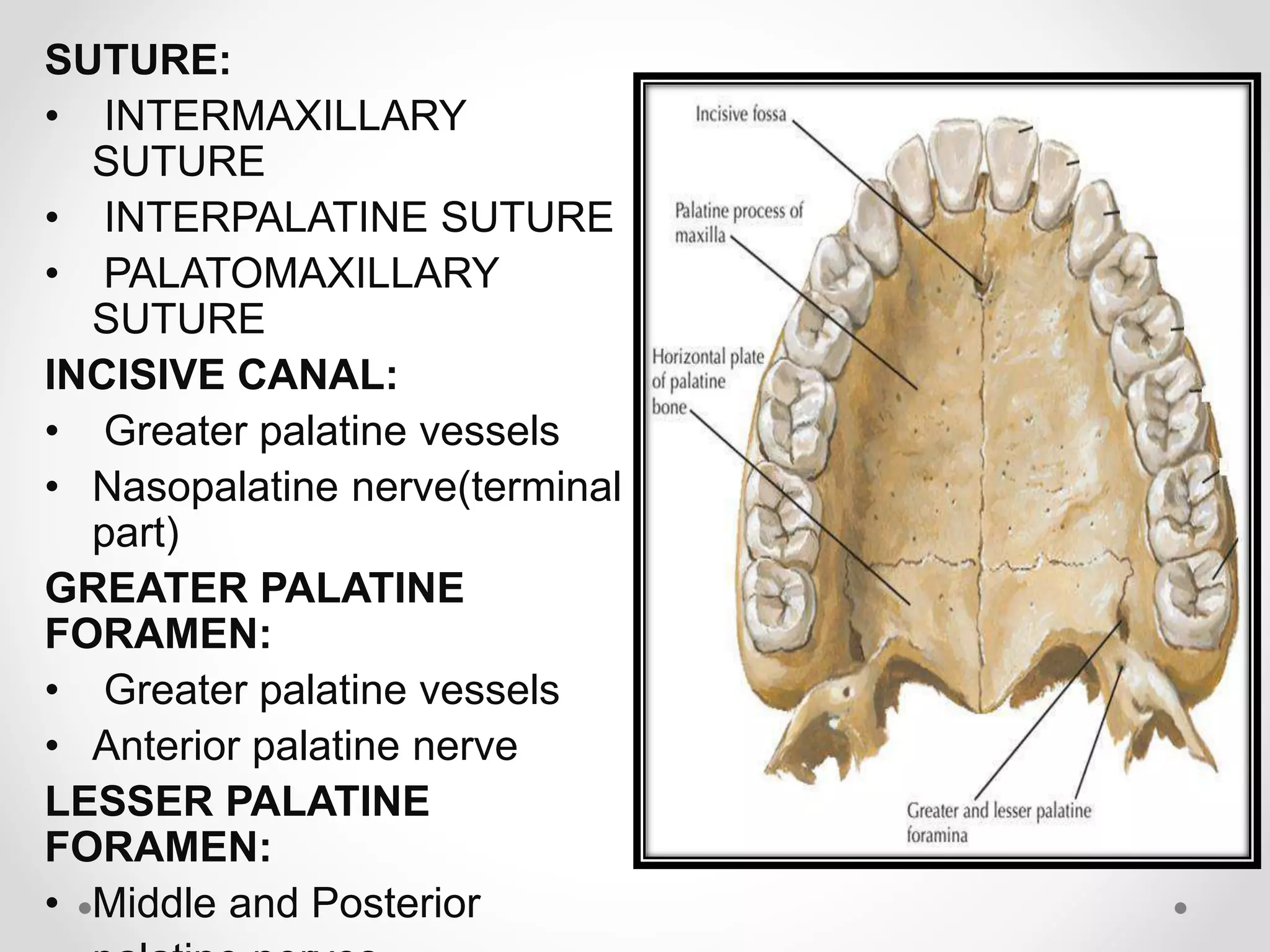
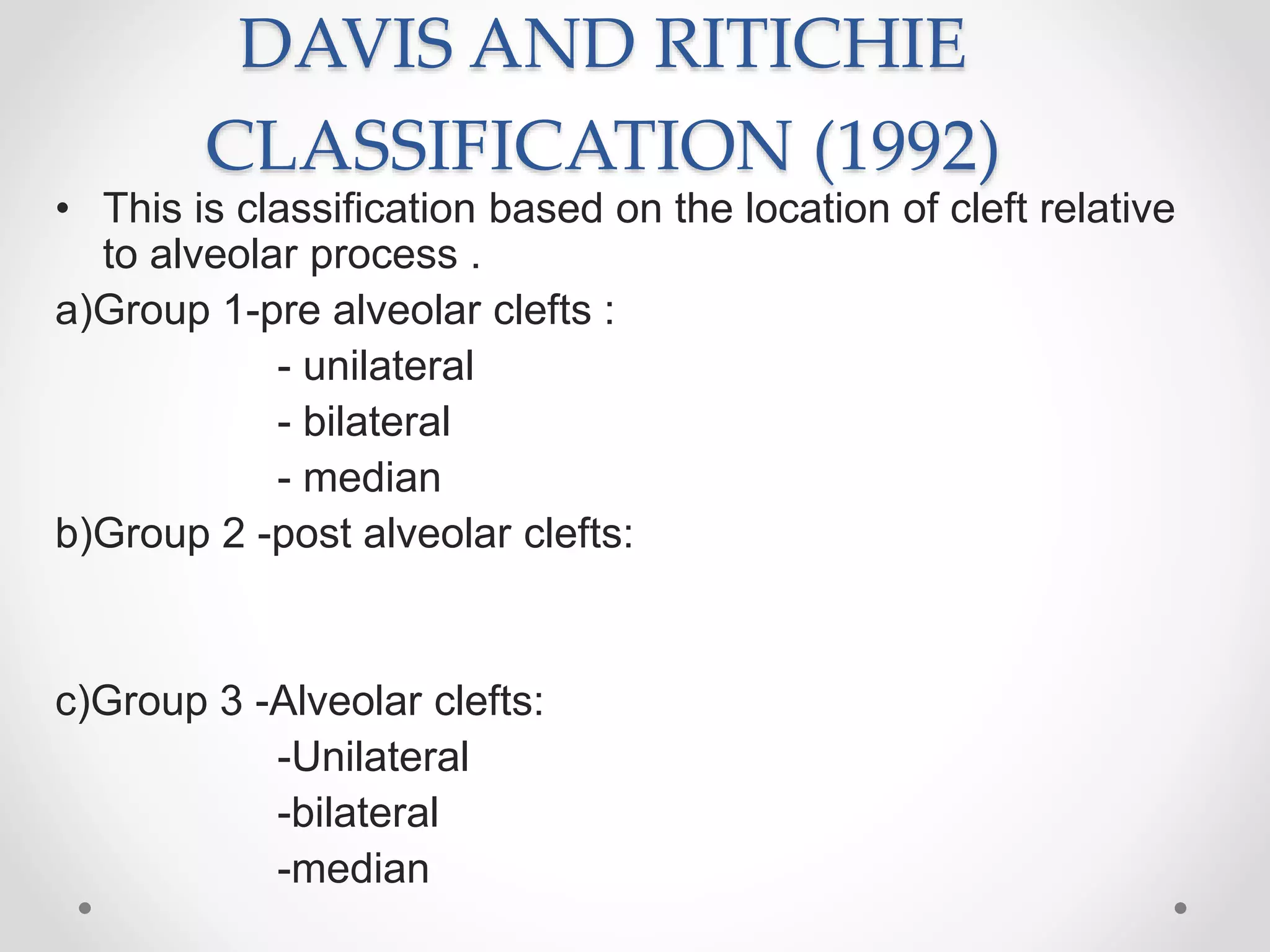
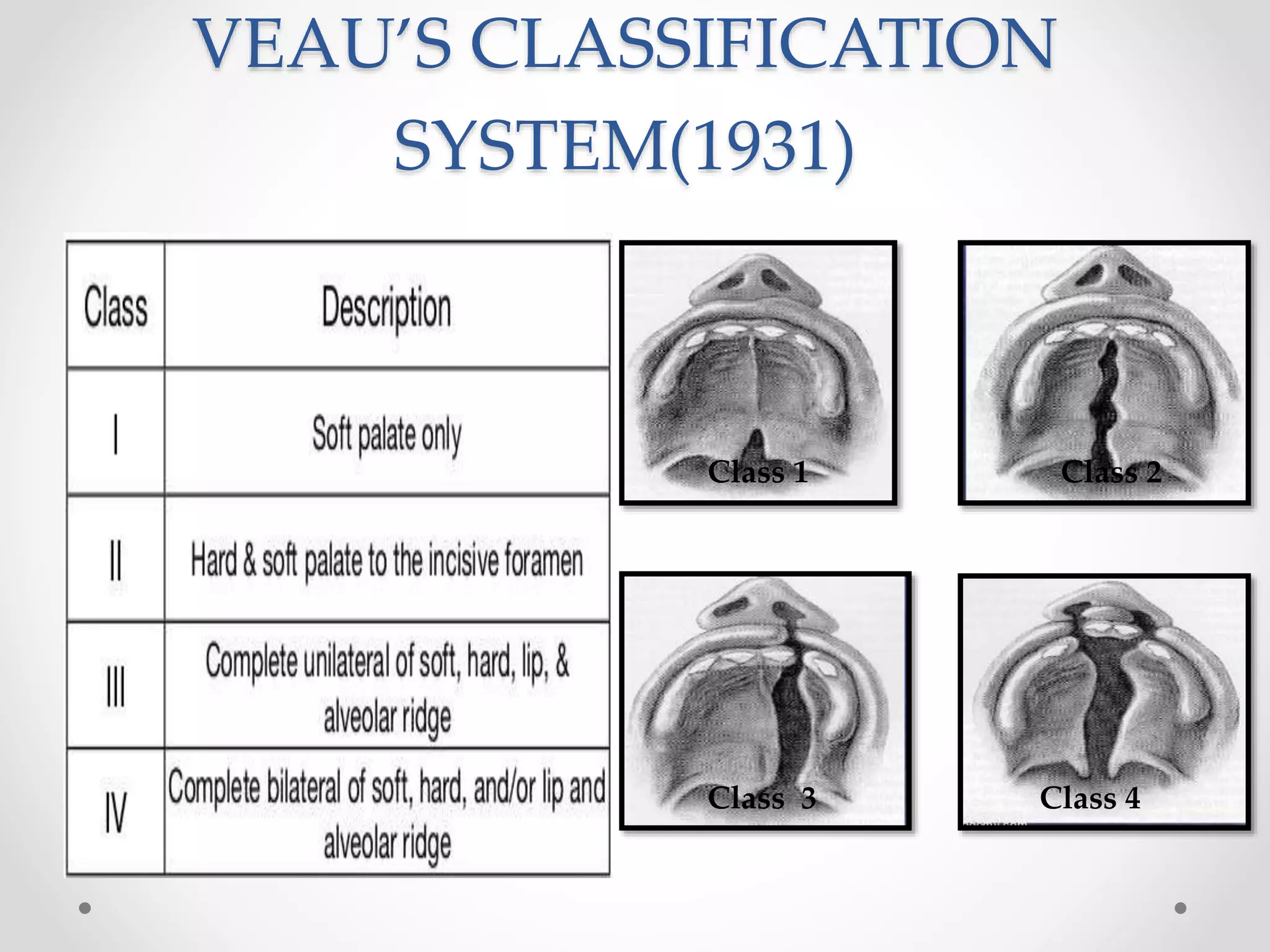
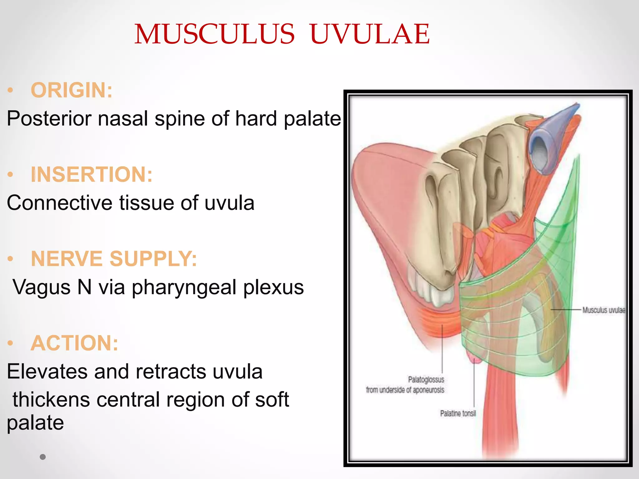
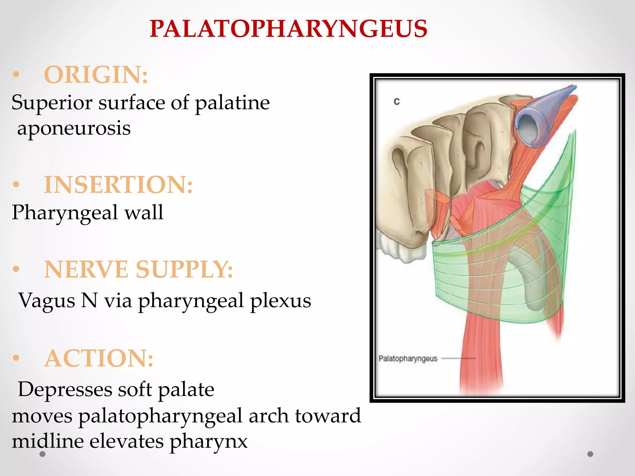
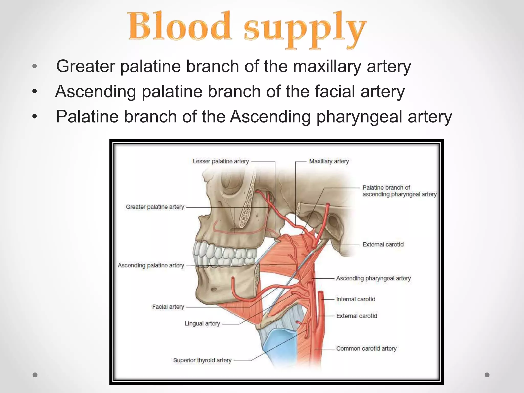
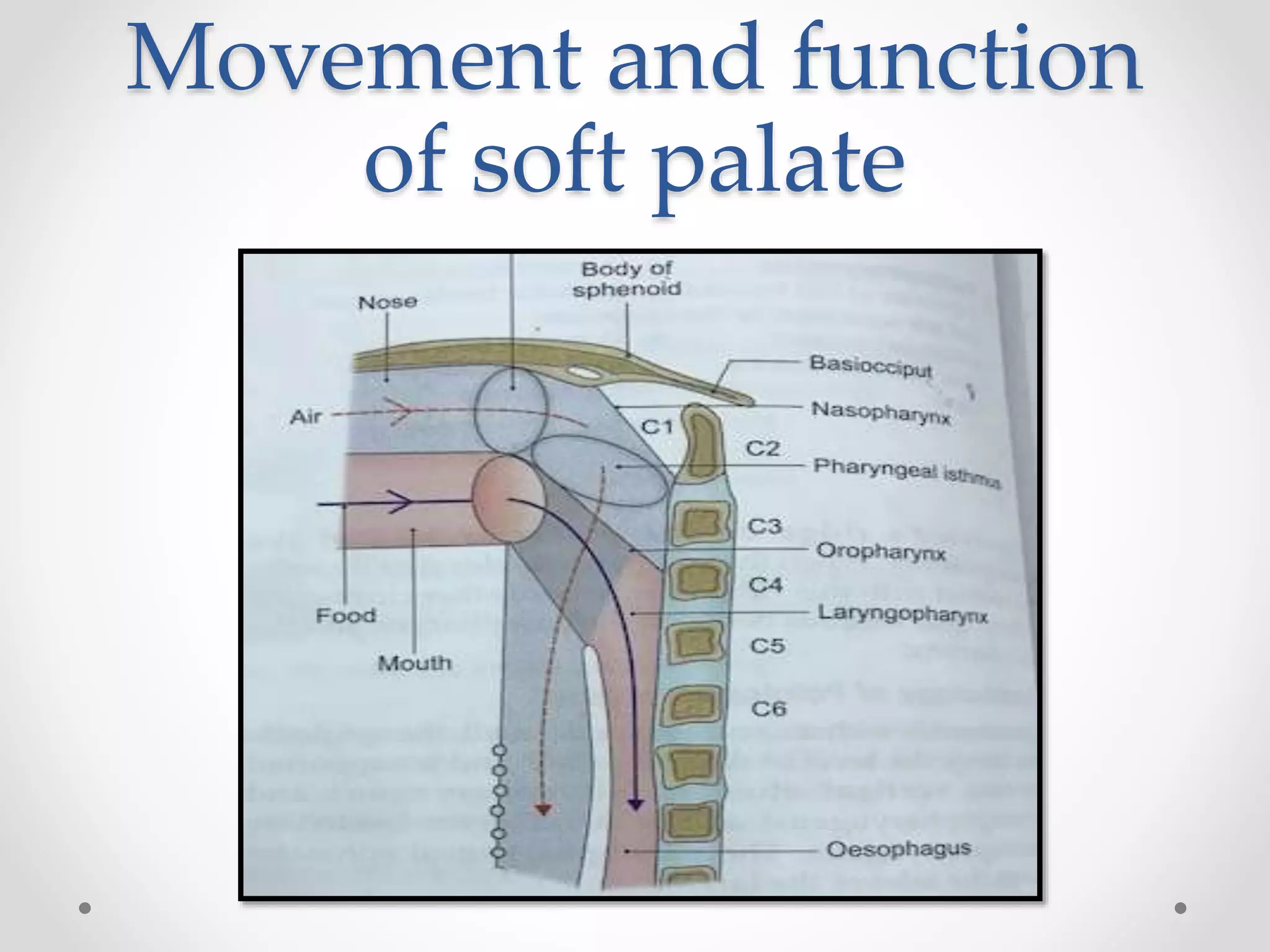
Presentation by:
Madhushree patil
1st year PG student
Dept of prosthodontics
content
• Introduction
• Hard palate
• Development and embryology
• Anatomy
• Developmental anomalies
• Prosthodontics consideration
• Soft palate
• Anatomy
• Developmental anomalies
• Prosthodontic consideration
INTRODUCTION
• Palate : Roof of the oral cavity.
• It has two parts
–an anterior hard palate
–a posterior soft palate
Palate is a bony plate covered above and below by
Mucosa.
Above:
covered by respiratory mucosa and forms floor of nasal cavity
Below:
covered by oral mucosa and forms much of the roof of oral
cavity
Development and embryology
Development of palate
Constituent of
development of palate
Embryological subdivision
Anatomy and osteology
• The anteriolateral -margins of the palate are
continuous with the alveolar arches and gums.
• The posterior- margin gives attachment to the
soft palate.
• The superior -surface forms the floor of the
nose.
• The inferior- surface forms the roof of the oral
cavity
• Palatine processes of the maxillae form the anterior 3/4 of
the hard palate
• Horizontal plates of the palatine bones form the posterior
1/4
SUTURE:
• INTERMAXILLARY
SUTURE
• INTERPALATINE SUTURE
• PALATOMAXILLARY
SUTURE
INCISIVE CANAL:
• Greater palatine vessels
• Nasopalatine nerve(terminal
part)
GREATER PALATINE
FORAMEN:
• Greater palatine vessels
• Anterior palatine nerve
LESSER PALATINE
FORAMEN:
• Middle and Posterior
Developmental defects of
palate
• Diagnosis generally unilateral and bilateral clefts
in the palate is classified in 3 groups :
• Clefts of anterior (primary)palate (i.e clefts ant.
To incisive fossa)results from failure of
mesenchymal masses in lateral palatine
processes to meet and fuse with mesenchyme in
primary palate
DAVIS AND RITICHIE
CLASSIFICATION (1992)
• This is classification based on the location of cleft relative
to alveolar process .
a)Group 1-pre alveolar clefts :
- unilateral
- bilateral
- median
b)Group 2 -post alveolar clefts:
c)Group 3 -Alveolar clefts:
-Unilateral
-bilateral
-median
VEAU’S CLASSIFICATION
SYSTEM(1931)
Class 1 Class 2
Class 3 Class 4
FOGH ANDERSONS
CLASSIFICATON (1942)
• Group 1: They are clefts of lips
-single-unilateral
-double –bilateral clefts
• Group 2: They are the clefts of lip and palate
-single –unilateral
-double – bilateral clefts.
• Group 3: They are cleft of palate extending up
to Incisive formane
SCHUCHARDT AND PFEIFERS
SYMBOLIC CLASSIFICATION
KERNAHAN’S STRIPPED
(Y)CLASSIFICATION
• Obturators
A prosthesis used to close a congenital or acquired
tissues opening ,primarily of hard palate and contiguous
alveolar structures. Prosthetic restoration of defects often
includes use of surgical obturators ,interim obturators, and
definitive obturators .
TORUS PALATINUS
• Localized nodular enlargement (exostosis) of the cortical
bone
• Usually – midline of the palate
• Pose a mechanical problem in the construction of
denture
INFLAMMATORY PAPILLARY HYPERPLASIA
• Common lesion that develops on the central hard palate
• in response to chronic denture irritation
Soft palate
• Movable, muscular fold,
suspended from the posterior
border of the hard palate.
• It separates the nasopharynx
from the oropharynx. Acts as a
valve that can be:
 depressed to help close the
oropharyngeal isthmus;
 elevated to separate the
nasopharynx from the
oropharynx.
MUSCLES OF SOFT PALATE
• Tensor veli palatini
• Levator veli palatini
• Musculus uvulae
• Palato pharyngeus
• Palatoglossus
• ORIGIN:
Lateral side of auditary tube
Scaphoid fossa of sphenoid
bone
• INSERTION:
Palatine aponeurosis
• NERVE SUPPLY:
Mandibular nerve to
medial pterygoid muscle
• ACTION:
Tightens the soft palate
Opens the auditory tube
TENSOR VELI PALATINI
• ORIGIN:
Petrous temporal bone
Inferior aspect of auditory tube
• INSERTION:
Upper surface of palatine
aponeurosis
• NERVE SUPPLY:
Vagus N via pharyngeal plexus
• ACTION:
Elevates the soft palate
LEVATORE VELI PALATINI
• ORIGIN:
Posterior nasal spine of hard palate
• INSERTION:
Connective tissue of uvula
• NERVE SUPPLY:
Vagus N via pharyngeal plexus
• ACTION:
Elevates and retracts uvula
thickens central region of soft
palate
MUSCULUS UVULAE
• ORIGIN:
Inferior surface of palatine
aponeurosis
• INSERTION:
Lateral margin of tongue
• NERVE SUPPLY:
Vagus N via pharyngealn plexus
• ACTION:
Depresses palate Moves palatoglossal
arch toward midline elevates back of
the tongue
PALATOGLOSSUS
• ORIGIN:
Superior surface of palatine
aponeurosis
• INSERTION:
Pharyngeal wall
• NERVE SUPPLY:
Vagus N via pharyngeal plexus
• ACTION:
Depresses soft palate
moves palatopharyngeal arch toward
midline elevates pharynx
PALATOPHARYNGEUS
• Greater palatine branch of the maxillary artery
• Ascending palatine branch of the facial artery
• Palatine branch of the Ascending pharyngeal artery
VEINS:
• Pterygoid plexuses
• tonsillar plexuses of veins.
LYMPHATICS:
• Upper deep cervical
• retropharyngeal lymph
nodes.
NERVE SUPPLY
• Supplied by the greater and lesser palatine nerves and the
nasopalatine nerve
• General sensory fibers carried in all these nerves originate in the
pterygopalatine fossa from the maxillary nerve
• Special sensory and scretomotor nerves are contained in lesser palatine
nerves.
Movement and function
of soft palate
• The anatomy of the soft palate determines the location of
the distal border of maxillary denture base and posterior
palatal seal
• The posterior extention of the maxillary denture base lies
in soft palate i.e, in palatal aponeurosis and overlying
mucosa
• Palatine muscles and contour of the soft palate
determines the extent and contour of the soft palate
• The seal should follow the contour of the palatine bones
and extends from hammular notch to hammular notch
Prosthodontic consideretion
• House classification of palatal throat forms
Found on line drawn between to hammular notches
Class I- more than 5 mm of tissue available for post
damming Ideal for retention
• Class II – 3-5 mm distal about 1-5 mm tissue available
• Class III – 3-5 mm anterior less than 1 mm tissue
available for post damming
• The slender tendon of tensor palatinae could
influence the denture contour when tout in
hammular notch area
• Vibrating line determined by the elevation of soft
palate during contraction of levator palatinae
• When 2 palatoglossi contract they draw the tongue
and soft palate together and close the isthumus of
fauces and bring lateral pressure ligual to the
extension of the mandibular denture base
summary
• Before construction of complete denture prostheses is
begun,the oral tissues and oral environment should be
assessed to ascertain that the denture bearing tissues
will accept the prosthesis and support it in comfort
• proper border seal will ensure a more retentive
prosthesis for patient,whose satisfaction is the dentists
main concern if anatomy and physiology of area is
understood
Refrences
• B D Chaurasia’s human anatomy 5th edition
• Human emnryology – inderbir singh 11th edition
• Burket’s oral medicine 11th edition
• Orban’ s oral histology and embryology
• Shafers textbook of oral pathology 7th edition

hard and soft palate

  • 1.
    Presentation by: Madhushree patil 1styear PG student Dept of prosthodontics
  • 2.
    content • Introduction • Hardpalate • Development and embryology • Anatomy • Developmental anomalies • Prosthodontics consideration • Soft palate • Anatomy • Developmental anomalies • Prosthodontic consideration
  • 3.
    INTRODUCTION • Palate :Roof of the oral cavity. • It has two parts –an anterior hard palate –a posterior soft palate
  • 4.
    Palate is abony plate covered above and below by Mucosa. Above: covered by respiratory mucosa and forms floor of nasal cavity Below: covered by oral mucosa and forms much of the roof of oral cavity
  • 5.
  • 6.
  • 7.
  • 8.
  • 9.
    Anatomy and osteology •The anteriolateral -margins of the palate are continuous with the alveolar arches and gums. • The posterior- margin gives attachment to the soft palate. • The superior -surface forms the floor of the nose. • The inferior- surface forms the roof of the oral cavity
  • 10.
    • Palatine processesof the maxillae form the anterior 3/4 of the hard palate • Horizontal plates of the palatine bones form the posterior 1/4
  • 11.
    SUTURE: • INTERMAXILLARY SUTURE • INTERPALATINESUTURE • PALATOMAXILLARY SUTURE INCISIVE CANAL: • Greater palatine vessels • Nasopalatine nerve(terminal part) GREATER PALATINE FORAMEN: • Greater palatine vessels • Anterior palatine nerve LESSER PALATINE FORAMEN: • Middle and Posterior
  • 12.
    Developmental defects of palate •Diagnosis generally unilateral and bilateral clefts in the palate is classified in 3 groups : • Clefts of anterior (primary)palate (i.e clefts ant. To incisive fossa)results from failure of mesenchymal masses in lateral palatine processes to meet and fuse with mesenchyme in primary palate
  • 13.
    DAVIS AND RITICHIE CLASSIFICATION(1992) • This is classification based on the location of cleft relative to alveolar process . a)Group 1-pre alveolar clefts : - unilateral - bilateral - median b)Group 2 -post alveolar clefts: c)Group 3 -Alveolar clefts: -Unilateral -bilateral -median
  • 14.
  • 15.
    FOGH ANDERSONS CLASSIFICATON (1942) •Group 1: They are clefts of lips -single-unilateral -double –bilateral clefts • Group 2: They are the clefts of lip and palate -single –unilateral -double – bilateral clefts. • Group 3: They are cleft of palate extending up to Incisive formane
  • 16.
  • 17.
  • 18.
    • Obturators A prosthesisused to close a congenital or acquired tissues opening ,primarily of hard palate and contiguous alveolar structures. Prosthetic restoration of defects often includes use of surgical obturators ,interim obturators, and definitive obturators .
  • 19.
    TORUS PALATINUS • Localizednodular enlargement (exostosis) of the cortical bone • Usually – midline of the palate • Pose a mechanical problem in the construction of denture
  • 20.
    INFLAMMATORY PAPILLARY HYPERPLASIA •Common lesion that develops on the central hard palate • in response to chronic denture irritation
  • 21.
    Soft palate • Movable,muscular fold, suspended from the posterior border of the hard palate. • It separates the nasopharynx from the oropharynx. Acts as a valve that can be:  depressed to help close the oropharyngeal isthmus;  elevated to separate the nasopharynx from the oropharynx.
  • 22.
    MUSCLES OF SOFTPALATE • Tensor veli palatini • Levator veli palatini • Musculus uvulae • Palato pharyngeus • Palatoglossus
  • 23.
    • ORIGIN: Lateral sideof auditary tube Scaphoid fossa of sphenoid bone • INSERTION: Palatine aponeurosis • NERVE SUPPLY: Mandibular nerve to medial pterygoid muscle • ACTION: Tightens the soft palate Opens the auditory tube TENSOR VELI PALATINI
  • 24.
    • ORIGIN: Petrous temporalbone Inferior aspect of auditory tube • INSERTION: Upper surface of palatine aponeurosis • NERVE SUPPLY: Vagus N via pharyngeal plexus • ACTION: Elevates the soft palate LEVATORE VELI PALATINI
  • 25.
    • ORIGIN: Posterior nasalspine of hard palate • INSERTION: Connective tissue of uvula • NERVE SUPPLY: Vagus N via pharyngeal plexus • ACTION: Elevates and retracts uvula thickens central region of soft palate MUSCULUS UVULAE
  • 26.
    • ORIGIN: Inferior surfaceof palatine aponeurosis • INSERTION: Lateral margin of tongue • NERVE SUPPLY: Vagus N via pharyngealn plexus • ACTION: Depresses palate Moves palatoglossal arch toward midline elevates back of the tongue PALATOGLOSSUS
  • 27.
    • ORIGIN: Superior surfaceof palatine aponeurosis • INSERTION: Pharyngeal wall • NERVE SUPPLY: Vagus N via pharyngeal plexus • ACTION: Depresses soft palate moves palatopharyngeal arch toward midline elevates pharynx PALATOPHARYNGEUS
  • 28.
    • Greater palatinebranch of the maxillary artery • Ascending palatine branch of the facial artery • Palatine branch of the Ascending pharyngeal artery
  • 29.
    VEINS: • Pterygoid plexuses •tonsillar plexuses of veins. LYMPHATICS: • Upper deep cervical • retropharyngeal lymph nodes.
  • 30.
    NERVE SUPPLY • Suppliedby the greater and lesser palatine nerves and the nasopalatine nerve • General sensory fibers carried in all these nerves originate in the pterygopalatine fossa from the maxillary nerve • Special sensory and scretomotor nerves are contained in lesser palatine nerves.
  • 31.
  • 32.
    • The anatomyof the soft palate determines the location of the distal border of maxillary denture base and posterior palatal seal • The posterior extention of the maxillary denture base lies in soft palate i.e, in palatal aponeurosis and overlying mucosa • Palatine muscles and contour of the soft palate determines the extent and contour of the soft palate • The seal should follow the contour of the palatine bones and extends from hammular notch to hammular notch Prosthodontic consideretion
  • 33.
    • House classificationof palatal throat forms Found on line drawn between to hammular notches Class I- more than 5 mm of tissue available for post damming Ideal for retention
  • 34.
    • Class II– 3-5 mm distal about 1-5 mm tissue available • Class III – 3-5 mm anterior less than 1 mm tissue available for post damming
  • 35.
    • The slendertendon of tensor palatinae could influence the denture contour when tout in hammular notch area • Vibrating line determined by the elevation of soft palate during contraction of levator palatinae • When 2 palatoglossi contract they draw the tongue and soft palate together and close the isthumus of fauces and bring lateral pressure ligual to the extension of the mandibular denture base
  • 36.
    summary • Before constructionof complete denture prostheses is begun,the oral tissues and oral environment should be assessed to ascertain that the denture bearing tissues will accept the prosthesis and support it in comfort • proper border seal will ensure a more retentive prosthesis for patient,whose satisfaction is the dentists main concern if anatomy and physiology of area is understood
  • 37.
    Refrences • B DChaurasia’s human anatomy 5th edition • Human emnryology – inderbir singh 11th edition • Burket’s oral medicine 11th edition • Orban’ s oral histology and embryology • Shafers textbook of oral pathology 7th edition