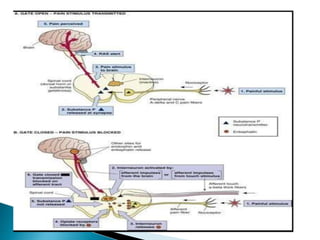
Prof. (Dr.) Nidhi Sharma
Professor, MMIPR, MM (DU),
Mullana-Ambala, Haryana
 According to this Gate-Control Theory of Pain
proposed by Ronald Melzack and Patrick Wall
in 1965.
 Activity in large diameter low threshold
mechanoreceptive (touch-related) nerve
fibers could inhibit the transmission of action
potentials from small diameter higher
threshold nociceptive (pain-related) fibers
through pre and post synaptic inhibition in
the dorsal horn of spinal cord.
 https://www.youtube.com/watch?v=oQLFfvG
M7nI
Modulation of pain transmission
At the level of spinal cord
 As already noted primary nociceptive afferent terminate at
the 2nd order neuron or T cells. The excitability of this
pathway can be altered by other inter neurons present in the
dorsal horn. The cells of Substantia gelatinosa have an
inhibitory influence on the transmission cell by Presynaptic
Inhibition of the nociceptive afferent terminals at the point
where they synapse with transmission cells.
 Substantia Gelatinosa cells are inhibited when the
nociceptive afferents are activated, this reduces the
presynaptic inhibition and allows nociceptive information to
be passed to higher center.
 Substantia Gelatinosa cells are stimulated by the activation
of low threshold large diameter A beta mechanosensitive
afferents. This leads to increase in presynaptic inhibition and
prevents the transmission of nociceptive information to be
passed to higher centers.
 The Gate Control Theory of Pain is a mechanism, in the
spinal cord, in which pain signals can be sent up to the
brain to be processed to accentuate the possible perceived
pain, or attenuate it at the spinal cord itself.
 The 'gate' is the mechanism where pain signals can be let
through or restricted. One of two things can happen, the
gate can be 'open' or the gate can be 'closed':
 If the gate is open, pain signals can pass through and
will be sent to the brain to perceive the pain.
 If the gate is closed, pain signals will be restricted from
travelling up to the brain, and the sensation of pain won't
be perceived.
 The pain gate mechanism is located in the dorsal horn of
the spinal cord, specifically in the Substantia gelatinosa.
 The interneurons within the Substantia gelatinosa are
synapse to the primary afferent neurons, and are where
the gate mechanism occurs.
 Thus, the substantia gelatinosa modulates the sensory
information that is coming in from the primary afferent
neurons.
 Primary neurons come in three different types:
 A-β fibers, large diameter fibers, have a quick
transmission of impulses, due to their myelination-
these type of fibers are activated by non-noxious
stimuli, such as light touch, pressure, and hair
movement.
 A-δ fibers, a smaller diameter fiber- they are thinly
myelinated, and are stimulated by noxious stimuli, such
as pain and temperature, specifically sharp, intense,
tingling sensations.
 C fibers, similar to A-δ fibers, have the slowest
transmission of impulse since they are not myelinated-
these type of fibers are activated by pain and
temperature, namely prolonged burning sensations.
 If the interneurons in the substantia gelatinosa are
stimulated by the non-noxious large diameter A-β fibers, an
inhibitory response is produced and there are no pain
signals sent to the brain, and in this instance the 'pain gate'
is closed.
 When the interneurons are stimulated by the smaller
diameter A-δ or C fibers, an excitatory response is
produced. In this case, pain signals are sent to the brain,
these can be modulated, sent back down through
descending modulation, and perceived as varying amounts
of pain.
 The activation of the large diameter A-β fibers also can help
reduce and inhibit the transmission of the small diameter A-
δ and C fibers.
 At the spinal cord-
 The primary afferent neurons come from the periphery
and synapse with the second order neurons in the
dorsal horn in the spinal cord, and release respective
neurotransmitters or neuropeptides.
 Possible neurotransmitters or neuropeptides that can be
released are:
 Glutamate, which is excitatory - the activation of NMDA
receptors by glutamate decreases activation threshold,
and extends depolarization, which leads to activation of
the dorsal horn neurons.
 Glycine and gamma-amino-butyric-acid (GABA),
which are inhibitory - glycine can bind onto NMDA,
while GABA has its own specific receptors.
 Substance P is an excitatory neuropeptide - these
are found in C-fibers in the periphery, and respond to
tissue damage by causing vasodilation,
inflammation, and/or pain.
 Endorphins and serotonin are released in the
descending pathway to also help with gate control
and the modulation of pain.
 There are two types of second order neurons: wide
dynamic range (WDR) neurons, and nociceptive specific
(NS) range neurons.
 The WDR neurons synapse to A-β, A-δ, and C fibers,
and therefore are activated by noxious and non-noxious
stimuli.
 The NS neurons, on the other hand, only synapse to A-δ
and C fibers, thus are activated by noxious stimuli.
 Third-order neurons, which are located in the brainstem
and diencephalon, transmit the pain signal to the cerebral
cortex, where the pain signal, from the A-δ and C fibers,
can be further modulated.
Pain modulation at Higher Centers: Gate control theory of
pain
 If the nociceptive information is allowed through the gate
according to gate control theory of pain, then the traffic will
continue up the lateral spinothalamic tract of the spinal cord
through the thalamus and to the cerebral cortex.
 As the stimulus passes through brain stem, it may cause an
interaction between Periaqueductal grey matter and
the raphe nucleus in the mid brain.
 These nuclei form part of the Descending pain
suppression system and their neurons lead to the
excitation of Substantia Gelatinosa cells and thus will cause
inhibition of the pain transmission.
 The endogenous opioids the enkephelins, endorphins
and dianorphins are involved in pain modulation at
this level.
 They are thought to be associated to produce
analgesia related only to prolonged pain rather than
initial fast pain, produced when an injury first occurs.
 Therefore the inhibitory effect of higher centers
influence the pain transmission mediated through C
fibres.
Modulation of pain by Physical Therapy
At the level of spinal cord The large diameter
mechanosensitive afferents can be stimulated by large
number of modalities. They can be stimulated by direct
simple mechanical stimulation of receptors in skin, muscles
and joints. Techniques used include- massage, joint
mobilisation, traction, compression, thermal stimulation,
TENS, IFT, electrical stimulation of muscles.
At the level of Higher Centres The Physio therapeutic
agents which cause stimuli to flow along nociceptive fibres
can stimulate the higher centers to inhibit to inhibit the pain
transmission. Modalities used include- ice, friction,
Ultrasonic therapy, UVR, low TENS, thermotherapy.
At the Peripheral level i) Chemical released by the tissue
injury may stimulate the nociceptive nerve ending. Degree of
stimulation depends on the amount of chemical present.
Therefore removal of these chemicals by the physio
therapeutic agents affecting the circulation may help to
reduce the level of nociceptive stimulation. Example- HEAT,
ICE, CONTRAST BATH.
ii) Nociceptive fibres have a maximum frequency at which
they can conduct. C fibres- 15 pulse/sec and A delta fibres-
40 pulse/sec. If higher frequency of stimulation is applied, a
physiologic block to conduction might occur. Example- TENS
and IFT.
Pain Gate theory.pptx

Pain Gate theory.pptx

  • 1.
    Prof. (Dr.) NidhiSharma Professor, MMIPR, MM (DU), Mullana-Ambala, Haryana
  • 3.
     According tothis Gate-Control Theory of Pain proposed by Ronald Melzack and Patrick Wall in 1965.  Activity in large diameter low threshold mechanoreceptive (touch-related) nerve fibers could inhibit the transmission of action potentials from small diameter higher threshold nociceptive (pain-related) fibers through pre and post synaptic inhibition in the dorsal horn of spinal cord.  https://www.youtube.com/watch?v=oQLFfvG M7nI
  • 4.
    Modulation of paintransmission At the level of spinal cord  As already noted primary nociceptive afferent terminate at the 2nd order neuron or T cells. The excitability of this pathway can be altered by other inter neurons present in the dorsal horn. The cells of Substantia gelatinosa have an inhibitory influence on the transmission cell by Presynaptic Inhibition of the nociceptive afferent terminals at the point where they synapse with transmission cells.  Substantia Gelatinosa cells are inhibited when the nociceptive afferents are activated, this reduces the presynaptic inhibition and allows nociceptive information to be passed to higher center.  Substantia Gelatinosa cells are stimulated by the activation of low threshold large diameter A beta mechanosensitive afferents. This leads to increase in presynaptic inhibition and prevents the transmission of nociceptive information to be passed to higher centers.
  • 5.
     The GateControl Theory of Pain is a mechanism, in the spinal cord, in which pain signals can be sent up to the brain to be processed to accentuate the possible perceived pain, or attenuate it at the spinal cord itself.  The 'gate' is the mechanism where pain signals can be let through or restricted. One of two things can happen, the gate can be 'open' or the gate can be 'closed':  If the gate is open, pain signals can pass through and will be sent to the brain to perceive the pain.  If the gate is closed, pain signals will be restricted from travelling up to the brain, and the sensation of pain won't be perceived.
  • 6.
     The paingate mechanism is located in the dorsal horn of the spinal cord, specifically in the Substantia gelatinosa.  The interneurons within the Substantia gelatinosa are synapse to the primary afferent neurons, and are where the gate mechanism occurs.  Thus, the substantia gelatinosa modulates the sensory information that is coming in from the primary afferent neurons.  Primary neurons come in three different types:  A-β fibers, large diameter fibers, have a quick transmission of impulses, due to their myelination- these type of fibers are activated by non-noxious stimuli, such as light touch, pressure, and hair movement.
  • 7.
     A-δ fibers,a smaller diameter fiber- they are thinly myelinated, and are stimulated by noxious stimuli, such as pain and temperature, specifically sharp, intense, tingling sensations.  C fibers, similar to A-δ fibers, have the slowest transmission of impulse since they are not myelinated- these type of fibers are activated by pain and temperature, namely prolonged burning sensations.
  • 8.
     If theinterneurons in the substantia gelatinosa are stimulated by the non-noxious large diameter A-β fibers, an inhibitory response is produced and there are no pain signals sent to the brain, and in this instance the 'pain gate' is closed.  When the interneurons are stimulated by the smaller diameter A-δ or C fibers, an excitatory response is produced. In this case, pain signals are sent to the brain, these can be modulated, sent back down through descending modulation, and perceived as varying amounts of pain.  The activation of the large diameter A-β fibers also can help reduce and inhibit the transmission of the small diameter A- δ and C fibers.
  • 9.
     At thespinal cord-  The primary afferent neurons come from the periphery and synapse with the second order neurons in the dorsal horn in the spinal cord, and release respective neurotransmitters or neuropeptides.  Possible neurotransmitters or neuropeptides that can be released are:  Glutamate, which is excitatory - the activation of NMDA receptors by glutamate decreases activation threshold, and extends depolarization, which leads to activation of the dorsal horn neurons.
  • 10.
     Glycine andgamma-amino-butyric-acid (GABA), which are inhibitory - glycine can bind onto NMDA, while GABA has its own specific receptors.  Substance P is an excitatory neuropeptide - these are found in C-fibers in the periphery, and respond to tissue damage by causing vasodilation, inflammation, and/or pain.  Endorphins and serotonin are released in the descending pathway to also help with gate control and the modulation of pain.
  • 11.
     There aretwo types of second order neurons: wide dynamic range (WDR) neurons, and nociceptive specific (NS) range neurons.  The WDR neurons synapse to A-β, A-δ, and C fibers, and therefore are activated by noxious and non-noxious stimuli.  The NS neurons, on the other hand, only synapse to A-δ and C fibers, thus are activated by noxious stimuli.  Third-order neurons, which are located in the brainstem and diencephalon, transmit the pain signal to the cerebral cortex, where the pain signal, from the A-δ and C fibers, can be further modulated.
  • 12.
    Pain modulation atHigher Centers: Gate control theory of pain  If the nociceptive information is allowed through the gate according to gate control theory of pain, then the traffic will continue up the lateral spinothalamic tract of the spinal cord through the thalamus and to the cerebral cortex.  As the stimulus passes through brain stem, it may cause an interaction between Periaqueductal grey matter and the raphe nucleus in the mid brain.  These nuclei form part of the Descending pain suppression system and their neurons lead to the excitation of Substantia Gelatinosa cells and thus will cause inhibition of the pain transmission.
  • 13.
     The endogenousopioids the enkephelins, endorphins and dianorphins are involved in pain modulation at this level.  They are thought to be associated to produce analgesia related only to prolonged pain rather than initial fast pain, produced when an injury first occurs.  Therefore the inhibitory effect of higher centers influence the pain transmission mediated through C fibres.
  • 14.
    Modulation of painby Physical Therapy At the level of spinal cord The large diameter mechanosensitive afferents can be stimulated by large number of modalities. They can be stimulated by direct simple mechanical stimulation of receptors in skin, muscles and joints. Techniques used include- massage, joint mobilisation, traction, compression, thermal stimulation, TENS, IFT, electrical stimulation of muscles. At the level of Higher Centres The Physio therapeutic agents which cause stimuli to flow along nociceptive fibres can stimulate the higher centers to inhibit to inhibit the pain transmission. Modalities used include- ice, friction, Ultrasonic therapy, UVR, low TENS, thermotherapy.
  • 15.
    At the Peripherallevel i) Chemical released by the tissue injury may stimulate the nociceptive nerve ending. Degree of stimulation depends on the amount of chemical present. Therefore removal of these chemicals by the physio therapeutic agents affecting the circulation may help to reduce the level of nociceptive stimulation. Example- HEAT, ICE, CONTRAST BATH. ii) Nociceptive fibres have a maximum frequency at which they can conduct. C fibres- 15 pulse/sec and A delta fibres- 40 pulse/sec. If higher frequency of stimulation is applied, a physiologic block to conduction might occur. Example- TENS and IFT.